

The DOI matches its Crossref record; 44 of the 45 references it cites were found in a registry or an index.
Regulation of nucleotide excision repair activity by transcriptional and post-transcriptional control of the XPA protein
Open accessAbstract
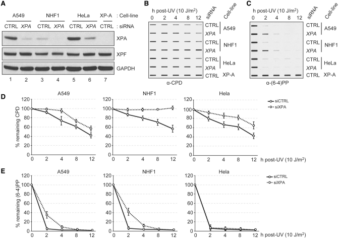
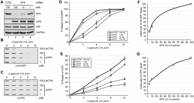
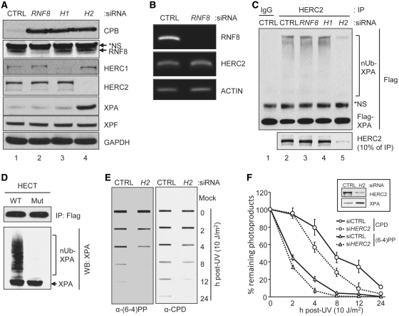
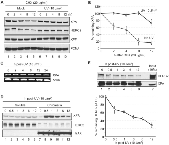
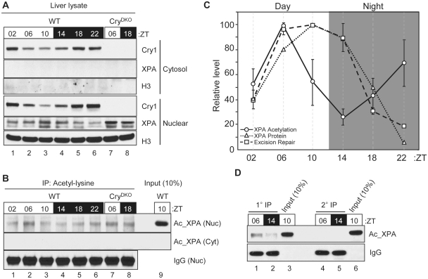
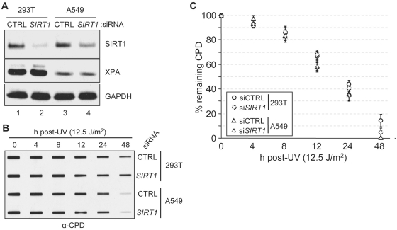
The XPA (Xeroderma pigmentosum A) protein is one of the six core factors of the human nucleotide excision repair system. In this study we show that XPA is a rate-limiting factor in all human cell lines tested, including a normal human fibroblast cell line. The level of XPA is controlled at the transcriptional level by the molecular circadian clock and at the post-translational level by a HECT domain family E3 ubiquitin ligase called HERC2. Stabilization of XPA by downregulation of HERC2 moderately enhances excision repair activity. Conversely, downregulation of XPA by siRNA reduces excision repair activity in proportion to the level of XPA. Ubiquitination and proteolysis of XPA are inhibited by DNA damage that promotes tight association of the protein with chromatin and its dissociation from the HERC2 E3 ligase. Finally, in agreement with a recent report, we find that XPA is post-translationally modified by acetylation. However, contrary to the previous claim, we find that in mouse liver only a small fraction of XPA is acetylated and that downregulation of SIRT1 deacetylase in two human cell lines does not affect the overall repair rate. Collectively, the data reveal that XPA is a limiting factor in excision repair and that its level is coordinately regulated by the circadian clock, the ubiquitin-proteasome system and DNA damage.
Overview
The claims and statistics of this paper haven’t been checked yet.
Run the check — free, no account needed. It reads the open-access text and checks the sources it cites.
Integrity signals
Registry
Cites
Statistics
Figures
Verification
Claims the paper makes
The claims and statistics of this paper haven’t been checked yet.
Run the check — free, no account needed. It reads the open-access text and checks the sources it cites.
Claims other papers make about this one
Each is checked against this paper’s own text: does it say what the citing paper says it does?
In the citing paper
“Because of XPA’s crucial functions in NER, the activity of NER and sensitization of cancer cells to chemotherapy can be regulated by transcriptional and post-transcriptional control of the XPA protein [19], [20], [21], [22], [23], [24], [25], [26].”
From UV-Induced Nuclear Import of XPA Is Mediated by Importin-α4 in An ATR-Dependent Manner (2013) · cites this paper as [21]
In the citing paper
“XPA may serve as a potential target for sensitization to cancer chemotherapy (e.g. cisplatin) via manipulation of available nuclear XPA, either at the transcriptional or post-transcriptional level [53], [54], [55].”
From UV-Induced Nuclear Import of XPA Is Mediated by Importin-α4 in An ATR-Dependent Manner (2013) · cites this paper as [55]
In the citing paper
“Xeroderma pigmentosum complement group A (XPA), a gene frequently mutated in XP patients, is part of the core incision complex of NER and interacts with DNA as well as many other NER and damage response proteins”
From RETRACTED: PKA-Mediated Phosphorylation of ATR Promotes Recruitment of XPA to UV-Induced DNA Damage (2014) · cites this paper as Kang et al., 2011
Self-check
Percentages, sums and reported differences re-computed from the paper’s own numbers.
The self-check needs the paper’s full text.
Citations
Cites
Molecular Mechanisms of Mammalian DNA Repair and the DNA Damage Checkpoints
Sancar, A., Lindsey-Boltz, L. A., Ünsal-Kaçmaz, K., & Linn, S. (2004) · Annual Review of Biochemistry · doi:10.1146/annurev.biochem.73.011303.073723 · Cited by 3,317
Self-citationNakahata, Y., Kaluzova, M., Grimaldi, B., Sahar, S., Hirayama, J., Chen, D., Guarente, L. P., & Sassone-Corsi, P. (2008) · Cell · doi:10.1016/j.cell.2008.07.002 · Cited by 1,427
SIRT1 regulates circadian clock gene expression through PER2 deacetylation
Asher, G., Gatfield, D., Stratmann, M., Reinke, H., Dibner, C., Kreppel, F., Mostoslavsky, R., Alt, F. W., & Schibler, U. (2008) · Cell · doi:10.1016/j.cell.2008.06.050 · Cited by 1,376
Circadian Control of the NAD + Salvage Pathway by CLOCK-SIRT1
Nakahata, Y., Sahar, S., Astarita, G., Kaluzova, M., & Sassone-Corsi, P. (2009) · Science · doi:10.1126/science.1170803 · Cited by 1,198
Sancar, A. (1996) · Annual Review of Biochemistry · doi:10.1146/annurev.bi.65.070196.000355 · Cited by 1,131
Self-citationCircadian Clock Feedback Cycle Through NAMPT-Mediated NAD + Biosynthesis
Ramsey, K. M., Yoshino, J., Brace, C. S., Abrassart, D., Kobayashi, Y., Marcheva, B., Hong, H.-K., Chong, J. L., Buhr, E. D., Lee, C., et al. (2009) · Science · doi:10.1126/science.1171641 · Cited by 1,126
Physiological functions of the HECT family of ubiquitin ligases
Rotin, D., & Kumar, S. (2009) · Nature Reviews Molecular Cell Biology · doi:10.1038/nrm2690 · Cited by 1,105
Transcription-coupled DNA repair: two decades of progress and surprises
Hanawalt, P. C., & Spivak, G. (2008) · Nature Reviews Molecular Cell Biology · doi:10.1038/nrm2549 · Cited by 1,081
Differential regulation of mammalian Period genes and circadian rhythmicity by cryptochromes 1 and 2
Vitaterna, M. H., Selby, C. P., Todo, T., Niwa, H., Thompson, C., Fruechte, E. M., Hitomi, K., Thresher, R. J., Ishikawa, T., Miyazaki, J., et al. (1999) · Proceedings of the National Academy of Sciences · doi:10.1073/pnas.96.21.12114 · Cited by 690
Self-citationCircadian Rhythms: Mechanisms and Therapeutic Implications
Levi, F., & Schibler, U. (2007) · Annual Review of Pharmacology and Toxicology · doi:10.1146/annurev.pharmtox.47.120505.105208 · Cited by 645
Research works printed but not linked: 2
- Boyer, 1991, Cancer Research, 51, 2960
- Cleaver, 1995, Cancer Research, 55, 6152
Cited by
Sorted by citations
Nucleotide Excision Repair in EukaryotesPreprint
Schärer, O. D. (2013) · Cold Spring Harbor Perspectives in Biology · doi:10.1101/cshperspect.a012609 · Cited by 863
Mukherji, A., Kobiita, A., Ye, T., & Chambon, P. (2013) · Cell · doi:10.1016/j.cell.2013.04.020 · Cited by 560
Nucleotide excision repair in humans
Spivak, G. (2015) · DNA Repair · doi:10.1016/j.dnarep.2015.09.003 · Cited by 354
Mammalian HECT ubiquitin-protein ligases: Biological and pathophysiological aspects
Scheffner, M., & Kumar, S. (2013) · Biochimica et Biophysica Acta (BBA) - Molecular Cell Research · doi:10.1016/j.bbamcr.2013.03.024 · Cited by 290
Mechanisms of DNA Repair by Photolyase and Excision Nuclease (Nobel Lecture)
Sancar, A. (2016) · Angewandte Chemie International Edition · doi:10.1002/anie.201601524 · Cited by 283
Self-citationCancer and the Circadian Clock
Shafi, A. A., & Knudsen, K. E. (2019) · Cancer Research · doi:10.1158/0008-5472.can-19-0566 · Cited by 264
E3 ubiquitin ligases in cancer and implications for therapies
Wang, D., Ma, L., Wang, B., Liu, J., & Wei, W. (2017) · Cancer and Metastasis Reviews · doi:10.1007/s10555-017-9703-z · Cited by 174
Circadian Clock, Cancer, and Chemotherapy
Sancar, A., Lindsey-Boltz, L. A., Gaddameedhi, S., Selby, C. P., Ye, R., Chiou, Y.-Y., Kemp, M. G., Hu, J., Lee, J. H., & Ozturk, N. (2015) · Biochemistry · doi:10.1021/bi5007354 · Cited by 170
Self-citationThe impact of environmental metals in young urbanites’ brains
Calderón‐Garcidueñas, L., Serrano-Sierra, A., Torres‐Jardón, R., Hongtu Zhu, B., Yuan, Y., Smith, D. A., Delgado‐Chávez, R., Cross, J. V., Medina‐Cortina, H., Kavanaugh, M., et al. (2012) · Experimental and Toxicologic Pathology · doi:10.1016/j.etp.2012.02.006 · Cited by 159
Roles of UVA radiation and DNA damage responses in melanoma pathogenesis
Khan, A. Q., Travers, J. B., & Kemp, M. G. (2018) · Environmental and Molecular Mutagenesis · doi:10.1002/em.22176 · Cited by 152
Li, H., Li, J., Chen, L., Qi, S., Yu, S., Weng, Z., Hu, Z., Zhou, Q., Xin, Z., Shi, L., et al. (2019) · Clinical Cancer Research · doi:10.1158/1078-0432.ccr-18-3791 · Cited by 126
CREBBP and p300 lysine acetyl transferases in the DNA damage response
Dutto, I., Scalera, C., & Prosperi, E. (2017) · Cellular and Molecular Life Sciences · doi:10.1007/s00018-017-2717-4 · Cited by 118
Functional and pathological relevance of HERC family proteins: a decade later
Sánchez‐Tena, S., Cubillos‐Rojas, M., Schneider, T., & Rosa, J. L. (2016) · Cellular and Molecular Life Sciences · doi:10.1007/s00018-016-2139-8 · Cited by 118
Disruption of the circadian clock drives Apc loss of heterozygosity to accelerate colorectal cancer
Chun, S. K., Fortin, B. M., Fellows, R. C., Habowski, A. N., Verlande, A., Song, W. A., Mahieu, A. L., Lefebvre, A. E. Y. T., Sterrenberg, J. N., Vélez, L. M., et al. (2022) · Science Advances · doi:10.1126/sciadv.abo2389 · Cited by 112
Dakup, P., Porter, K. I., Little, A., Gajula, R. P., Zhang, H., Skornyakov, E., Kemp, M. G., Dongen, H. P. A. V., & Gaddameedhi, S. (2018) · Oncotarget · doi:10.18632/oncotarget.24539 · Cited by 101
RETRACTED: PKA-Mediated Phosphorylation of ATR Promotes Recruitment of XPA to UV-Induced DNA Damage
Jarrett, S. G., Horrell, E. M. W., Christian, P. A., Vanover, J. C., Boulanger, M. C., Zou, Y., & D’Orazio, J. A. (2014) · Molecular Cell · doi:10.1016/j.molcel.2014.05.030 · Cited by 85
RetractedModulation of ATR-mediated DNA damage checkpoint response by cryptochrome 1
Kang, T., & Leem, S.-H. (2014) · Nucleic Acids Research · doi:10.1093/nar/gku094 · Cited by 91
Self-citationHERC2-USP20 axis regulates DNA damage checkpoint through Claspin
Yuan, J., Luo, K., Deng, M., Li, Y., Yin, P., Gao, B., Fang, Y., Wu, P., Liu, T., & Lou, Z. (2014) · Nucleic Acids Research · doi:10.1093/nar/gku1034 · Cited by 78
The Circadian Clock Controls Sunburn Apoptosis and Erythema in Mouse Skin
Gaddameedhi, S., Selby, C. P., Kemp, M. G., Ye, R., & Sancar, A. (2014) · Journal of Investigative Dermatology · doi:10.1038/jid.2014.508 · Cited by 81
Self-citationAberrations in DNA repair pathways in cancer and therapeutic significances
Motegi, A., Masutani, M., Yoshioka, K., & Bessho, T. (2019) · Seminars in Cancer Biology · doi:10.1016/j.semcancer.2019.02.005 · Cited by 76
Temozolomide chronotherapy in patients with glioblastoma: a retrospective single-institute study
Damato, A. R., Luo, J. R., Katumba, R. G. N., Talcott, G. R., Rubin, J. B., Herzog, E. D., & Campian, J. L. (2021) · Neuro-Oncology Advances · doi:10.1093/noajnl/vdab041 · Cited by 77
Skin, Reactive Oxygen Species, and Circadian Clocks
Ndiaye, M. A., Nihal, M., Wood, G. S., & Ahmad, N. (2013) · Antioxidants and Redox Signaling · doi:10.1089/ars.2013.5645 · Cited by 72
Gilljam, K. M., Müller, R., Liabakk, N. B., & Otterlei, M. (2012) · PLoS ONE · doi:10.1371/journal.pone.0049199 · Cited by 69
King, B. S., Cooper, K. L., Liu, K. J., & Hudson, L. G. (2012) · Journal of Biological Chemistry · doi:10.1074/jbc.m112.393504 · Cited by 68
XPA: DNA Repair Protein of Significant Clinical Importance
Pulzová, L., Ward, T. A., & Chovanec, M. (2020) · International Journal of Molecular Sciences · doi:10.3390/ijms21062182 · Cited by 70
Roles of Farnesyl-Diphosphate Farnesyltransferase 1 in Tumour and Tumour Microenvironments
Hà, N. T. T., & Lee, C. H. (2020) · Cells · doi:10.3390/cells9112352 · Cited by 73
Sirtuin-dependent clock control
Masri, S. (2015) · Current Opinion in Clinical Nutrition & Metabolic Care · doi:10.1097/mco.0000000000000219 · Cited by 63
Circadian clock disruption improves the efficacy of chemotherapy through p73-mediated apoptosis
Lee, J. H., & Sancar, A. (2011) · Proceedings of the National Academy of Sciences · doi:10.1073/pnas.1106284108 · Cited by 65
Self-citationCoordinated regulation of XPA stability by ATR and HERC2 during nucleotide excision repair
Lee, T.-H., Park, J.-M., Leem, S.-H., & Kang, T.-H. (2014) · Oncogene · doi:10.1038/onc.2012.539 · Cited by 61
Self-citationTsutakawa, S. E., Tsai, C.-L., Yan, C., Bralić, A., Chazin, W., Hamdan, S. M., Schärer, O. D., Ivanov, I. N., & Tainer, J. A. (2020) · DNA repair · doi:10.1016/j.dnarep.2020.102972 · Cited by 59
Proteomic Analysis and Identification of Cellular Interactors of the Giant Ubiquitin Ligase HERC2
Galligan, J. T., Martínez-Noël, G., Arndt, V., Hayes, S., Chittenden, T. W., Harper, J. W., & Howley, P. M. (2014) · Journal of Proteome Research · doi:10.1021/pr501005v · Cited by 62
UV-Induced RPA1 Acetylation Promotes Nucleotide Excision Repair
He, H., Wang, J., & Liu, T. (2017) · Cell Reports · doi:10.1016/j.celrep.2017.08.016 · Cited by 57
Jarrett, S. G., Wolf Horrell, E. M., Boulanger, M. C., & D'Orazio, J. A. (2015) · Journal of Investigative Dermatology · doi:10.1038/jid.2015.280 · Cited by 54
Feltes, B. C., & Bonatto, D. (2014) · Mutation Research/Reviews in Mutation Research · doi:10.1016/j.mrrev.2014.12.002 · Cited by 53
Interstrand Crosslink Repair as a Target for HDAC Inhibition
Nikolova, T., Kiweler, N., & Krämer, O. H. (2017) · Trends in Pharmacological Sciences · doi:10.1016/j.tips.2017.05.009 · Cited by 52
Circadian rhythms and cancer: implications for timing in therapy
El‐Tanani, M., Rabbani, S. A., Ali, A. A., Alfaouri, I. G. A., Nsairat, H., Al-Ani, I. H., Aljabali, A. A. A., Rizzo, M., Patoulias, D., Khan, M. A., et al. (2024) · Discover Oncology · doi:10.1007/s12672-024-01643-4 · Cited by 53
The aging clock and circadian control of metabolism and genome stability
Belancio, V. P., Blask, D. E., Deininger, P. L., Hill, S. M., & Jazwinski, S. M. (2015) · Frontiers in Genetics · doi:10.3389/fgene.2014.00455 · Cited by 47
HERC2 Interacts with Claspin and Regulates DNA Origin Firing and Replication Fork Progression
Izawa, N., Wu, W., Sato, K., Nishikawa, H., Kato, A., Boku, N., Itoh, F., & Ohta, T. (2011) · Cancer Research · doi:10.1158/0008-5472.can-11-0385 · Cited by 45
Sarkar, S., & Gaddameedhi, S. (2020) · Environmental and Molecular Mutagenesis · doi:10.1002/em.22370 · Cited by 48
Park, J.-M., & Kang, T.-H. (2016) · International Journal of Molecular Sciences · doi:10.3390/ijms17111840 · Cited by 44
Self-citationp300/CBP acetyl transferases interact with and acetylate the nucleotide excision repair factor XPG
Tillhon, M., Cazzalini, O., Nardò, T., Necchi, D., Sommatis, S., Stivala, L. A., Scovassi, A. I., & Prosperi, E. (2012) · DNA repair · doi:10.1016/j.dnarep.2012.08.001 · Cited by 43
HERCing: Structural and Functional Relevance of the Large HERC Ubiquitin Ligases
García‐Cano, J., Martinez-Martinez, A., Sala-Gaston, J., Pedrazza, L., & Rosa, J. L. (2019) · Frontiers in Physiology · doi:10.3389/fphys.2019.01014 · Cited by 43
Complete loss of function of the ubiquitin ligase HERC2 causes a severe neurodevelopmental phenotype
Morice‐Picard, F., Bénard, G., Rezvani, H. R., Lasseaux, E., Simon, D., Moutton, S., Rooryck, C., Lacombe, D., Baumann, C., & Arveiler, B. (2016) · European Journal of Human Genetics · doi:10.1038/ejhg.2016.139 · Cited by 41
Lehmann, R., Childs, L. H., Thomas, P. E., Abreu, M., Fuhr, L., Herzel, H., Leser, U., & Relógio, A. (2015) · PLoS ONE · doi:10.1371/journal.pone.0126283 · Cited by 43
Abreu, M., Basti, A., Генов, Н., Mazzoccoli, G., & Relógio, A. (2018) · Scientific Reports · doi:10.1038/s41598-018-29847-z · Cited by 41
Li, Z., Musich, P. R., Serrano, M. A., Dong, Z., & Zou, Y. (2011) · PLoS ONE · doi:10.1371/journal.pone.0028326 · Cited by 38
XPA, XPC, and XPD Modulate Sensitivity in Gastric Cisplatin Resistance Cancer Cells
Pajuelo‐Lozano, N., Bargiela-Iparraguirre, J., Domínguez, G., Quiroga, A. G., Perona, R., & Sánchez‐Pérez, I. (2018) · Frontiers in Pharmacology · doi:10.3389/fphar.2018.01197 · Cited by 34
AHR-mediated DNA damage contributes to BaP-induced cardiac malformations in zebrafish
Zou, H., Zhang, M., Chen, J., Aniagu, S. O., Jiang, Y., & Chen, T. (2023) · The Science of The Total Environment · doi:10.1016/j.scitotenv.2023.167636 · Cited by 33
Circadian clock protein BMAL1 regulates melanogenesis through MITF in melanoma cells
Sarkar, S., Porter, K. I., Dakup, P., Gajula, R. P., Koritala, B. S. C., Hylton, R. K., Kemp, M. G., Wakamatsu, K., & Gaddameedhi, S. (2021) · Pigment Cell & Melanoma Research · doi:10.1111/pcmr.12998 · Cited by 31
Ubiquitin at work: The ubiquitous regulation of the damage recognition step of NER
van Cuijk, L., Vermeulen, W., & Marteijn, J. A. F. (2014) · Experimental Cell Research · doi:10.1016/j.yexcr.2014.07.018 · Cited by 32
Holcomb, N., Goswami, M., Han, S. G., Clark, S., Orren, D. K., Gairola, C. G., & Mellon, I. (2016) · PLoS ONE · doi:10.1371/journal.pone.0158858 · Cited by 32
Jarrett, S. G., Carter, K. M., Bautista, R., He, D., Wang, C., & D'Orazio, J. A. (2018) · Journal of Biological Chemistry · doi:10.1074/jbc.ra118.003940 · Cited by 28
RetractedZhuo, M., Gorgun, M. F., & Englander, E. W. (2017) · Molecular Neurobiology · doi:10.1007/s12035-017-0507-5 · Cited by 30
A Rapid Assay for Measuring Nucleotide Excision Repair by Oligonucleotide Retrieval
Shen, J.-C., Fox, E., Ahn, E. H., & Loeb, L. A. (2014) · Scientific Reports · doi:10.1038/srep04894 · Cited by 27
Nucleotide excision repair efficiency in quiescent human fibroblasts is modulated by circadian clock
Bee, L., Marini, S., Pontarin, G., Ferraro, P., Costa, R., Albrecht, U., & Celotti, L. (2015) · Nucleic Acids Research · doi:10.1093/nar/gkv081 · Cited by 27
Kobaisi, F., Fayyad, N., Rezvani, H. R., Fayyad‐Kazan, M., Sulpice, E., Badran, B., Fayyad‐Kazan, H., Gidrol, X., & Rachidi, W. (2019) · Oxidative Medicine and Cellular Longevity · doi:10.1155/2019/4654206 · Cited by 26
UV-Induced Nuclear Import of XPA Is Mediated by Importin-α4 in An ATR-Dependent Manner
Li, Z., Musich, P. R., Cartwright, B. M., Wang, H., & Zou, Y. (2013) · PLoS ONE · doi:10.1371/journal.pone.0068297 · Cited by 26
“[53,54,55].”
Espina, M., Corte‐Rodríguez, M., Aguado, L., Montes‐Bayón, M., Sierra, M., Martínez‐Camblor, P., Blanco‐González, E., & Sierra, L. M. (2017) · Metallomics · doi:10.1039/c7mt00014f · Cited by 26
Yu, S., Tang, S., Mayer, G. D., Cobb, G. P., & Maul, J. D. (2014) · Aquatic Toxicology · doi:10.1016/j.aquatox.2014.12.004 · Cited by 26
Choi, J. Y., Park, J., Yi, J. M., Leem, S., & Kang, T. (2015) · Oncotarget · doi:10.18632/oncotarget.4610 · Cited by 25
Self-citationCircadian Rhythm of NER and ATR Pathways
Kang, T. (2021) · Biomolecules · doi:10.3390/biom11050715 · Cited by 24
Self-citationYeom, M., Lee, H., Shin, S., Park, D., & Jung, E. (2018) · Molecules · doi:10.3390/molecules23040745 · Cited by 24
Retracted: AKAP12 mediates PKA-induced phosphorylation of ATR to enhance nucleotide excision repair
Jarrett, S. G., Wolf Horrell, E. M., & D'Orazio, J. A. (2016) · Nucleic Acids Research · doi:10.1093/nar/gkw871 · Cited by 23
RetractedOestergaard, V. H., Pentzold, C., Pedersen, R. T., Iosif, S., Alpi, A. F., Bekker‐Jensen, S., Mailand, N., & Lisby, M. (2012) · DNA repair · doi:10.1016/j.dnarep.2012.08.005 · Cited by 23
Mohiuddin, M., Kobayashi, S., Keka, I. S., Guilbaud, G., Sale, J. E., Narita, T., Abdel-Aziz, H. I., Wang, X., Ogawa, S., Sasanuma, H., et al. (2016) · DNA repair · doi:10.1016/j.dnarep.2016.02.002 · Cited by 21
Transcriptional consequences of XPA disruption in human cell lines
Manandhar, M., Lowery, M. G., Boulware, K. S., Lin, K., Lu, Y., & Wood, R. D. (2017) · DNA repair · doi:10.1016/j.dnarep.2017.06.028 · Cited by 24
Chang, Y., Lee, W.-Y., Lin, Y.-J., & Hsu, T. (2017) · Aquatic Toxicology · doi:10.1016/j.aquatox.2017.09.001 · Cited by 18
Cellular and Molecular Mechanisms of Oxidative DNA Damage and Repair
Ayna, A., Çağlayan, C., & Tayşi, S. (2025) · Medicina · doi:10.3390/medicina61112013 · Cited by 24
Epigenetic Regulation of Nucleotide Excision Repair
Li, W., Jones, K., Burke, T. J., Hossain, M. A., & Lariscy, L. (2022) · Frontiers in Cell and Developmental Biology · doi:10.3389/fcell.2022.847051 · Cited by 17
Manganaro, S., Loddenkemper, T., & Rotenberg, A. (2017) · Journal of Central Nervous System Disease · doi:10.1177/1179573516685883 · Cited by 16
Barve, A., Ghaskadbi, S., & Ghaskadbi, S. (2013) · BioMed Research International · doi:10.1155/2013/854745 · Cited by 15
Anabtawi, N., Cvammen, W., & Kemp, M. G. (2021) · Scientific Reports · doi:10.1038/s41598-021-97603-x · Cited by 20
Hirobe, T. (2024) · International Journal of Molecular Sciences · doi:10.3390/ijms25084560 · Cited by 15
Nucleotide Excision Repair and Response and Survival to Chemotherapy in Colorectal Cancer Patients
Kap, E. J., Popanda, O., & Chang-Claude, J. C. (2016) · Pharmacogenomics · doi:10.2217/pgs-2015-0017 · Cited by 14
Jarrett, S. G., Carter, K. M., Shelton, B. J., & D’Orazio, J. A. (2017) · Scientific Reports · doi:10.1038/s41598-017-12056-5 · Cited by 13
RetractedProfile of Tomas Lindahl, Paul Modrich, and Aziz Sancar, 2015 Nobel Laureates in Chemistry
Cleaver, J. E. (2015) · Proceedings of the National Academy of Sciences · doi:10.1073/pnas.1521829112 · Cited by 13
The XPA Protein—Life under Precise Control
Krasikova, Y. S., Lavrik, O. I., & Rechkunova, N. I. (2022) · Cells · doi:10.3390/cells11233723 · Cited by 12
Guthrie, O. W., & Xu, H. Z. (2012) · Hearing Research · doi:10.1016/j.heares.2012.09.001 · Cited by 12
Guthrie, O. W. (2012) · International Journal of Neuroscience · doi:10.3109/00207454.2012.721828 · Cited by 12
DNA Repair Repertoire of the Enigmatic Hydra
Barve, A., Galande, A. A., Ghaskadbi, S., & Ghaskadbi, S. (2021) · Frontiers in Genetics · doi:10.3389/fgene.2021.670695 · Cited by 11
Cross Talk between the Circadian Clock Proteins and TP53 in Cancer and Therapeutic Significance
Valafar, B., Zaravinos, A., & Bonavida, B. (2021) · Critical Reviews™ in Oncogenesis · doi:10.1615/critrevoncog.2022042860 · Cited by 11
Horrell, E. M. W., Jarrett, S. G., Carter, K. M., & D'Orazio, J. A. (2017) · Experimental Dermatology · doi:10.1111/exd.13291 · Cited by 11
Mechanismen der DNA‐Reparatur durch Photolyasen und Exzisionsnukleasen (Nobel‐Aufsatz)
Sancar, A. (2016) · Angewandte Chemie · doi:10.1002/ange.201601524 · Cited by 10
Self-citationEffects of in vivo treatment of mice with sulforaphane on repair of DNA pyridyloxylbutylation
Harris, C. M., Zamperoni, K. E., Sernoskie, S. C., Chow, N., & Massey, T. E. (2021) · Toxicology · doi:10.1016/j.tox.2021.152753 · Cited by 10
Rhythms of risk: the intersection of clocks, cancer, and chronotherapy
Mello, R. M., Masri, S., & Lamia, K. A. (2026) · Journal of Clinical Investigation · doi:10.1172/jci198780 · Cited by 10
Nishinaga, M., Kurata, R., ONISHI, K., Kuriyama, K., Wakasugi, M., & Matsunaga, T. (2012) · Photochemistry and Photobiology · doi:10.1111/j.1751-1097.2012.01073.x · Cited by 8
Lawania, S., Singh, N., Behera, D., & Sharma, S. (2018) · Biochemical Genetics · doi:10.1007/s10528-018-9850-0 · Cited by 8
Cvammen, W., & Kemp, M. G. (2022) · Photochemistry and Photobiology · doi:10.1111/php.13625 · Cited by 6
The REV‐ERB antagonist SR8278 modulates keratinocyte viability in response to UVA and UVB radiation
Cvammen, W., & Kemp, M. G. (2024) · Photochemistry and Photobiology · doi:10.1111/php.13930 · Cited by 6
Guthrie, O. W. (2015) · Acta Histochemica · doi:10.1016/j.acthis.2015.10.001 · Cited by 7
Park, J., & Kang, T. (2015) · BIO-PROTOCOL · doi:10.21769/bioprotoc.1453 · Cited by 6
Self-citationShen, Y., Hsu, T., Ling, L.-B., You, W.-C., & Liu, C. (2017) · Fish Physiology and Biochemistry · doi:10.1007/s10695-016-0321-4 · Cited by 7
Pradhan, S., Sarma, H., & Mattaparthi, V. S. K. (2018) · Journal of Biomolecular Structure and Dynamics · doi:10.1080/07391102.2018.1517051 · Cited by 6
Circadian control of cisplatin-DNA adduct repair and apoptosis in culture cells
Jeon, J., Lee, S.-G., Park, J., Lee, T.-H., & Kang, T. (2023) · The International Journal of Biochemistry & Cell Biology · doi:10.1016/j.biocel.2023.106454 · Cited by 4
Self-citationA molecular assembly that crawls on a solid substrate with a metabolic-like process
Nakada, M., Fujikami, Y., Kawaguchi, M., Yamamoto, D., & Shioi, A. (2016) · Molecular Systems Design & Engineering · doi:10.1039/c5me00012b · Cited by 4
Epidermal Pigmentation, Nucleotide Excision Repair and Risk of Skin Cancer
(2012) · Journal of Carcinogenesis & Mutagenesis · doi:10.4172/2157-2518.s4-001 · Cited by 4
Pradhan, S., Das, P., & Mattaparthi, V. S. K. (2018) · ACS Omega · doi:10.1021/acsomega.8b01793 · Cited by 4
The role of HECT-type E3 ubiquitin ligases in DNA damage response and repair
Giovannini, S., Fiorilli, C., Moriconi, V., Shi, Y., Candi, E., Melino, G., & Bernassola, F. (2025) · Cell Death Discovery · doi:10.1038/s41420-025-02911-0 · Cited by 3
Houten, B. V. (2016) · DNA repair · doi:10.1016/j.dnarep.2015.12.002 · Cited by 3
Metadata
- Authors
- Kang, T.-H., Reardon, J. T., & Sancar, A.
- Affiliations
- University of North Carolina at Chapel Hill (US)
- Corresponding author
- Sancar, A.
- Venue
- Nucleic Acids Research
- Publisher
- Oxford University Press (OUP)
- Published
- 2010-12-30
- Volume
- 39
- Issue
- 8
- Pages
- 3176-3187
- ISSN
- 0305-1048 (ISSN-L)
- 1362-4962
- PMC
- PMC3082913
- OpenAlex
- W2116594363
- Cited by
- 124
- References
- 45
- Citations by year
- 2026: 4 · 2025: 4 · 2024: 5 · 2023: 2 · 2022: 4 · 2021: 15 · 2020: 6 · 2019: 5 · 2018: 9 · 2017: 13 · 2016: 11 · 2015: 11 · 2014: 12 · 2013: 7 · 2012: 11
- FWCI
- 2.99
- Type
- journal article
- Language
- en
- Open access
- gold
- doi.org
- Licence
- Locations
- Nucleic Acids Research (publishedVersion, cc-by-nc)
- Europe PMC (PubMed Central) (publishedVersion, cc-by-nc)
- PubMed Central (publishedVersion, cc-by-nc)
- PubMed (publishedVersion)
- Carolina Digital Repository (University of North Carolina at Chapel Hill) (submittedVersion)
- UNC Libraries
- Funders
- National Institutes of Health
- National Institute of General Medical Sciences (R01GM032833, R01GM031082, GM310.82, R37GM031082, GM32833)
- National Institute of Environmental Health Sciences (P30ES010126)
- Topics
- Life Sciences › Biochemistry, Genetics and Molecular Biology › Molecular Biology › DNA Repair Mechanisms
- Life Sciences › Biochemistry, Genetics and Molecular Biology › Molecular Biology › Ubiquitin and proteasome pathways
- Life Sciences › Biochemistry, Genetics and Molecular Biology › Molecular Biology › Nuclear Structure and Function
- Keywords
- XPA, nucleotide excision repair, xeroderma pigmentosum, HERC2, E3 ubiquitin ligase, ubiquitination, protein stability, circadian clock, SIRT1, protein acetylation, DNA damage response, chromatin association, transcriptional regulation, post-transcriptional regulation, mouse liver
- MeSH terms
- Acetylation, Animals, Cell Line, DNA Damage, DNA Repair, Fibroblasts, Humans, Transcription, Genetic, Ubiquitin-Protein Ligases, Mice, Xeroderma Pigmentosum Group A Protein, Ubiquitination, Circadian Clocks
About these checks
Registry, retraction, Cites and statistics results are computed, not judged, with one exception: a printed reference that no index lists is read once by a model before it counts as a citation error. Claims are extracted by a model and kept only when their quote appears verbatim in the text; each cited source is then checked, with a model’s help, for whether it says what the claim relies on. Those verdicts can be wrong — read the claim’s quote and the cited source before relying on one.
Statistics and arithmetic are recomputed, not judged.
Confidence bands are shown only for fields calibrated on our annotated test set. None is calibrated yet, so no band is shown.
What counts as a claim ›How we measure confidence ›How we check this ›
Are you an author, or do you see a problem with this page? Request a review
