

The DOI matches its Crossref record; 82 of the 83 references it cites were found in a registry or an index.
Visualization of self-delivering hydrophobically modified siRNA cellular internalization
Open accessAbstract
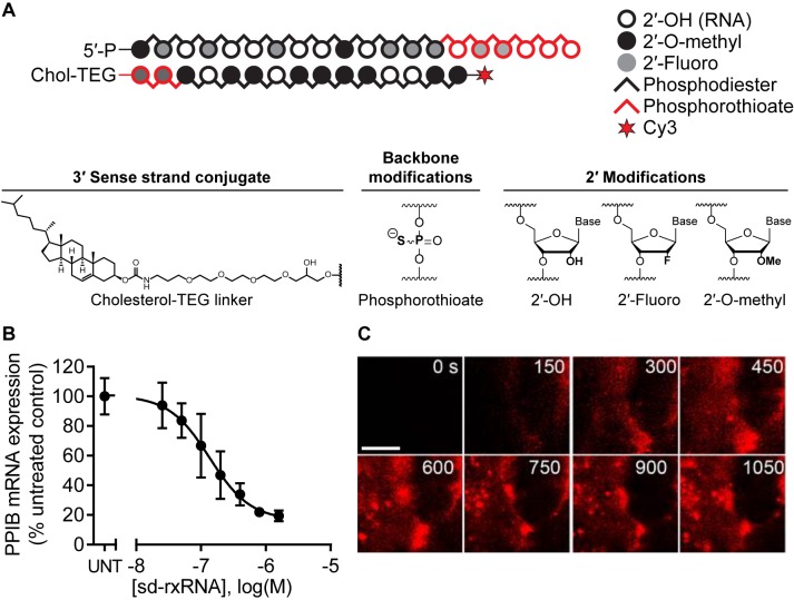
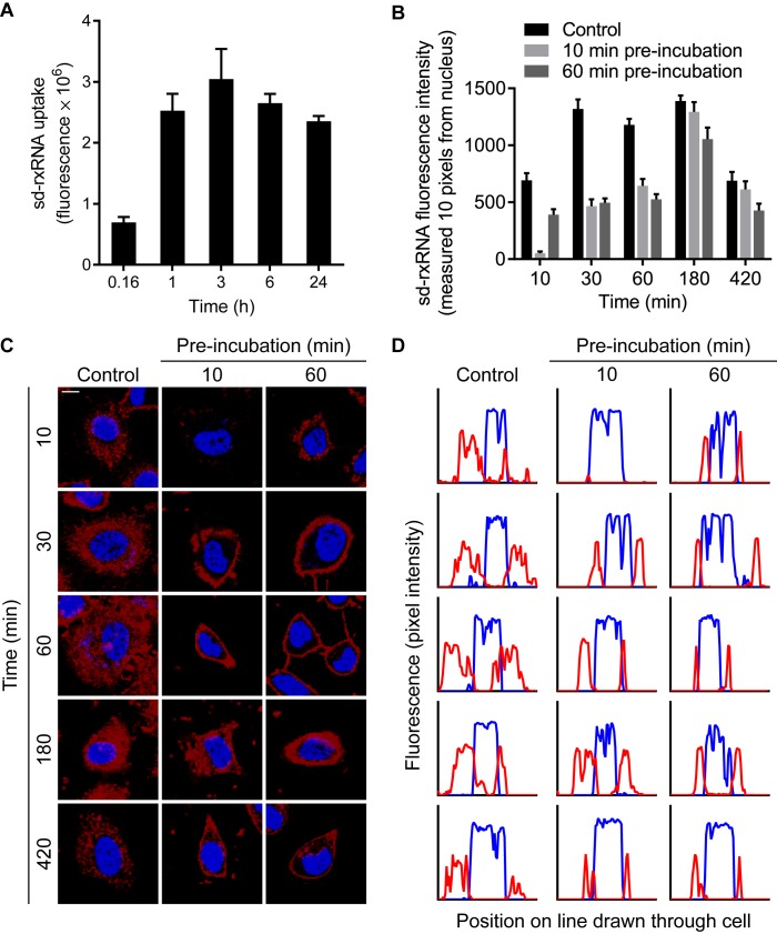
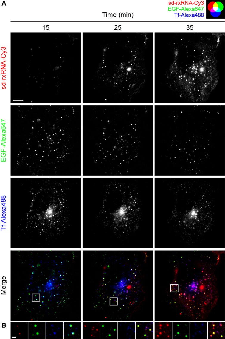
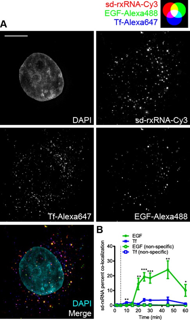
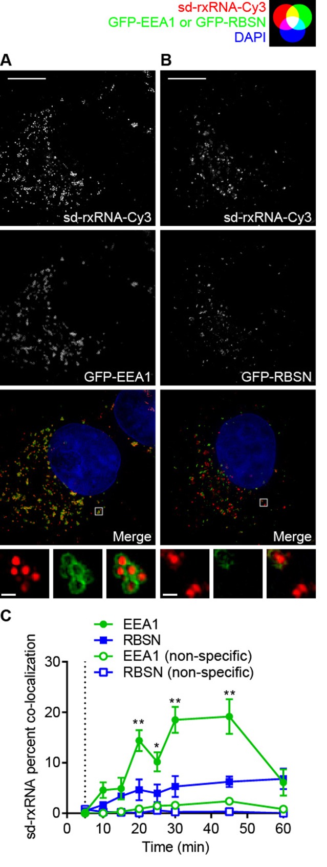
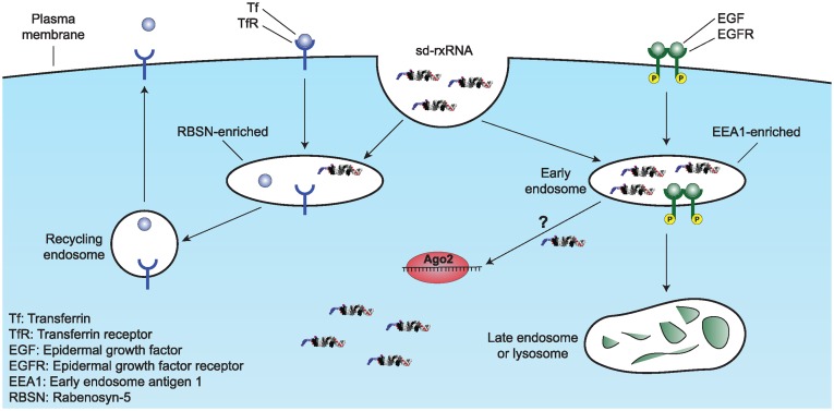
siRNAs are a new class of therapeutic modalities with promising clinical efficacy that requires modification or formulation for delivery to the tissue and cell of interest. Conjugation of siRNAs to lipophilic groups supports efficient cellular uptake by a mechanism that is not well characterized. Here we study the mechanism of internalization of asymmetric, chemically stabilized, cholesterol-modified siRNAs (sd-rxRNAs®) that efficiently enter cells and tissues without the need for formulation. We demonstrate that uptake is rapid with significant membrane association within minutes of exposure followed by the formation of vesicular structures and internalization. Furthermore, sd-rxRNAs are internalized by a specific class of early endosomes and show preferential association with epidermal growth factor (EGF) but not transferrin (Tf) trafficking pathways as shown by live cell TIRF and structured illumination microscopy (SIM). In fixed cells, we observe ∼25% of sd-rxRNA co-localizing with EGF and <5% with Tf, which is indicative of selective endosomal sorting. Likewise, preferential sd-rxRNA co-localization was demonstrated with EEA1 but not RBSN-containing endosomes, consistent with preferential EGF-like trafficking through EEA1-containing endosomes. sd-rxRNA cellular uptake is a two-step process, with rapid membrane association followed by internalization through a selective, saturable subset of the endocytic process. However, the mechanistic role of EEA1 is not yet known. This method of visualization can be used to better understand the kinetics and mechanisms of hydrophobic siRNA cellular uptake and will assist in further optimization of these types of compounds for therapeutic intervention.
Overview
The claims and statistics of this paper haven’t been checked yet.
Run the check — free, no account needed. It reads the open-access text and checks the sources it cites.
Integrity signals
Registry
Cites
Statistics
Figures
Verification
Claims the paper makes
The claims and statistics of this paper haven’t been checked yet.
Run the check — free, no account needed. It reads the open-access text and checks the sources it cites.
Claims other papers make about this one
Each is checked against this paper’s own text: does it say what the citing paper says it does?
The source backs part of the claim, not all of it — or backs it for a different population, condition or comparison.
In the citing paper
“Intracellular distribution followed a perinuclear localization pattern that is characteristic of oligonucleotides11.”
From A divalent siRNA chemical scaffold for potent and sustained modulation of gene expression throughout the central nervous system (2019) · cites this paper as [11]
Experimental — accuracy not yet measured
Self-check
Percentages, sums and reported differences re-computed from the paper’s own numbers.
The self-check needs the paper’s full text.
Citations
Cites
Huang, D. W., Sherman, B. T., & Lempicki, R. A. (2009) · Nucleic Acids Research · doi:10.1093/nar/gkn923 · Cited by 14,108
Knocking down barriers: advances in siRNA delivery
Whitehead, K. A., Langer, R., & Anderson, D. G. (2009) · Nature Reviews Drug Discovery · doi:10.1038/nrd2742 · Cited by 3,115
Therapeutic silencing of an endogenous gene by systemic administration of modified siRNAs
Soutschek, J., Akinc, A., Bramlage, B., Charissé, K., Constien, R., Donoghue, M., Elbashir, S. M., Geick, A., Hadwiger, P., Harborth, J., et al. (2004) · Nature · doi:10.1038/nature03121 · Cited by 2,206
Rational design of cationic lipids for siRNA delivery
Semple, S. C., Akinc, A., Chen, J., Sandhu, A. P., Mui, B. L., Cho, C. K., Sah, D. W. Y., Stebbing, D., Crosley, E. J., Yaworski, E., et al. (2010) · Nature Biotechnology · doi:10.1038/nbt.1602 · Cited by 1,822 · cited 1×
“Despite years of exploration, only a limited number of formulations and conjugates have progressed to the clinic: lipid nanoparticles (LNPs) (16,17), dynamic polyconjugates (18), dextrins (19), N-acetylgalactosamine (GalNAc) (8) and cholesterol (9).”
BackgroundDelivery materials for siRNA therapeutics
Kanasty, R., Dorkin, J. R., Vegas, A., & Anderson, D. (2013) · Nature Materials · doi:10.1038/nmat3765 · Cited by 1,800
Gilleron, J., Querbes, W., Zeigerer, A., Borodovsky, A., Marsico, G., Schubert, U., Manygoats, K., Seifert, S., Andree, C., Stöter, M., et al. (2013) · Nature Biotechnology · doi:10.1038/nbt.2612 · Cited by 1,681
Endosomal escape pathways for delivery of biologicals
Varkouhi, A. K., Scholte, M., Storm, G., & Haisma, H. J. (2011) · Journal of Controlled Release · doi:10.1016/j.jconrel.2010.11.004 · Cited by 1,561
A new vital stain for visualizing vacuolar membrane dynamics and endocytosis in yeast.
Vida, T. A., & Emr, S. D. (1995) · The Journal of Cell Biology · doi:10.1083/jcb.128.5.779 · Cited by 1,364
Nair, J. K., Willoughby, J. L. S., Chan, A., Charisse, K., Alam, M. R., Wang, Q., Hoekstra, M., Kandasamy, P., Kel’in, A. V., Milstein, S., et al. (2014) · Journal of the American Chemical Society · doi:10.1021/ja505986a · Cited by 1,141
Gibbings, D., Ciaudo, C., Erhardt, M., & Voinnet, O. (2009) · Nature Cell Biology · doi:10.1038/ncb1929 · Cited by 1,037
Mechanisms and optimization of in vivo delivery of lipophilic siRNAs
Wolfrum, C., Shi, S., Jayaprakash, K. N., Jayaraman, M., Wang, G., Pandey, R. K., Rajeev, K. G., Nakayama, T., Charrise, K., Ndungo, E. M., et al. (2007) · Nature Biotechnology · doi:10.1038/nbt1339 · Cited by 974
Lipid-like materials for low-dose, in vivo gene silencing
Love, K. T., Mahon, K. P., Levins, C. G., Whitehead, K. A., Querbes, W., Dorkin, J. R., Qin, J., Cantley, W., Qin, L. L., Racie, T., et al. (2010) · Proceedings of the National Academy of Sciences · doi:10.1073/pnas.0910603106 · Cited by 969
Efficiency of siRNA delivery by lipid nanoparticles is limited by endocytic recycling
Sahay, G., Querbes, W., Alabi, C., Eltoukhy, A., Sarkar, S., Zurenko, C., Karagiannis, E., Love, K., Chen, D., Zoncu, R., et al. (2013) · Nature Biotechnology · doi:10.1038/nbt.2614 · Cited by 948
Sönnichsen, B., De Renzis, S., Nielsen, E., Rietdorf, J., & Zerial, M. (2000) · The Journal of Cell Biology · doi:10.1083/jcb.149.4.901 · Cited by 919
Pharmacokinetics, biodistribution and cell uptake of antisense oligonucleotides
Geary, R. S., Norris, D. A., Yu, R. Z., & Bennett, C. F. (2015) · Advanced Drug Delivery Reviews · doi:10.1016/j.addr.2015.01.008 · Cited by 870
Knocking down disease: a progress report on siRNA therapeutics
Wittrup, A., & Lieberman, J. (2015) · Nature Reviews Genetics · doi:10.1038/nrg3978 · Cited by 827
Endocytosis and intracellular trafficking of ErbBs
Sorkin, A. D., & Goh, L. K. (2008) · Experimental Cell Research · doi:10.1016/j.yexcr.2008.07.029 · Cited by 776
Visualizing lipid-formulated siRNA release from endosomes and target gene knockdown
Wittrup, A., Ai, A., Liu, X., Hamar, P., Trifonova, R., Charisse, K., Manoharan, M., Kirchhausen, T., & Lieberman, J. (2015) · Nature Biotechnology · doi:10.1038/nbt.3298 · Cited by 693
Dynamic PolyConjugates for targeted in vivo delivery of siRNA to hepatocytes
Rozema, D. B., Lewis, D. L., Wakefield, D. H., Wong, S. C., Klein, J. J., Roesch, P. L., Bertin, S. L., Reppen, T. W., Chu, Q., Blokhin, A. V., et al. (2007) · Proceedings of the National Academy of Sciences · doi:10.1073/pnas.0703778104 · Cited by 667
The endocytic pathway mediates cell entry of dsRNA to induce RNAi silencing
Saleh, M., van Rij, R. P., Hekele, A., Gillis, A. N., Foley, E., O’Farrell, P. H., & Andino, R. (2006) · Nature Cell Biology · doi:10.1038/ncb1439 · Cited by 561
Spatial control of EGF receptor activation by reversible dimerization on living cells
Chung, I., Akita, R. W., VANDLEN, R. L., Toomre, D. K., Schlessinger, J., & Mellman, I. (2010) · Nature · doi:10.1038/nature08827 · Cited by 538
Lakadamyali, M., Rust, M. J., & Zhuang, X. (2006) · Cell · doi:10.1016/j.cell.2005.12.038 · Cited by 508
Hrs regulates multivesicular body formation via ESCRT recruitment to endosomes
Bache, K. G., Brech, A., Mehlum, A., & Stenmark, H. (2003) · The Journal of Cell Biology · doi:10.1083/jcb.200302131 · Cited by 465
Laukaitis, C. M., Webb, D. J., Donais, K., & Horwitz, A. F. (2001) · The Journal of Cell Biology · doi:10.1083/jcb.153.7.1427 · Cited by 455
Systems survey of endocytosis by multiparametric image analysis
Collinet, C., Stöter, M., Bradshaw, C. R., Samusik, N., Rink, J. C., Kenski, D. M., Habermann, B. H., Buchholz, F., Henschel, R., Mueller, M., et al. (2010) · Nature · doi:10.1038/nature08779 · Cited by 453
Chemically modified siRNA: tools and applications
Watts, J. K., Deleavey, G. F., & Damha, M. J. (2008) · Drug Discovery Today · doi:10.1016/j.drudis.2008.05.007 · Cited by 440
DiFiglia, M., Sena‐Esteves, M., Chase, K. O., Sapp, E., Pfister, E., Sass, M., Yoder, J., Reeves, P., Pandey, R. K., Rajeev, K. G., et al. (2007) · Proceedings of the National Academy of Sciences · doi:10.1073/pnas.0708285104 · Cited by 433
Hrs recruits clathrin to early endosomes
Raiborg, C., Bache, K. G., Mehlum, A., Stang, E., & Stenmark, H. (2001) · The EMBO Journal · doi:10.1093/emboj/20.17.5008 · Cited by 403
Nielsen, E., Christoforidis, S., Uttenweiler‐Joseph, S., Miączyńska, M., Dewitte, F., Wilm, M. S., Hoflack, B., & Zerial, M. (2000) · The Journal of Cell Biology · doi:10.1083/jcb.151.3.601 · Cited by 383
Patel, P. C., Giljohann, D. A., Daniel, W. L., Zheng, D., Prigodich, A. E., & Mirkin, C. A. (2010) · Bioconjugate Chemistry · doi:10.1021/bc1002423 · Cited by 366
A Phosphoinositide Switch Controls the Maturation and Signaling Properties of APPL Endosomes
Zoncu, R., Perera, R. M., Balkin, D. M., Pirruccello, M., Toomre, D. K., & Camilli, P. V. D. (2009) · Cell · doi:10.1016/j.cell.2009.01.032 · Cited by 347
Silencing by small RNAs is linked to endosomal trafficking
Lee, Y. S., Pressman, S., Andress, A. P., Kim, K., White, J. L., Cassidy, J. J., Li, X., Lubell, K., Lim, D., Cho, I. S., et al. (2009) · Nature Cell Biology · doi:10.1038/ncb1930 · Cited by 346
Daro, E., van der Sluijs, P., Galli, T., & Mellman, I. (1996) · Proceedings of the National Academy of Sciences · doi:10.1073/pnas.93.18.9559 · Cited by 343
Sehgal, A., Barros, S. A., Ivanciu, L., Cooley, B. C., Qin, J., Racie, T., Hettinger, J. L., Carioto, M., Jiang, Y., Brodsky, J., et al. (2015) · Nature Medicine · doi:10.1038/nm.3847 · Cited by 315
A status report on RNAi therapeutics
Vaishnaw, A. K., Gollob, J., Gamba‐Vitalo, C., Hutabarat, R. M., Sah, D. W. Y., Meyers, R. E., de Fougerolles, T., & Maraganore, J. M. (2010) · Silence · doi:10.1186/1758-907x-1-14 · Cited by 305
Koller, E., Vincent, T. M., Chappell, A., De, S., Manoharan, M., & Bennett, C. F. (2011) · Nucleic Acids Research · doi:10.1093/nar/gkr089 · Cited by 281
A distinct class of endosome mediates clathrin-independent endocytosis to the Golgi complex
Nichols, B. J. (2002) · Nature Cell Biology · doi:10.1038/ncb787 · Cited by 258
Distinct Roles for Tsg101 and Hrs in Multivesicular Body Formation and Inward Vesiculation
Razi, M., & Futter, C. E. (2006) · Molecular Biology of the Cell · doi:10.1091/mbc.e05-11-1054 · Cited by 255
The rough endoplasmatic reticulum is a central nucleation site of siRNA‐mediated RNA silencing
Stalder, L., Heusermann, W., Sokol, L., Trojer, D., Wirz, J., Hean, J., Fritzsche, A., Aeschimann, F., Pfanzagl, V., Basselet, P., et al. (2013) · The EMBO Journal · doi:10.1038/emboj.2013.52 · Cited by 229
Matsuda, S., Keiser, K., Nair, J. K., Charissé, K., Manoharan, R. M., Kretschmer, P., Peng, C. G., Kel’in, A. V., Pachamuthu, K., Willoughby, J. L. S., et al. (2015) · ACS Chemical Biology · doi:10.1021/cb501028c · Cited by 228
Pharmacokinetic properties of several novel oligonucleotide analogs in mice.
Crooke, S. T., Graham, M. J., Zuckerman, J. E., Brooks, D., Conklin, B., Cummins, L. L., Greig, M. J., Guinosso, C. J., Kornbrust, D. J., Manoharan, M., et al. (1996) · Journal of Pharmacology and Experimental Therapeutics · doi:10.1016/s0022-3565(25)12951-0 · Cited by 227
A Novel Mechanism Is Involved in Cationic Lipid-Mediated Functional siRNA Delivery
Lu, J. J.-Q., Langer, R., & Chen, J. (2009) · Molecular Pharmaceutics · doi:10.1021/mp900023v · Cited by 222
Hau, P., Jachimczak, P., Schlingensiepen, R., Schulmeyer, F. J., Jauch, T., Steinbrecher, A., Brawanski, A. T., Proescholdt, M. A., Schlaier, J. R., Buchroithner, J., et al. (2007) · Oligonucleotides · doi:10.1089/oli.2006.0053 · Cited by 215
Epidermal Growth Factor and Membrane Trafficking
Barbieri, M. A., Roberts, R. L., Gumusboga, A., Highfield, H., Alvarez-Dominguez, C., Wells, A., & Stahl, P. D. (2000) · The Journal of Cell Biology · doi:10.1083/jcb.151.3.539 · Cited by 204
Receptor-mediated endocytosis: The intracellular journey of transferrin and its receptor
Dautry‐Varsat, A. (1986) · Biochimie · doi:10.1016/s0300-9084(86)80004-9 · Cited by 201
KRIEG, A. M., Tonkinson, J. L., Matson, S., Zhao, Q., Saxon, M. E., Zhang, L. M., Bhanja, U., Yakubov, L. A., & Stein, C. (1993) · Proceedings of the National Academy of Sciences · doi:10.1073/pnas.90.3.1048 · Cited by 201
EGF induces macropinocytosis and SNX1-modulated recycling of E-cadherin
Bryant, D. M., Kerr, M. C., Hammond, L. A., Joseph, S. R., Mostov, K. E., Teasdale, R. D., & Stow, J. L. (2007) · Journal of Cell Science · doi:10.1242/jcs.000653 · Cited by 193
Rajeev, K. G., Nair, J. K., Jayaraman, M., Charissé, K., Taneja, N., O′Shea, J., Willoughby, J. L. S., Yucius, K., Nguyen, T. M., Shulga‐Morskaya, S., et al. (2015) · ChemBioChem · doi:10.1002/cbic.201500023 · Cited by 190
Sequential signaling in plasma-membrane domains during macropinosome formation in macrophages
Yoshida, S., Hoppe, A. D., Araki, N., & Swanson, J. A. (2009) · Journal of Cell Science · doi:10.1242/jcs.053207 · Cited by 168
Madshus, I. H., & Stang, E. (2009) · Journal of Cell Science · doi:10.1242/jcs.050260 · Cited by 167
Biochemical analysis of distinct Rab5- and Rab11-positive endosomes along the transferrin pathway
Trischler, M., Stoorvogel, W., & Ullrich, O. (1999) · Journal of Cell Science · doi:10.1242/jcs.112.24.4773 · Cited by 162
Imaging individual microRNAs in single mammalian cells in situ
Lü, J., & Tsourkas, A. (2009) · Nucleic Acids Research · doi:10.1093/nar/gkp482 · Cited by 160
Hydrophobically Modified siRNAs Silence Huntingtin mRNA in Primary Neurons and Mouse Brain
Alterman, J. F., Hall, L. M., Coles, A. H., Hassler, M. R., Didiot, M.-C., Chase, K., Abraham, J., Sottosanti, E., Johnson, E., Sapp, E., et al. (2015) · Molecular Therapy - Nucleic Acids · doi:10.1038/mtna.2015.38 · Cited by 158
Self-citationCao, T. T., Mays, R. W., & von Zastrow, M. (1998) · Journal of Biological Chemistry · doi:10.1074/jbc.273.38.24592 · Cited by 138
Grewal, P. K. (2010) · Methods in enzymology on CD-ROM/Methods in enzymology · doi:10.1016/s0076-6879(10)79013-3 · Cited by 136
Transferrin receptor recycling in the absence of perinuclear recycling endosomes
Sheff, D. R., Pelletier, L., O’Connell, C. B., Warren, G., & Mellman, I. (2002) · The Journal of Cell Biology · doi:10.1083/jcb.20111048 · Cited by 135
Eyster, C. A., Cole, N. B., Harrington, S. P., Viswanathan, K., Früh, K., & Donaldson, J. G. (2011) · Molecular Biology of the Cell · doi:10.1091/mbc.e10-11-0874 · Cited by 130
DNA/RNA heteroduplex oligonucleotide for highly efficient gene silencing
Nishina, K., Piao, W., Yoshida‐Tanaka, K., Sujino, Y., Nishina, T., Yamamoto, T., Nitta, K., Yoshioka, K., Kuwahara, H., Yasuhara, H., et al. (2015) · Nature Communications · doi:10.1038/ncomms8969 · Cited by 128
Leonard, D. M., Hayakawa, A., Lawe, D., Lambright, D., Bellve, K., Standley, C., Lifshitz, L. M., Fogarty, K., & Corvera, S. (2008) · Journal of Cell Science · doi:10.1242/jcs.031484 · Cited by 123
Self-citationChen, Y., Deng, L., Maeno-Hikichi, Y., Lai, M., Chang, S., Chen, G., & Zhang, J.-F. (2003) · Cell · doi:10.1016/s0092-8674(03)00726-8 · Cited by 122
Dynamics of Tubulovesicular Recycling Endosomes in Hippocampal Neurons
Prekeris, R., Foletti, D. L., & Scheller, R. H. (1999) · Journal of Neuroscience · doi:10.1523/jneurosci.19-23-10324.1999 · Cited by 108
Hopkins, C. R., Miller, K., & Beardmore, J. M. (1985) · Journal of Cell Science · doi:10.1242/jcs.1985.supplement_3.17 · Cited by 104
Petrova, N. S., Chernikov, I. V., Meschaninova, M. I., Dovydenko, I. S., Venyaminova, A. G., Аркадьевна, З. М., Vlassov, V. V., & Chernolovskaya, E. L. (2011) · Nucleic Acids Research · doi:10.1093/nar/gkr1002 · Cited by 96
Rabenosyn-5 defines the fate of the transferrin receptor following clathrin-mediated endocytosis
Navaroli, D. M., Bellve, K. D., Standley, C., Lifshitz, L. M., Cardia, J. P., Lambright, D. G., Leonard, D. M., Fogarty, K. E., & Corvera, S. (2012) · Proceedings of the National Academy of Sciences · doi:10.1073/pnas.1115495109 · Cited by 87
Self-citationQuantitative evaluation of siRNA delivery in vivo
Pei, Y., Hancock, P. J., Zhang, H., Bartz, R., Cherrin, C., Innocent, N., Pomerantz, C. J., Seitzer, J. L., Koser, M. L., Abrams, M. T., et al. (2010) · RNA · doi:10.1261/rna.2255810 · Cited by 82
Byrne, M., Tzekov, R. T., Wang, Y., Rodgers, A., Cardia, J. P., Ford, G., Holton, K., Pandarinathan, L., Lapierre, J., Stanney, W., et al. (2013) · Journal of Ocular Pharmacology and Therapeutics · doi:10.1089/jop.2013.0148 · Cited by 78
Self-citationPlasma Membrane Domains Specialized for Clathrin-mediated Endocytosis in Primary Cells*
Bellve, K., Leonard, D. M., Standley, C., Lifshitz, L. M., Tuft, R., Hayakawa, A., Corvera, S., & Fogarty, K. (2006) · Journal of Biological Chemistry · doi:10.1074/jbc.m511370200 · Cited by 74
Self-citationRegarding the Amazing Choreography of Clathrin Coats
Traub, L. M. (2011) · PLoS Biology · doi:10.1371/journal.pbio.1001037 · Cited by 73
Kuwahara, H., Nishina, K., Yoshida, K., Nishina, T., Yamamoto, M., Saito, Y., Piao, W., Yoshida, M., Mizusawa, H., & Yokota, T. (2011) · Molecular Therapy · doi:10.1038/mt.2011.186 · Cited by 71
Mipomersen and other therapies for the treatment of severe familial hypercholesterolemia
Burnett, J., Bell, D. A., Hooper, & Watts, G. F. (2012) · Vascular Health and Risk Management · doi:10.2147/vhrm.s28581 · Cited by 69
Cyclodextrin-Containing Polymers: Versatile Platforms of Drug Delivery Materials
Heidel, J. D., & Schluep, T. (2012) · Journal of Drug Delivery · doi:10.1155/2012/262731 · Cited by 65
Videira, M., Arranja, A., Rafael, D. F. D. S., & Gaspar, R. S. (2013) · Nanomedicine Nanotechnology Biology and Medicine · doi:10.1016/j.nano.2013.11.018 · Cited by 61
Hayakawa, A., Leonard, D. M., Murphy, S. T., Hayes, S. J., Soto, M. C., Fogarty, K., Standley, C., Bellve, K., Lambright, D., Mello, C., et al. (2006) · Proceedings of the National Academy of Sciences of the United States of America · doi:10.1073/pnas.0508832103 · Cited by 58
Self-citationGLUT4 and Transferrin Receptor Are Differentially Sorted Along the Endocytic Pathway in CHO Cells
Wei, M. L., Bonzelius, F., Scully, R., Kelly, R. B., & Herman, G. A. (1998) · The Journal of Cell Biology · doi:10.1083/jcb.140.3.565 · Cited by 58
Analysis of synaptic vesicle endocytosis in synaptosomes by high-content screening
Daniel, J. A., Malladi, C. S., Kettle, E., McCluskey, A., & Robinson, P. J. (2012) · Nature Protocols · doi:10.1038/nprot.2012.070 · Cited by 57
ARAP1 Regulates Endocytosis of EGFR
Yoon, H., Lee, J., & Randazzo, P. A. (2008) · Traffic · doi:10.1111/j.1600-0854.2008.00839.x · Cited by 52
cAMP-sensitive endocytic trafficking in A6 epithelia
Butterworth, M. B., Helman, S. I., & Els, W. J. (2001) · American Journal of Physiology-Cell Physiology · doi:10.1152/ajpcell.2001.280.4.c752 · Cited by 52
Rahajeng, J., Caplan, S., & Naslavsky, N. (2009) · Experimental Cell Research · doi:10.1016/j.yexcr.2009.11.007 · Cited by 51
Beas, A. O., Taupin, V., Teodorof‐Diedrich, C., Nguyen, L. T., Garcia‐Marcos, M., & Farquhar, M. G. (2012) · Molecular Biology of the Cell · doi:10.1091/mbc.e12-02-0133 · Cited by 48
Effect of cycloheximide on epidermal growth factor receptor trafficking and signaling
Oksvold, M. P., Pedersen, N. M., Forfang, L., & Smeland, E. B. (2012) · FEBS Letters · doi:10.1016/j.febslet.2012.08.022 · Cited by 29
Felber, A. E., Bayó‐Puxán, N., Deleavey, G. F., Castagner, B., Damha, M. J., & Leroux, J. (2012) · Biomaterials · doi:10.1016/j.biomaterials.2012.05.019 · Cited by 20
Dye selection for live cell imaging of intact siRNA
Hirsch, M., Strand, D. J., & Helm, M. (2011) · Biological Chemistry · doi:10.1515/bc-2011-256 · Cited by 13
Cited by
Sorted by citations
The current state and future directions of RNAi-based therapeutics
Setten, R. L., Rossi, J. J., & Han, S.-P. (2019) · Nature Reviews Drug Discovery · doi:10.1038/s41573-019-0017-4 · Cited by 1,443
Detection of immunogenic cell death and its relevance for cancer therapy
Fucikova, J., Kepp, O., Kasikova, L., Petroni, G., Yamazaki, T., Liu, P., Zhao, L., Spisek, R., Kroemer, G., & Galluzzi, L. (2020) · Cell Death & Disease · doi:10.1038/s41419-020-03221-2 · Cited by 1,138
Translation and functional roles of circular RNAs in human cancer
Lei, M., Zheng, G., Ning, Q., Zheng, J., & Dong, D. (2020) · Molecular Cancer · doi:10.1186/s12943-020-1135-7 · Cited by 814
Chemistry, mechanism and clinical status of antisense oligonucleotides and duplex RNAs
Shen, X., & Corey, D. R. (2017) · Nucleic Acids Research · doi:10.1093/nar/gkx1239 · Cited by 654
Emerging role of tumor-related functional peptides encoded by lncRNA and circRNA
Wu, P., Mo, Y., Miao, P., Tang, T. F., Zhong, Y. M., Deng, X., Xiong, F., Guo, C. J., Wu, X., Li, Y., et al. (2020) · Molecular Cancer · doi:10.1186/s12943-020-1147-3 · Cited by 538
Metadata
- Authors
- Ly, S., Navaroli, D. M., Didiot, M.-C., Cardia, J., Pandarinathan, L., Alterman, J. F., Fogarty, K., Standley, C., Lifshitz, L. M., Bellve, K. D., Prot, M., Echeverria, D., Corvera, S., & Khvorova, A.
- Affiliations
- University of Massachusetts Chan Medical School (US)
- Phio Pharmaceuticals (United States) (US)
- Corresponding author
- Khvorova, Anastasia
- ORCID
- Venue
- Nucleic Acids Research
- Publisher
- Oxford University Press (OUP)
- Published
- 2016-11-28
- Volume
- 45
- Issue
- 1
- Pages
- 15-25
- ISSN
- 0305-1048 (ISSN-L)
- 1362-4962
- PMC
- PMC5224471
- OpenAlex
- W2557242880
- Cited by
- 132
- References
- 83
- Citations by year
- 2026: 4 · 2025: 13 · 2024: 13 · 2023: 14 · 2022: 9 · 2021: 9 · 2020: 19 · 2019: 22 · 2018: 21 · 2017: 8
- FWCI
- 8.84
- Type
- journal article
- Language
- en
- Open access
- gold
- doi.org
- Licence
- Locations
- Nucleic Acids Research (publishedVersion, cc-by)
- PubMed Central (publishedVersion, cc-by)
- PubMed (publishedVersion)
- The Journal of the American Medical Association (JAMA) Network (American Medical Association) (submittedVersion, cc-by)
- HighWire Press Open Archive (submittedVersion)
- Funders
- University of Massachusetts (DK32520)
- University of Oxford
- National Institutes of Health (1s10od020012-01)
- National Institute of General Medical Sciences (R01GM108803, R25GM113686)
- Topics
- Life Sciences › Biochemistry, Genetics and Molecular Biology › Molecular Biology › RNA Interference and Gene Delivery
- Life Sciences › Biochemistry, Genetics and Molecular Biology › Cell Biology › Cellular transport and secretion
- Physical Sciences › Materials Science › Biomaterials › Nanoparticle-Based Drug Delivery
- Keywords
- siRNA delivery, cellular uptake, endocytosis, early endosomes, transferrin receptor, EEA1, membrane association
- MeSH terms
- Animals, Biological Transport, Chlorocebus aethiops, Cholesterol, Endocytosis, Epidermal Growth Factor, HeLa Cells, Humans, Microscopy, Fluorescence, Endosomes, Transferrin, Gene Expression, COS Cells, Cyclophilins, Vesicular Transport Proteins, RNA, Small Interfering, Hydrophobic and Hydrophilic Interactions
About these checks
Registry, retraction, Cites and statistics results are computed, not judged, with one exception: a printed reference that no index lists is read once by a model before it counts as a citation error. Claims are extracted by a model and kept only when their quote appears verbatim in the text; each cited source is then checked, with a model’s help, for whether it says what the claim relies on. Those verdicts can be wrong — read the claim’s quote and the cited source before relying on one.
Statistics and arithmetic are recomputed, not judged.
Confidence bands are shown only for fields calibrated on our annotated test set. None is calibrated yet, so no band is shown.
What counts as a claim ›How we measure confidence ›How we check this ›
Are you an author, or do you see a problem with this page? Request a review
