

Decellularized extracellular matrix-based composite scaffolds for tissue engineering and regenerative medicine
Open accessThe DOI matches its Crossref record; 223 of the 226 references it cites were found in a registry or an index.
Abstract
Despite the considerable advancements in fabricating polymeric-based scaffolds for tissue engineering, the clinical transformation of these scaffolds remained a big challenge because of the difficulty of simulating native organs/tissues’ microenvironment. As a kind of natural tissue-derived biomaterials, decellularized extracellular matrix (dECM)-based scaffolds have gained attention due to their unique biomimetic properties, providing a specific microenvironment suitable for promoting cell proliferation, migration, attachment and regulating differentiation. The medical applications of dECM-based scaffolds have addressed critical challenges, including poor mechanical strength and insufficient stability. For promoting the reconstruction of damaged tissues or organs, different types of dECM-based composite platforms have been designed to mimic tissue microenvironment, including by integrating with natural polymer or/and syntenic polymer or adding bioactive factors. In this review, we summarized the research progress of dECM-based composite scaffolds in regenerative medicine, highlighting the critical challenges and future perspectives related to the medical application of these composite materials.
Overview
The claims and statistics of this paper haven’t been checked yet.
Free: it reads the open-access text and checks the first 5 claims that cite a source we can check. Create a free account to check every claim.
Integrity signals
Registry
Cites
Statistics
Figures


Verification
Claims the paper makes
The claims and statistics of this paper haven’t been checked yet.
Free: it reads the open-access text and checks the first 5 claims that cite a source we can check. Create a free account to check every claim.
Claims other papers make about this one
Each is checked against this paper’s own text: does it say what the citing paper says it does?
The source backs part of the claim, not all of it — or backs it for a different population, condition or comparison.
In the citing paper
“Inorganic materials such as hydroxyapatite(HA) can be added to PLCL to design a scaffold with higher hardness and mechanical strength, which can improve the mechanical properties of the osteochondral repair interface [195].”
From Application of PLCL as a biodegradable polymer in biomedical engineering (2026) · cites this paper as [195]
Experimental — accuracy not yet measuredThe cited source says what the claim attributes to it.
In the citing paper
“Poor mechanical properties; rapid degradation rate.”
From Constructing biomimetic microenvironments for liver regeneration (2025) · doi:10.1186/s12951-025-03729-9 · cites this paper as [134]
In the citing paper
“Imitates the liver’s complexity and exhibits excellent biocompatibility.”
From Constructing biomimetic microenvironments for liver regeneration (2025) · doi:10.1186/s12951-025-03729-9 · cites this paper as [134]
In the citing paper
“These materials are produced through specialized physical, chemical, or biological processing methods that remove all cellular components from native tissues or organs [149].”
From Constructing biomimetic microenvironments for liver regeneration (2025) · doi:10.1186/s12951-025-03729-9 · cites this paper as [149]
In the citing paper
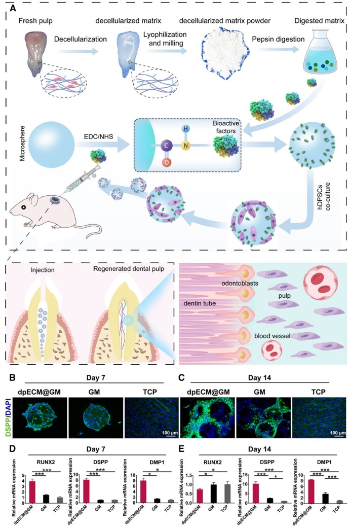
“Chen et al. further advanced the field by fabricating a bioactive dECM platform embedding dihydrolipoic acid–stabilised Pt nanoclusters doped with Cu nanozymes within cell-laden microgels [149].”
From Constructing biomimetic microenvironments for liver regeneration (2025) · doi:10.1186/s12951-025-03729-9 · cites this paper as [149]
Self-check
Percentages, sums and reported differences re-computed from the paper’s own numbers.
The self-check needs the paper’s full text.
Citations
Cites
1 retracted
Gholipourmalekabadi, M., Seifalian, A. M., Urbanska, A. M., Omrani, M. D., Hardy, J. G., Madjd, Z., Hashemi, S. M., Ghanbarian, H., Brouki Milan, P., Mozafari, M., et al. (2018) · Biomacromolecules · doi:10.1021/acs.biomac.7b01807 · Cited by 89 · cited 1×
“[143] designed electrospun nanofibrous silk fibroin that coated with human amniotic dECM membrane to seeded adipose-tissue-derived MSC, which could accelerate wound healing process regeneration and significantly reduces scars formation in burn wound.”
BackgroundRetractedMaterials design for bone-tissue engineering
Koons, G. L., Diba, M., & Mikos, A. G. (2020) · Nature Reviews Materials · doi:10.1038/s41578-020-0204-2 · Cited by 1,699
Injectable and biodegradable hydrogels: gelation, biodegradation and biomedical applications
Li, Y., Rodrigues, J., & Tomás, H. (2012) · Chem. Soc. Rev. · doi:10.1039/c1cs15203c · Cited by 1,445
Biomimetic materials for tissue engineering
Ma, P. X. (2008) · Advanced Drug Delivery Reviews · doi:10.1016/j.addr.2007.08.041 · Cited by 1,310
Alginate-based composite materials for wound dressing application:A mini review
Varaprasad, K., Jayaramudu, T., Kanikireddy, V., Toro, C., & Sadiku, E. R. (2020) · Carbohydrate Polymers · doi:10.1016/j.carbpol.2020.116025 · Cited by 691
Synthetic biodegradable functional polymers for tissue engineering: a brief review
Guo, B., & Ma, P. X. (2014) · Science China Chemistry · doi:10.1007/s11426-014-5086-y · Cited by 592
Bone tissue engineering via growth factor delivery: from scaffolds to complex matrices
De Witte, T.-M., Fratila-Apachitei, L. E., Zadpoor, A. A., & Peppas, N. A. (2018) · Regenerative Biomaterials · doi:10.1093/rb/rby013 · Cited by 513
Multifunctional GelMA platforms with nanomaterials for advanced tissue therapeutics
Kurian, A. G., Singh, R. K., Patel, K. D., Lee, J.-H., & Kim, H.-W. (2022) · Bioactive Materials · doi:10.1016/j.bioactmat.2021.06.027 · Cited by 444 · cited 1×
“GelMA maintained functional sequences of gelatin (Arg–Gly–Asp and the matrix metalloproteinase) and could form hydrogel with tunable physicochemical properties by photo-crosslinking [125].”
BackgroundExtracellular vesicle-loaded hydrogels for tissue repair and regeneration
Ju, Y., Hu, Y., Yang, P., Xie, X., & Fang, B. (2023) · Materials Today Bio · doi:10.1016/j.mtbio.2022.100522 · Cited by 440
Gelatin carriers for drug and cell delivery in tissue engineering
Santoro, M., Tatara, A. M., & Mikos, A. G. (2014) · Journal of Controlled Release · doi:10.1016/j.jconrel.2014.04.014 · Cited by 395 · cited 1×
“Gelatin is an essential protein obtained from native collagen by hydrolysis, which can be obtained by disrupting the intermolecular bonds and breaking the a -helix conformation of collagen [109, 110].”
Background
Cited by
Liu, Y., & Ge, L. (2025) · Bioengineering · doi:10.3390/bioengineering12111178 · Cited by 84
“Conductive polymers may contain residual monomers or dopants that could be inflammatory [87].”
Conductive hydrogels: intelligent dressings for monitoring and healing chronic wounds
Fang, Y. Z., Han, Y., Lu, Y., Kankala, R. K., Wang, S., Chen, A., & Fu, C. (2024) · Regenerative Biomaterials · doi:10.1093/rb/rbae127 · Cited by 72
Self-citationDesign and Applications of Extracellular Matrix Scaffolds in Tissue Engineering and Regeneration
Mangani, S., Vetoulas, M., Mineschou, K., Spanopoulos, K., Vivanco, M. D., Piperigkou, Z., & Karamanos, N. K. (2025) · Cells · doi:10.3390/cells14141076 · Cited by 77
“More-over, unresolved issues in appropriate sterilization and disinfection methods, cell sourcing, regulatory classification, cost-efficiency, and the lack of robust preclinical data—especially in large animal models—pose further barriers [115,117–120].”
Advances in Electrospun Poly(ε-caprolactone)-Based Nanofibrous Scaffolds for Tissue Engineering
Robles, K. N., Zahra, F. T., Mu, R. R., & Giorgio, T. D. (2024) · Polymers · doi:10.3390/polym16202853 · Cited by 67
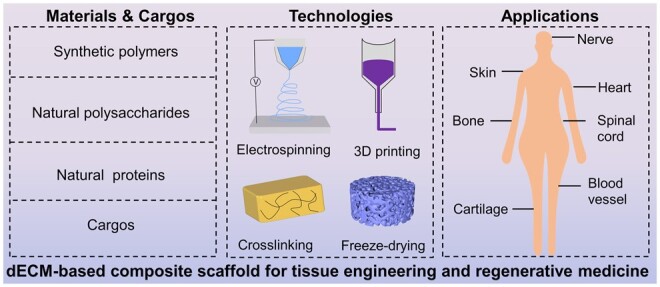
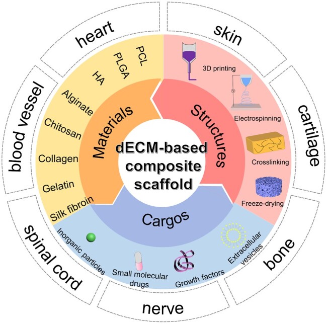
“Decellularized ECM (dECM) is biocompatible and can form 3D hydrogels; however, it lacks mechanical Decellularized ECM-based composite scaffolds have been fabricated with synthetic polymers, natural polysaccharides, and natural proteins for tissue engineering [2]. strength.”
BackgroundLiu, B., Alimi, O. A., Wang, Y., Kong, Y., Kuss, M. A., Krishnan, M. A., Hu, G., Xiao, Y., Dong, J., DiMaio, D. J., et al. (2024) · Journal of Controlled Release · doi:10.1016/j.jconrel.2024.02.019 · Cited by 48
Metadata
- Authors
- Xu, P., Kankala, R. K., Wang, S., & Chen, A.
- Affiliations
- Institute of Biomaterials and Tissue Engineering, Huaqiao University , Xiamen, Fujian 361021, PR China
- Huaqiao University (CN)
- Venue
- Regenerative Biomaterials
- Publisher
- Oxford University Press (OUP)
- Published
- 2023-12-01
- Volume
- 11
- Pages
- rbad107-rbad107
- ISSN
- 2056-3418 (ISSN-L)
- 2056-3426
- PMC
- PMC10761212
- OpenAlex
- W4389243078
- Cited by
- 129
- References
- 226
- Citations by year
- 2026: 56 · 2025: 45 · 2024: 28
- FWCI
- 8.33
- Type
- journal article
- Language
- en
- Open access
- gold
- doi.org
- Licence
- Locations
- Regenerative Biomaterials (publishedVersion, cc-by)
- PubMed Central (publishedVersion, cc-by)
- PubMed (publishedVersion)
- Funders
- National Natural Science Foundation of China (NSFC 32271410, 32071323, 81971734)
- Science and Technology Projects in Fujian Province (2022FX1, 2023Y4008)
- Scientific Research Funds of Huaqiao University (21BS113)
- Open Research Fund of Academy of Advanced Carbon Conversion Technology, Huaqiao University (AACCT0004)
- Topics
- Health Sciences › Medicine › Surgery › Tissue Engineering and Regenerative Medicine
- Physical Sciences › Materials Science › Biomaterials › Hydrogels: synthesis, properties, applications
- Physical Sciences › Engineering › Biomedical Engineering › 3D Printing in Biomedical Research
- Keywords
- decellularized extracellular matrix, composite scaffolds, tissue engineering, regenerative medicine, biomimetic microenvironment, natural polymers, bioactive agents, mechanical strength, tissue reconstruction
About these checks
Registry, retraction, Cites and statistics results are computed, not judged, with one exception: a printed reference that no index lists is read once by a model before it counts as a citation error. Claims are extracted by a model and kept only when their quote appears verbatim in the text; each cited source is then checked, with a model’s help, for whether it says what the claim relies on. Those verdicts can be wrong — read the claim’s quote and the cited source before relying on one.
Statistics and arithmetic are recomputed, not judged.
Confidence bands are shown only for fields calibrated on our annotated test set. None is calibrated yet, so no band is shown.
What counts as a claim ›How we measure confidence ›How we check this ›
Are you an author, or do you see a problem with this page? Request a review
How to cite
Cite CoChat for the checks on this page, and the paper for its own findings.
- This page
- CoChat. (2026). Citation check: Decellularized extracellular matrix-based composite scaffolds for tissue engineering and regenerative medicine. https://app.cochat.ai/papers/10.1093/rb/rbad107
- The paper
- Xu, P., Kankala, R. K., Wang, S., & Chen, A. (2024). Decellularized extracellular matrix-based composite scaffolds for tissue engineering and regenerative medicine. Regenerative Biomaterials. https://doi.org/10.1093/rb/rbad107
Data sources
Bibliographic data: Semantic Scholar, OpenAlex, Crossref.
Contains information from the Semantic Scholar Academic Graph Datasets, made available under the ODC Attribution License (ODC-BY 1.0).
Semantic Scholar’s data is described in: Kinney, R., et al. (2023). The Semantic Scholar Open Data Platform. arXiv:2301.10140.
