

The DOI matches its Crossref record; 98 of the 101 references it cites were found in a registry or an index.
A human coronavirus evolves antigenically to escape antibody immunity
Open accessPreprint — published as 10.1371/journal.ppat.1009453
Abstract
There is intense interest in antibody immunity to coronaviruses. However, it is unknown if coronaviruses evolve to escape such immunity, and if so, how rapidly. Here we address this question by characterizing the historical evolution of human coronavirus 229E. We identify human sera from the 1980s and 1990s that have neutralizing titers against contemporaneous 229E that are comparable to the anti-SARS-CoV-2 titers induced by SARS-CoV-2 infection or vaccination. We test these sera against 229E strains isolated after sera collection, and find that neutralizing titers are lower against these “future” viruses. In some cases, sera that neutralize contemporaneous 229E viral strains with titers >1:100 do not detectably neutralize strains isolated 8–17 years later. The decreased neutralization of “future” viruses is due to antigenic evolution of the viral spike, especially in the receptor-binding domain. If these results extrapolate to other coronaviruses, then it may be advisable to periodically update SARS-CoV-2 vaccines.
Overview
The claims and statistics of this paper haven’t been checked yet.
Run the check — free, no account needed. It reads the open-access text and checks the sources it cites.
Integrity signals
Registry
Cites
Statistics
Figures
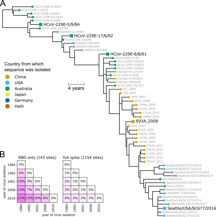
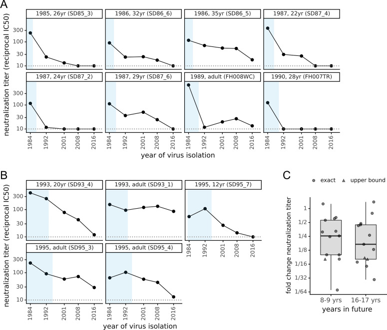
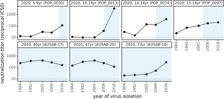
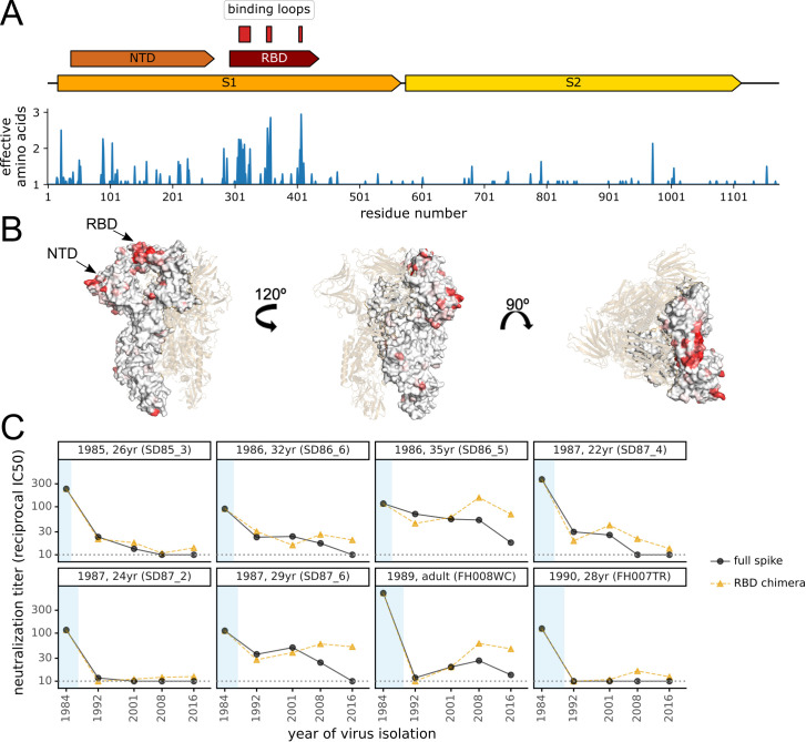
Verification
Claims the paper makes
The claims and statistics of this paper haven’t been checked yet.
Run the check — free, no account needed. It reads the open-access text and checks the sources it cites.
Claims other papers make about this one
Each is checked against this paper’s own text: does it say what the citing paper says it does?
The source backs part of the claim, not all of it — or backs it for a different population, condition or comparison.
In the citing paper
“In addition, similar results have been reported by others using vaccinee and convalescent plasmas and a variety of different pseudotype and authentic virus assays15,31,45–49.”
From mRNA vaccine-elicited antibodies to SARS-CoV-2 and circulating variants (2021) · cites this paper as [49]
Experimental — accuracy not yet measured
Self-check
Percentages, sums and reported differences re-computed from the paper’s own numbers.
The self-check needs the paper’s full text.
Citations
Cites
MAFFT Multiple Sequence Alignment Software Version 7: Improvements in Performance and Usability
Katoh, K., & Standley, D. M. (2013) · Molecular Biology and Evolution · doi:10.1093/molbev/mst010 · Cited by 46,209
IQ-TREE 2: New Models and Efficient Methods for Phylogenetic Inference in the Genomic Era
Minh, B. Q., Schmidt, H. A., Chernomor, O., Schrempf, D., Woodhams, M. D., von Haeseler, A., & Lanfear, R. (2020) · Molecular Biology and Evolution · doi:10.1093/molbev/msaa015 · Cited by 16,873
Safety and Efficacy of the BNT162b2 mRNA Covid-19 Vaccine
Polack, F. P., Thomas, S. J., Kitchin, N., Absalon, J., Gurtman, A., Lockhart, S., Perez, J. L., Pérez Marc, G., Moreira, E. D., Zerbini, C., et al. (2020) · New England Journal of Medicine · doi:10.1056/nejmoa2034577 · Cited by 14,592
Structure, function and antigenicity of the SARS-CoV-2 spike glycoprotein
Walls, A. C., Park, Y.-J., Tortorici, M. A., Wall, A., McGuire, A. T., & Veesler, D. (2020) · doi:10.1101/2020.02.19.956581 · Cited by 8,586
ETE 3: Reconstruction, Analysis, and Visualization of Phylogenomic Data
Huerta‐Cepas, J., Serra, F., & Bork, P. (2016) · Molecular Biology and Evolution · doi:10.1093/molbev/msw046 · Cited by 2,255
SARS-CoV-2 vaccines in development
Krammer, F. (2020) · Nature · doi:10.1038/s41586-020-2798-3 · Cited by 2,098
Starr, T. N., Greaney, A. J., Hilton, S. K., Ellis, D., Crawford, K. H. D., Dingens, A. S., Navarro, M. J., Bowen, J. E., Tortorici, M. A., Walls, A. C., et al. (2020) · Cell · doi:10.1016/j.cell.2020.08.012 · Cited by 1,956
Self-citationIdentification of a new human coronavirus
van der Hoek, L., Pyrć, K., Jebbink, M. F., Vermeulen-Oost, W., Berkhout, R. J. M., Wolthers, K. C., Dillen, P. M. E. W., Kaandorp, J. C., Spaargaren, J., & Berkhout, B. (2004) · Nature Medicine · doi:10.1038/nm1024 · Cited by 1,868
Mapping the Antigenic and Genetic Evolution of Influenza Virus
Smith, D. J., Lapedes, A. S., de Jong, J. C., Bestebroer, T. M., Rimmelzwaan, G. F., Osterhaus, A. D. M. E., & Fouchier, R. A. M. (2004) · Science · doi:10.1126/science.1097211 · Cited by 1,868
dendextend: an R package for visualizing, adjusting and comparing trees of hierarchical clustering
Galili, T. (2015) · Bioinformatics · doi:10.1093/bioinformatics/btv428 · Cited by 1,826
Research works printed but not linked: 6
- Crawford, K.H.D. , Dingens, A.S. , Eguia, R. , Wolf, C.R. , Wilcox, N. , Logue, J.K. , Shuey, K. , Casto, A.M. , Fiala, B. , Wrenn, S. , et al. (2020a). Dynamics of neutralizing antibody titers in the months after SARS-CoV-2 infection. J. Infect. Dis. · DOI
- Kemp, S. (2020). Neutralising antibodies drive Spike mediated SARS-CoV-2 evasion | medRxiv. BioRxiv.
- Kemp, S.A. , Collier, D.A. , Datir, R. , Gayed, S. , Jahun, A. , Hosmillo, M. , Ferreira, I. , Rees-Spear, C. , Mlcochova, P. , Lumb, I.U. , et al. (2020). Neutralising antibodies drive Spike mediated SARS-CoV-2 evasion. MedRxiv 2020.12.05.20241927.
- Lumley, S.F. , O’Donnell, D. , Stoesser, N.E. , Matthews, P.C. , Howarth, A. , Hatch, S.B. , Marsden, B.D. , Cox, S. , James, T. , Warren, F. , et al. (2020). Antibodies to SARS-CoV-2 are associated with protection against reinfection. MedRxiv 2020.11.18.20234369. · DOI
- Muñoz-Alía, M.Á. , Nace, R.A. , Zhang, L. , and Russell, S.J. (2020). Pathogenic measles viruses cannot evolve to bypass vaccine-induced neutralizing antibodies. BioRxiv 2020.10.22.351189. · DOI
- Thomson, E. (2020). The circulating SARS-CoV-2 spike variant N439K maintains fitness while evading antibody-mediated immunity | bioRxiv. BioRxiv.
Cited by
Sorted by citations
mRNA vaccine-elicited antibodies to SARS-CoV-2 and circulating variants
Wang, Z., Schmidt, F., Weisblum, Y., Muecksch, F., Barnes, C. O., Finkin, S., Schaefer-Babajew, D., Cipolla, M., Gaebler, C., Lieberman, J. A., et al. (2021) · Nature · doi:10.1038/s41586-021-03324-6 · Cited by 1,343
SARS-CoV-2 variant biology: immune escape, transmission and fitness
Carabelli, A. M., Peacock, T. P., Thorne, L., Harvey, W. T., Hughes, J., de Silva, T. I., Peacock, S. J., Barclay, W., de Silva, T. I., Towers, G. J., et al. (2023) · Nature Reviews Microbiology · doi:10.1038/s41579-022-00841-7 · Cited by 1,386
Greaney, A. J., Loes, A. N., Crawford, K. H. D., Starr, T. N., Malone, K. D., Chu, H. Y., & Bloom, J. D. (2021) · Cell Host & Microbe · doi:10.1016/j.chom.2021.02.003 · Cited by 1,178
Self-citationThe biological and clinical significance of emerging SARS-CoV-2 variants
Tao, K., Tzou, P. L., Nouhin, J., Gupta, R. K., de Oliveira, T., Kosakovsky Pond, S. L., Fera, D., & Shafer, R. W. (2021) · Nature Reviews Genetics · doi:10.1038/s41576-021-00408-x · Cited by 1,009
SARS-CoV-2 RBD antibodies that maximize breadth and resistance to escape
Starr, T. N., Czudnochowski, N., Liu, Z., Zatta, F., Park, Y., Addetia, A., Pinto, D., Beltramello, M., Hernandez, P., Greaney, A. J., et al. (2021) · Nature · doi:10.1038/s41586-021-03807-6 · Cited by 503
Self-citationStarr, T. N., Greaney, A. J., Dingens, A. S., & Bloom, J. D. (2021) · Cell Reports Medicine · doi:10.1016/j.xcrm.2021.100255 · Cited by 470
Self-citationThe coronavirus is here to stay — here’s what that means
Phillips, N. (2021) · Nature · doi:10.1038/d41586-021-00396-2 · Cited by 444
After the pandemic: perspectives on the future trajectory of COVID-19
Telenti, A., Arvin, A. M., Corey, L., Corti, D., Diamond, M., García‐Sastre, A., Garry, R. F., Holmes, E. C., Pang, P. S., & Virgin, H. W. (2021) · Nature · doi:10.1038/s41586-021-03792-w · Cited by 450
Immunological characteristics govern the transition of COVID-19 to endemicity
Lavine, J. S., Bjornstad, O. N., & Antia, R. (2021) · Science · doi:10.1126/science.abe6522 · Cited by 416
Structural and functional ramifications of antigenic drift in recent SARS-CoV-2 variants
Yuan, M., Huang, D., Lee, C.-C. D., Wu, N. C., Jackson, A. M., Zhu, X., Liu, H., Peng, L., van Gils, M. J., Sanders, R. W., et al. (2021) · doi:10.1101/2021.02.16.430500 · Cited by 364
The origins and potential future of SARS-CoV-2 variants of concern in the evolving COVID-19 pandemic
Otto, S. P., Day, T., Arino, J., Colijn, C., Dushoff, J. G., Li, M., Mechai, S., Domselaar, G. V., Wu, J., Earn, D. J. D., et al. (2021) · Current Biology · doi:10.1016/j.cub.2021.06.049 · Cited by 326
Kundu, R., Narean, J. S., Wang, L., Fenn, J., Pillay, T. D., Derqui, N., Conibear, E., Koycheva, A., Davies, M., Tolosa-Wright, M., et al. (2022) · Nature Communications · doi:10.1038/s41467-021-27674-x · Cited by 316
Shifting mutational constraints in the SARS-CoV-2 receptor-binding domain during viral evolution
Starr, T. N., Greaney, A. J., Hannon, W. W., Loes, A. N., Hauser, K., Dillen, J. R., Ferri, E., Farrell, A. G., Dadonaite, B., McCallum, M., et al. (2022) · Science · doi:10.1126/science.abo7896 · Cited by 298
Self-citationThe changing epidemiology of SARS-CoV-2
Koelle, K., Martin, M. A., Antia, R., Lopman, B. A., & Dean, N. E. (2022) · Science · doi:10.1126/science.abm4915 · Cited by 308
Greaney, A. J., Loes, A. N., Gentles, L. E., Crawford, K. H. D., Starr, T. N., Malone, K. D., Chu, H. Y., & Bloom, J. D. (2021) · Science Translational Medicine · doi:10.1126/scitranslmed.abi9915 · Cited by 244
Self-citationEpidemiological and evolutionary considerations of SARS-CoV-2 vaccine dosing regimes
Saad-Roy, C. M., Morris, S. E., Metcalf, C. J. E., Mina, M. J., Baker, R. E., Farrar, J., Holmes, E. C., Pybus, O. G., Graham, A. L., Levin, S. A., et al. (2021) · Science · doi:10.1126/science.abg8663 · Cited by 221
The SARS-CoV-2 mutations versus vaccine effectiveness: New opportunities to new challenges
Malik, J. A., Ahmed, S., Aroosa, M., Shinde, M., Bender, O., Alshammari, F., Ansari, M., & Anwar, S. (2022) · Journal of Infection and Public Health · doi:10.1016/j.jiph.2021.12.014 · Cited by 206
Beyond Omicron: what’s next for COVID’s viral evolution
Callaway, E. (2021) · Nature · doi:10.1038/d41586-021-03619-8 · Cited by 189
Implications of diversity in sperm size and function for sperm competition and fertility
Gomendio, M., & Roldán, E. R. S. (2008) · The International Journal of Developmental Biology · doi:10.1387/ijdb.082595mg · Cited by 187
Prolonged evolution of the human B cell response to SARS-CoV-2 infection
Sakharkar, M., Rappazzo, C. G., Wieland-Alter, W. F., Hsieh, C.-L., Wrapp, D., Esterman, E. S., Kaku, C. I., Wec, A. Z., Geoghegan, J. C., McLellan, J. S., et al. (2021) · Science Immunology · doi:10.1126/sciimmunol.abg6916 · Cited by 174
SARS-CoV-2 evolution and vaccines: cause for concern?
Williams, T., & Burgers, W. A. (2021) · The Lancet Respiratory Medicine · doi:10.1016/s2213-2600(21)00075-8 · Cited by 163
SARS-CoV-2 one year on: evidence for ongoing viral adaptation
Peacock, T. P., Penrice-Randal, R., Hiscox, J. A., & Barclay, W. (2021) · Journal of General Virology · doi:10.1099/jgv.0.001584 · Cited by 155
Focosi, D., & Maggi, F. (2021) · Reviews in Medical Virology · doi:10.1002/rmv.2231 · Cited by 150
An antibody-escape estimator for mutations to the SARS-CoV-2 receptor-binding domain
Greaney, A. J., Starr, T. N., & Bloom, J. D. (2022) · Virus Evolution · doi:10.1093/ve/veac021 · Cited by 147
Self-citationMoodley, A., Stegger, M., BAĞCIGİL, A. F., Baptiste, K. E., Loeffler, A., Lloyd, D. H., Williams, N. J., Leonard, N., Abbott, Y., Skov, R. L., et al. (2006) · Journal of Antimicrobial Chemotherapy · doi:10.1093/jac/dkl394 · Cited by 146
Antigenic drift: Understanding COVID-19
Yewdell, J. W. (2021) · Immunity · doi:10.1016/j.immuni.2021.11.016 · Cited by 145
Convergent Evolution in SARS-CoV-2 Spike Creates a Variant Soup from Which New COVID-19 Waves Emerge
Focosi, D., Quiroga, R., McConnell, S. A., Johnson, M. C., & Casadevall, A. (2023) · International Journal of Molecular Sciences · doi:10.3390/ijms24032264 · Cited by 132
SARS-CoV-2 evolution in the Omicron era
Roemer, C., Sheward, D. J., Hisner, R., Gueli, F., Sakaguchi, H., Frohberg, N., Schoenmakers, J., Sato, K., O’Toole, Á., Rambaut, A., et al. (2023) · Nature Microbiology · doi:10.1038/s41564-023-01504-w · Cited by 130
Mutations of SARS-CoV-2 spike protein: Implications on immune evasion and vaccine-induced immunity
Mengist, H. M., Kombe, A. J. K., Mekonnen, D. S., Abebaw, A., Getachew, M., & Jin, T. (2021) · Seminars in Immunology · doi:10.1016/j.smim.2021.101533 · Cited by 115
de Souza, W. M., Amorim, M. R., Sesti‐Costa, R., Coimbra, L. D., Brunetti, N. S., de Toledo-Teixeira, D. A., de Souza, G. F., Muraro, S. P., Parise, P. L., Barbosa, P. P., et al. (2021) · The Lancet Microbe · doi:10.1016/s2666-5247(21)00129-4 · Cited by 115
Impact of antigenic evolution and original antigenic sin on SARS-CoV-2 immunity
Aguilar-Bretones, M., Fouchier, R. A. M., Koopmans, M., & van Nierop, G. P. (2023) · Journal of Clinical Investigation · doi:10.1172/jci162192 · Cited by 108
Taft, J. M., Weber, C. R., Gao, B., Ehling, R. A., Han, J., Frei, L., Metcalfe, S. W., Overath, M. D., Yermanos, A., Kelton, W., et al. (2022) · Cell · doi:10.1016/j.cell.2022.08.024 · Cited by 119
COVID-19: Challenges of Viral Variants
Jacobs, J. L., Haidar, G., & Mellors, J. W. (2022) · Annual Review of Medicine · doi:10.1146/annurev-med-042921-020956 · Cited by 105
Rapid and Parallel Adaptive Mutations in Spike S1 Drive Clade Success in SARS-CoV-2
Kistler, K. E., Huddleston, J., & Bedford, T. (2021) · Cell Host & Microbe · doi:10.1016/j.chom.2022.03.018 · Cited by 102
Evolutionary Landscapes of Host-Virus Arms Races
Tenthorey, J. L., Emerman, M., & Malik, H. S. (2022) · Annual Review of Immunology · doi:10.1146/annurev-immunol-072621-084422 · Cited by 105
Tortorici, M. A., Walls, A. C., Joshi, A., Park, Y., Eguia, R. T., Miranda, M. C., Kepl, E., Dosey, A. M., Stevens‐Ayers, T., Boeckh, M. J., et al. (2022) · Cell · doi:10.1016/j.cell.2022.05.019 · Cited by 89
Self-citationThe success of SARS-CoV-2 vaccines and challenges ahead
Subbarao, K. (2021) · Cell Host & Microbe · doi:10.1016/j.chom.2021.06.016 · Cited by 92
Anand, U., Jakhmola, S., Indari, O., Jha, H. C., Chen, Z., Tripathi, V. S., & de la Lastra, J. M. P. (2021) · Frontiers in Immunology · doi:10.3389/fimmu.2021.658519 · Cited by 84
In search of a pan-coronavirus vaccine: next-generation vaccine design and immune mechanisms
Cankat, S., Demael, M. U., & Swadling, L. (2023) · Cellular & Molecular Immunology · doi:10.1038/s41423-023-01116-8 · Cited by 84
Structural definition of a pan-sarbecovirus neutralizing epitope on the spike S2 subunit
Hurlburt, N. K., Homad, L. J., Sinha, I. R., Jennewein, M. F., MacCamy, A. J., Wan, Y.-H., Boonyaratanakornkit, J. B., Sholukh, A. M., Jackson, A. M., Zhou, P.-P., et al. (2022) · Communications Biology · doi:10.1038/s42003-022-03262-7 · Cited by 80
The landscape of antibody binding in SARS-CoV-2 infection
Heffron, A. S., McIlwain, S. J., Amjadi, M. F., Baker, D. A., Khullar, S., Armbrust, T., Halfmann, P., Kawaoka, Y., Sethi, A. K., Palmenberg, A. C., et al. (2021) · PLoS Biology · doi:10.1371/journal.pbio.3001265 · Cited by 78
Instructing durable humoral immunity for COVID-19 and other vaccinable diseases
Bhattacharya, D. (2022) · Immunity · doi:10.1016/j.immuni.2022.05.004 · Cited by 64
A SARS-CoV-2 Nucleocapsid Variant that Affects Antigen Test Performance
Bourassa, L. A., Perchetti, G. A., Phung, Q., Lin, M. J., Mills, M. G., Roychoudhury, P., Harmon, K. G., Reed, J. C., & Greninger, A. L. (2021) · Journal of Clinical Virology · doi:10.1016/j.jcv.2021.104900 · Cited by 66
Self-citationAhn, J. Y., Lee, J., Suh, Y. S., Song, Y. G., Choi, Y.-J., Lee, K. H., Seo, S. H., Song, M., Oh, J., Kim, M., et al. (2022) · The Lancet Microbe · doi:10.1016/s2666-5247(21)00358-x · Cited by 67
A SARS-CoV-2 variant elicits an antibody response with a shifted immunodominance hierarchy
Greaney, A. J., Starr, T. N., Eguia, R. T., Loes, A. N., Khan, K., Karim, F., Cele, S., Bowen, J. E., Logue, J. K., Corti, D., et al. (2022) · PLoS Pathogens · doi:10.1371/journal.ppat.1010248 · Cited by 58
Self-citationAn atlas of continuous adaptive evolution in endemic human viruses
Kistler, K. E., & Bedford, T. (2023) · Cell Host & Microbe · doi:10.1016/j.chom.2023.09.012 · Cited by 65
Muñoz-Alía, M. Á., Nace, R. A., Zhang, L., & Russell, S. J. (2021) · Cell Reports Medicine · doi:10.1016/j.xcrm.2021.100225 · Cited by 60
The ins and outs of SARS-CoV-2 variants of concern (VOCs)
Salehi-Vaziri, ., Fazlalipour, M., Khorrami, S. M. S., Azadmanesh, K., Pouriayevali, M. H., Jalali, T., Shoja, Z., & Maleki, A. (2022) · Archives of Virology · doi:10.1007/s00705-022-05365-2 · Cited by 61
Pollett, S. D., Conte, M. A., Sanborn, M. A., Jarman, R. G., Lidl, G. M., Modjarrad, K., & Berry, I. M. (2021) · Scientific Reports · doi:10.1038/s41598-021-96626-8 · Cited by 59
The evolutionary dynamics of endemic human coronaviruses
Jo, W. K., Drosten, C., & Drexler, J. F. (2021) · Virus Evolution · doi:10.1093/ve/veab020 · Cited by 60
Wang, C., Hesketh, E. L., Shamorkina, T. M., Li, W., Franken, P. J., Drabek, D., van Haperen, R., Townend, S., van Kuppeveld, F. J. M., Grosveld, F. G., et al. (2022) · Nature Communications · doi:10.1038/s41467-022-30658-0 · Cited by 52
Deep mutational scanning for therapeutic antibody engineering
Hanning, K. R., Minot, M., Warrender, A. K., Kelton, W., & Reddy, S. T. (2021) · Trends in Pharmacological Sciences · doi:10.1016/j.tips.2021.11.010 · Cited by 53
Human coronavirus HKU1 recognition of the TMPRSS2 host receptor
McCallum, M., Park, Y., Stewart, C., Sprouse, K. R., Addetia, A., Brown, J. T., Tortorici, M. A., Gibson, C. A., Wong, E., Ieven, M. M., et al. (2024) · Cell · doi:10.1016/j.cell.2024.06.006 · Cited by 51
COVID-19, the first pandemic in the post-genomic era
van Dorp, L., Houldcroft, C. J., Richard, D., & Balloux, F. (2021) · Current Opinion in Virology · doi:10.1016/j.coviro.2021.07.002 · Cited by 49
SARS‐CoV‐2 virulence evolution: Avirulence theory, immunity and trade‐offs
Alizon, S., & Sofonea, M. T. (2021) · Journal of Evolutionary Biology · doi:10.1111/jeb.13896 · Cited by 53
Evolutionary implications of SARS-CoV-2 vaccination for the future design of vaccination strategies
Rouzine, I. M., & Rozhnova, G. (2023) · Communications Medicine · doi:10.1038/s43856-023-00320-x · Cited by 49
A biophysical model of viral escape from polyclonal antibodies
Yu, T. C., Thornton, Z., Hannon, W. W., DeWitt, W. S., Radford, C. E., Matsen, F. A., IV, & Bloom, J. D. (2022) · Virus Evolution · doi:10.1093/ve/veac110 · Cited by 42
Self-citationAntigenic escape selects for the evolution of higher pathogen transmission and virulence
Sasaki, A., Lion, S., & Boots, M. (2021) · Nature Ecology & Evolution · doi:10.1038/s41559-021-01603-z · Cited by 42
Rapid escape of new SARS-CoV-2 Omicron variants from BA.2-directed antibody responses
Dijokaite-Guraliuc, A., Das, R., Zhou, D., Ginn, H. M., Liu, C., Duyvesteyn, H. M. E., Huo, J., Nutalai, R., Supasa, P., Selvaraj, M., et al. (2023) · Cell Reports · doi:10.1016/j.celrep.2023.112271 · Cited by 46
The past, current and future epidemiological dynamic of SARS-CoV-2
Balloux, F., Tan, C. C. S., Swadling, L., Richard, D., Jenner, C., Maini, M. K., & van Dorp, L. (2022) · Oxford Open Immunology · doi:10.1093/oxfimm/iqac003 · Cited by 41
Anderson, E. M., Li, S. H., Awofolaju, M., Eilola, T., Goodwin, E. C., Bolton, M. J., Gouma, S., Manzoni, T. B., Hicks, P. D., Goel, R. R., et al. (2022) · Cell Reports · doi:10.1016/j.celrep.2022.111496 · Cited by 37
Herd immunity, vaccination and moral obligation
Bullen, M., Heriot, G., & Jamrozik, E. (2023) · Journal of Medical Ethics · doi:10.1136/jme-2022-108485 · Cited by 50
Investigating the evolutionary origins of the first three SARS-CoV-2 variants of concern
Ghafari, M., Liu, Q., Dhillon, A., Katzourakis, A., & Weissman, D. B. (2022) · Frontiers in Virology · doi:10.3389/fviro.2022.942555 · Cited by 35
Al‐Tawfiq, J. A., Hoang, V. T., Bui, N. L., Chu, D., & Memish, Z. A. (2022) · Journal of Epidemiology and Global Health · doi:10.1007/s44197-022-00032-w · Cited by 34
Immuno-epidemiology and the predictability of viral evolution
Saad-Roy, C. M., Metcalf, C. J. E., & Grenfell, B. T. (2022) · Science · doi:10.1126/science.abn9410 · Cited by 33
PARIS and SPARTA: Finding the Achilles’ Heel of SARS-CoV-2
Simon, V. R., Kota, V. K., Bloomquist, R. F., Hanley, H. B., Forgacs, D., Pahwa, S. G., Pallikkuth, S., Miller, L. G., Schaenman, J. M. D., Yeaman, M. R., et al. (2022) · mSphere · doi:10.1128/msphere.00179-22 · Cited by 28
Shao, N., Zhang, C., Dong, J., Sun, L., Chen, X., Xie, Z., Xu, B., An, S., Zhang, T., & Yang, F. (2022) · Frontiers in Microbiology · doi:10.3389/fmicb.2022.1023847 · Cited by 32
An overview of current drugs and prophylactic vaccines for coronavirus disease 2019 (COVID-19)
Bahrami, A. A., Azargoonjahromi, A., Sadraei, S., Aarabi, A., Payandeh, Z., & Rajabibazl, M. (2022) · Cellular & Molecular Biology Letters · doi:10.1186/s11658-022-00339-3 · Cited by 33
Cheng, Z., Elmes, M. J., Kirkup, S. E., Abayasekara, D. R. E., & Wathes, D. C. (2004) · Journal of Endocrinology · doi:10.1677/joe.0.1820249 · Cited by 31
Garrett, M. E., Galloway, J. G., Wolf, C., Logue, J. K., Franko, N., Chu, H. Y., Matsen, F. A., IV, & Overbaugh, J. (2022) · eLife · doi:10.7554/elife.73490 · Cited by 32
Tsai, K., Lee, Y.-C., & Tseng, T.-S. (2021) · Frontiers in Microbiology · doi:10.3389/fmicb.2021.698365 · Cited by 32
Adaptation of the endemic coronaviruses HCoV-OC43 and HCoV-229E to the human host
Forni, D., Cagliani, R., Arrigoni, F., Benvenuti, M., Mozzi, A., Pozzoli, U., Clerici, M., GIOIA, L. D., & Sironi, M. (2021) · Virus Evolution · doi:10.1093/ve/veab061 · Cited by 30
Shooting at a Moving Target—Effectiveness and Emerging Challenges for SARS-CoV-2 Vaccine Development
Günl, F., Mecate-Zambrano, A., Rehländer, S., Hinse, S., Ludwig, S., & Brunotte, L. (2021) · Vaccines · doi:10.3390/vaccines9101052 · Cited by 27
Rattanapisit, K., Bulaon, C. J. I., Khorattanakulchai, N., Shanmugaraj, B., Wangkanont, K., & Phoolcharoen, W. (2021) · PLoS ONE · doi:10.1371/journal.pone.0253574 · Cited by 26
How immunity from and interaction with seasonal coronaviruses can shape SARS-CoV-2 epidemiology
Waterlow, N. R., van Leeuwen, E., Davies, N. G., CMMID COVID-19 Working Group, Flasche, S., & Eggo, R. M. (2021) · Proceedings of the National Academy of Sciences · doi:10.1073/pnas.2108395118 · Cited by 26
Lawrenz, J., Xie, Q., Zech, F., Weil, T., Seidel, A., Krnavek, D., van der Hoek, L., Münch, J., Müller, J. A., & Kirchhoff, F. (2022) · Clinical Infectious Diseases · doi:10.1093/cid/ciac057 · Cited by 25
The challenge of emerging SARS-CoV-2 mutants to vaccine development
Li, R., Liu, J., & Zhang, H. (2021) · Journal of genetics and genomics/Journal of Genetics and Genomics · doi:10.1016/j.jgg.2021.03.001 · Cited by 25
Loss of FCoV-23 spike domain 0 enhances fusogenicity and entry kinetics
Tortorici, M. A., Choi, A., Gibson, C. A., Lee, J., Brown, J. T., Stewart, C., Joshi, A., Harari, S., Willoughby, I., Treichel, C., et al. (2025) · Nature · doi:10.1038/s41586-025-09155-z · Cited by 24
Self-citationCicaloni, V., Costanti, F., Pasqui, A., Bianchini, M., Niccolai, N., & Bongini, P. (2022) · Frontiers in Genetics · doi:10.3389/fgene.2022.891418 · Cited by 25
Kumar, S., Delipan, R., Chakraborty, D., Kanjo, K., Singh, R., Singh, N., Siddiqui, S., Tyagi, A., Jha, V., Thakur, K. G., et al. (2023) · Journal of Virology · doi:10.1128/jvi.00922-23 · Cited by 23
Which ‘imperfect vaccines’ encourage the evolution of higher virulence?
Bull, J. J., & Antia, R. (2022) · Evolution Medicine and Public Health · doi:10.1093/emph/eoac015 · Cited by 23
Fischer, R. J., Port, J. R., Holbrook, M. G., Yinda, C. K., Creusen, M., ter Stege, J., de Samber, M., & Munster, V. J. (2022) · Environmental Science & Technology · doi:10.1021/acs.est.2c02822 · Cited by 22
Weil, T., Lawrenz, J., Seidel, A., Münch, J., & Müller, J. A. (2022) · Antiviral Research · doi:10.1016/j.antiviral.2022.105343 · Cited by 20
Bolton, M. J., Santos, J. J. D. S., Arevalo, C. P., Griesman, T., Watson, M., Li, S. H., Bates, P. F., Ramage, H. R., Wilson, P. C., & Hensley, S. E. (2023) · Proceedings of the National Academy of Sciences · doi:10.1073/pnas.2216521120 · Cited by 22
Starr, T. N., Czudnochowski, N., Zatta, F., Park, Y., Liu, Z., Addetia, A., Pinto, D., Beltramello, M., Hernandez, P., Greaney, A. J., et al. (2021) · bioRxiv (Cold Spring Harbor Laboratory) · doi:10.1101/2021.04.06.438709 · Cited by 24
Self-citationHeterologous humoral immunity to human and zoonotic coronaviruses: Aiming for the achilles heel
Ng, K. W., Faulkner, N., Wrobel, A. G., Gamblin, S. J., & Kassiotis, G. (2021) · Seminars in Immunology · doi:10.1016/j.smim.2021.101507 · Cited by 19
Richard, D., Shaw, L. P., Lanfear, R., Corbett-Detig, R. B., Hinrichs, A. S., McBroome, J., Turakhia, Y., Acman, M., Owen, C. J., Tan, C. C. S., et al. (2021) · bioRxiv (Cold Spring Harbor Laboratory) · doi:10.1101/2021.05.06.442903 · Cited by 20
Persistence of immunity to SARS-CoV-2 over time in the ski resort Ischgl
Borena, W., Bánki, Z., Bates, K., Winner, H., Riepler, L., Rössler, A., Pipperger, L., Theurl, I., Falkensammer, B., Ulmer, H., et al. (2021) · EBioMedicine · doi:10.1016/j.ebiom.2021.103534 · Cited by 18
Uncertainty around the Long-Term Implications of COVID-19
Desforges, M., Gurdasani, D., Hamdy, A., & Leonardi, A. J. (2021) · Pathogens · doi:10.3390/pathogens10101267 · Cited by 18
Vermeulen, C. J., Dijkman, R., de Wit, S. J., Bosch, B., Heesterbeek, H., & Schaik, G. V. (2023) · Avian Pathology · doi:10.1080/03079457.2023.2177140 · Cited by 19
Rational design of a booster vaccine against COVID-19 based on antigenic distance
Hu, Y., Yuen, T. T., Gong, H.-R., Hu, B., Hu, J., Lin, X., Li, R., Zhou, C. L., Chen, L., Wang, X., et al. (2023) · Cell Host & Microbe · doi:10.1016/j.chom.2023.07.004 · Cited by 22
Shared N417-Dependent Epitope on the SARS-CoV-2 Omicron, Beta, and Delta Plus Variants
Moyo-Gwete, T., Madzivhandila, M., Mkhize, N. N., Kgagudi, P., Ayres, F., Lambson, B. E., Manamela, N. P., Richardson, S. I., Makhado, Z., van der Mescht, M. A., et al. (2022) · Journal of Virology · doi:10.1128/jvi.00558-22 · Cited by 18
SARS-CoV-2: The Interplay Between Evolution and Host Immunity
Case, J. B., Jain, S., Suthar, M. S., & Diamond, M. (2024) · Annual Review of Immunology · doi:10.1146/annurev-immunol-083122-043054 · Cited by 26
Structural Dynamics and Molecular Evolution of the SARS-CoV-2 Spike Protein
Wolf, K., Kwan, J. C., & Kamil, J. P. (2022) · mBio · doi:10.1128/mbio.02030-21 · Cited by 17
Omicron’s message on vaccines: Boosting begets breadth
Wesemann, D. R. (2022) · Cell · doi:10.1016/j.cell.2022.01.006 · Cited by 16
Laroche, A., Delgado, M. L. O., Chalopin, B., Cuniasse, P., DuBois, S. G., Sierocki, R., Gallais, F., Debroas, S., Bellanger, L., Simon, S., et al. (2022) · mAbs · doi:10.1080/19420862.2022.2076775 · Cited by 21
Reduced B cell antigenicity of Omicron lowers host serologic response
Tubiana, J., Xiang, Y., Fan, L., Wolfson, H. J., Chen, K., Schneidman‐Duhovny, D., & Shi, Y. (2022) · Cell Reports · doi:10.1016/j.celrep.2022.111512 · Cited by 18
Logue, J., Johnson, R. M., Patel, N., Zhou, B., Maciejewski, S. M., Foreman, B. M., Zhou, H., Portnoff, A. D., Tian, J.-H., Rehman, A., et al. (2023) · Nature Communications · doi:10.1038/s41467-022-35606-6 · Cited by 21
Immunisation, asymptomatic infection, herd immunity and the new variants of COVID 19Preprint
Grant, A., & Hunter, P. R. (2021) · medRxiv · doi:10.1101/2021.01.16.21249946 · Cited by 18
Waning immunity drives respiratory virus evolution and reinfection
Bull, J. J., Koelle, K., & Antia, R. (2025) · Evolution Medicine and Public Health · doi:10.1093/emph/eoaf002 · Cited by 16
Metadata
- Authors
- Eguia, R., Crawford, K. H. D., Stevens-Ayers, T., Kelnhofer-Millevolte, L., Greninger, A. L., Englund, J. A., Boeckh, M. J., & Bloom, J. D.
- Affiliations
- Fred Hutch Cancer Center (US)
- University of Washington (US)
- Howard Hughes Medical Institute (US)
- Corresponding author
- Bloom, Jesse D.
- ORCID
- Venue
- PLoS Pathogens
- Publisher
- openRxiv
- Published
- 2020-12-18
- Volume
- 17
- Issue
- 4
- Pages
- e1009453-e1009453
- ISSN
- 1553-7366 (ISSN-L)
- PMC
- PMC8031418
- OpenAlex
- W3144378969
- Cited by
- 258
- References
- 101
- Citations by year
- 2026: 7 · 2025: 21 · 2024: 11 · 2023: 33 · 2022: 69 · 2021: 103 · 2020: 2
- FWCI
- 17.09
- Type
- journal article
- Language
- en
- Open access
- gold
- doi.org
- Licence
- cc-by
- Locations
- PLoS Pathogens (publishedVersion, cc-by)
- bioRxiv (Cold Spring Harbor Laboratory) (acceptedVersion, cc-by)
- PubMed Central (publishedVersion, cc-by)
- PubMed (publishedVersion)
- DOAJ (DOAJ: Directory of Open Access Journals) (submittedVersion, cc-by-sa)
- Funders
- Howard Hughes Medical Institute
- Pfizer
- AstraZeneca
- GlaxoSmithKline
- Sanofi
- Fred Hutchinson Cancer Research Center
- Regeneron Pharmaceuticals
- Moderna
- National Institutes of Health
- Sanofi Pasteur
- National Institute of General Medical Sciences (T32GM007266)
- National Institute of Allergy and Infectious Diseases (R01AI127893, F30AI149928, R01AI141707)
- Topics
- Health Sciences › Medicine › Infectious Diseases › SARS-CoV-2 and COVID-19 Research
- Life Sciences › Agricultural and Biological Sciences › Animal Science and Zoology › Animal Virus Infections Studies
- Life Sciences › Biochemistry, Genetics and Molecular Biology › Molecular Biology › vaccines and immunoinformatics approaches
- Keywords
- HCoV-229E, antigenic evolution, antibody escape, neutralizing antibodies, spike protein, receptor binding domain, coronavirus evolution
- MeSH terms
- SARS-CoV-2, Antibodies, Viral, Humans, Coronavirus 229E, Human, Immune Evasion, Antibodies, Neutralizing
About these checks
Registry, retraction, Cites and statistics results are computed, not judged, with one exception: a printed reference that no index lists is read once by a model before it counts as a citation error. Claims are extracted by a model and kept only when their quote appears verbatim in the text; each cited source is then checked, with a model’s help, for whether it says what the claim relies on. Those verdicts can be wrong — read the claim’s quote and the cited source before relying on one.
Statistics and arithmetic are recomputed, not judged.
Confidence bands are shown only for fields calibrated on our annotated test set. None is calibrated yet, so no band is shown.
What counts as a claim ›How we measure confidence ›How we check this ›
Are you an author, or do you see a problem with this page? Request a review
