PreprintCited by 2Protein Degradation and InhibitorsUbiquitin and proteasome pathways14-3-3 protein interactions

The DOI matches its Crossref record; 1 of the 1 references it cites were found in a registry or an index.

A ubiquitination-mediated degradation system to target phospho-14-3-3-binding-motif embedded proteins

Open access

Preprint — published as 10.1016/j.heliyon.2023.e16318 · Paper page

Abstract

The phosphorylation of 14-3-3 binding motif is involved in many cellular processes. A strategy that enables targeted degradation of phospho-14-3-3-binding-motif embedded (P-14-3-3-BME) proteins for studying their functions is highly desirable for basic research. Here, we report a phosphorylation-induced, ubiquitin-proteasome-system-mediated targeted protein degradation (TPD) strategy that allows specific degradation of P-14-3-3-BME proteins. Specifically, by ligating a modified von Hippel-Lindau E3-ligase with an engineered 14-3-3 bait, we generated a protein chimera referred to as Targeted Degradation of P-14-3-3-BME Protein (TDPP). TDPP can serve as a universal degrader for P-14-3-3-BME proteins based on the specific recognition of the phosphorylation in 14-3-3 binding motifs. TDPP shows high efficiency and specificity to a difopein-EGFP reporter, general and specific P-14-3-3-BME proteins. TDPP can also be applied for the validation of P-14-3-3-BME proteins. These results strongly support TDPP as a powerful tool for 14-3-3 related research.

Overview

The claims and statistics of this paper haven’t been checked yet.

Run the check — free, no account needed. It reads the open-access text and checks the sources it cites.

A few free checks a day · papers already verified open instantly · results are public

Integrity signals

Registry

Crossref · 10.1101/2022.11.22.517526

Cites

1 of 1 references verified0 citation errors · 0 not checkable

Statistics

Figures

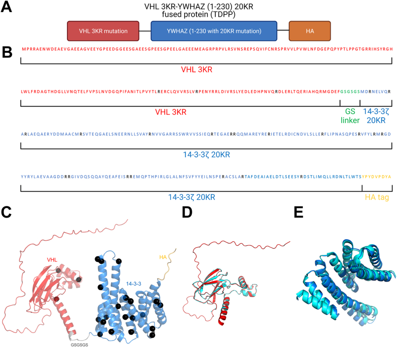
Fig. 1 Illustration and de novo structure modeling of a degrader of 14-3-3-BPPs. A. TDPP fused protein design. B. Sequence partition information of TDPP. C. Full-length structure model generated by ColabFold [26]. KR mutation sites are highlighted in black spheres. D. Comparison of predicted VHL 3 KR model and experimental VHL structure. Red: predicted VHL (extracted from full-length model); cyan: experimental VHL structure (PDB ID: 4WQO). E. Comparison of predicted 14-3-3ζ 20 KR model and experimental 14-3-3ζ structure. Blue: predicted 14-3-3ζ (extracted from full-length model); cyan: experimental 14-3-3ζ structure (PDB ID: 2O02). See also Fig. S1.
Fig. 1 Illustration and de novo structure modeling of a degrader of 14-3-3-BPPs. A. TDPP fused protein design. B. Sequence partition information of TDPP. C. Full-length structure model generated by ColabFold [26]. KR mutation sites are highlighted in black spheres. D. Comparison of predicted VHL 3 KR model and experimental VHL structure. Red: predicted VHL (extracted from full-length model); cyan: experimental VHL structure (PDB ID: 4WQO). E. Comparison of predicted 14-3-3ζ 20 KR model and experimental 14-3-3ζ structure. Blue: predicted 14-3-3ζ (extracted from full-length model); cyan: experimental 14-3-3ζ structure (PDB ID: 2O02). See also Fig. S1.CC BY-NC-ND · PubMed Central
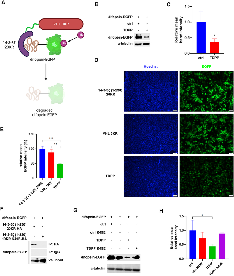
Fig. 2 TDPP degrades difopein-EGFP. A. Working model of TDPP-induced degradation of difopein-EGFP. Undegraded difopein-EGFP protein chimera has a green fluorescent ability from the EGFP tag. Since the 14-3-3 bait in TDPP could specifically interact with the difopein tag in difopein-EGFP, TDPP will provide the proximity of UPS to difopein-EGFP and degrade it, in which condition the green fluorescent intensity will decrease. B, C. Western blot result (B) and quantification of band intensity (C) of difopein-EGFP in HEK293T/17 cells co-transfected with expression plasmids of 14-3-3ζ (1–230) 20 KR-HA (referred as ctrl) or VHL 3 KR-14-3-3ζ (1–230) 20 KR-HA (referred as TDPP). D, E. Fluorescence assay result (D) and fluorescence intensity quantification (E) of HEK293T/17 cells transfected with difopein-EGFP vector, along with control vectors (14-3-3ζ (1–230) 20 KR or VHL 3 KR) or TDPP vector. F. Immunoprecipitation result of interaction between 14-3-3ζ (1–230) 20 KR-HA/14-3-3ζ (1–230) 19 KR K49E-HA and difopein-EGFP. IP: 14-3-3ζ (1–230) 20 KR-HA/14-3-3ζ (1–230) 19 KR K49E-HA. IB: difopein-EGFP. G, H. Western blot result (G) and quantification of band intensity (H) of difopein-EGFP in HEK293T/17 cells co-transfected with expression plasmids of 14-3-3ζ (1–230) 20 KR-HA (referred as ctrl), 14-3-3ζ (1–230) 19 KR K49E-HA (referred as ctrl K49E), VHL 3 KR-14-3-3ζ (1–230) 20 KR-HA (referred as TDPP), or VHL 3 KR-14-3-3ζ (1–230) 19 KR K49E-HA (referred as TDPP K49E). Data are represented as mean ± SEM.
Fig. 2 TDPP degrades difopein-EGFP. A. Working model of TDPP-induced degradation of difopein-EGFP. Undegraded difopein-EGFP protein chimera has a green fluorescent ability from the EGFP tag. Since the 14-3-3 bait in TDPP could specifically interact with the difopein tag in difopein-EGFP, TDPP will provide the proximity of UPS to difopein-EGFP and degrade it, in which condition the green fluorescent intensity will decrease. B, C. Western blot result (B) and quantification of band intensity (C) of difopein-EGFP in HEK293T/17 cells co-transfected with expression plasmids of 14-3-3ζ (1–230) 20 KR-HA (referred as ctrl) or VHL 3 KR-14-3-3ζ (1–230) 20 KR-HA (referred as TDPP). D, E. Fluorescence assay result (D) and fluorescence intensity quantification (E) of HEK293T/17 cells transfected with difopein-EGFP vector, along with control vectors (14-3-3ζ (1–230) 20 KR or VHL 3 KR) or TDPP vector. F. Immunoprecipitation result of interaction between 14-3-3ζ (1–230) 20 KR-HA/14-3-3ζ (1–230) 19 KR K49E-HA and difopein-EGFP. IP: 14-3-3ζ (1–230) 20 KR-HA/14-3-3ζ (1–230) 19 KR K49E-HA. IB: difopein-EGFP. G, H. Western blot result (G) and quantification of band intensity (H) of difopein-EGFP in HEK293T/17 cells co-transfected with expression plasmids of 14-3-3ζ (1–230) 20 KR-HA (referred as ctrl), 14-3-3ζ (1–230) 19 KR K49E-HA (referred as ctrl K49E), VHL 3 KR-14-3-3ζ (1–230) 20 KR-HA (referred as TDPP), or VHL 3 KR-14-3-3ζ (1–230) 19 KR K49E-HA (referred as TDPP K49E). Data are represented as mean ± SEM.CC BY-NC-ND · PubMed Central
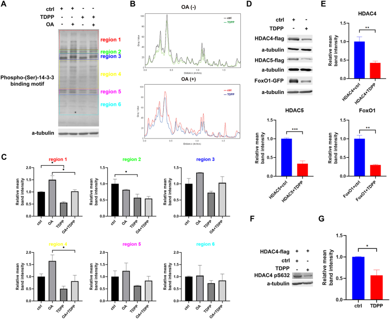
Fig. 3 TDPP degrades 14-3-3-BPPs. A. Western blot of phospho-(Ser)-14-3-3-binding-motif treated with control/TDPP vector with/without 10 nM OA. Different molecular weight ranges were grouped as different regions and marked as different colors, which were roughly grouped by similar band intensity and reflect the difference among treatments. The grey value of bands and regions were quantified and further analyzed in panel B and C, respectively. B. Illustration of the grey value distribution of phospho-(Ser)-14-3-3-binding-motif in each treatment in panel A. X axis represents the distance to the top edge of the blot results. Upper panel: lane 1 (black) vs. lane 3 (green). Lower panel: lane 2 (red) vs. lane 4 (blue). C. Quantification of phospho-(Ser)-14-3-3-binding-motifembedded proteins with different molecular weight in panel A. D and E. Western blot results (D) and the quantifications (E) of several different phospho-14-3-3-binding-motif embedded proteins (HDAC4, HDAC5, FoxO1) in HEK293T/17 cells treated with control/TDPP vectors. F and G. Western blot result (F) and the quantification (G) of HDAC4 phospho-S632 in a HDAC4 wildtype vectors transfected HEK293T/17 cells treated with control/TDPP vector. Data are represented as mean ± SEM. See also Fig. S2.
Fig. 3 TDPP degrades 14-3-3-BPPs. A. Western blot of phospho-(Ser)-14-3-3-binding-motif treated with control/TDPP vector with/without 10 nM OA. Different molecular weight ranges were grouped as different regions and marked as different colors, which were roughly grouped by similar band intensity and reflect the difference among treatments. The grey value of bands and regions were quantified and further analyzed in panel B and C, respectively. B. Illustration of the grey value distribution of phospho-(Ser)-14-3-3-binding-motif in each treatment in panel A. X axis represents the distance to the top edge of the blot results. Upper panel: lane 1 (black) vs. lane 3 (green). Lower panel: lane 2 (red) vs. lane 4 (blue). C. Quantification of phospho-(Ser)-14-3-3-binding-motifembedded proteins with different molecular weight in panel A. D and E. Western blot results (D) and the quantifications (E) of several different phospho-14-3-3-binding-motif embedded proteins (HDAC4, HDAC5, FoxO1) in HEK293T/17 cells treated with control/TDPP vectors. F and G. Western blot result (F) and the quantification (G) of HDAC4 phospho-S632 in a HDAC4 wildtype vectors transfected HEK293T/17 cells treated with control/TDPP vector. Data are represented as mean ± SEM. See also Fig. S2.CC BY-NC-ND · PubMed Central
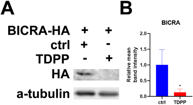
Fig. 4 TDPP validates BICRA as a new 14-3-3 interaction target protein. A. Western blot of BICRA-HA level in its expression HEK293T/17 cells transfected with control/TDPP vectors. B. Relative mean band intensity of BICRA-HA Western blot. Data are represented as mean ± SEM.
Fig. 4 TDPP validates BICRA as a new 14-3-3 interaction target protein. A. Western blot of BICRA-HA level in its expression HEK293T/17 cells transfected with control/TDPP vectors. B. Relative mean band intensity of BICRA-HA Western blot. Data are represented as mean ± SEM.CC BY-NC-ND · PubMed Central
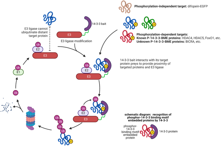
Fig. 5 Working model of TDPP, a ubiquitination-mediated degradation system to target 14-3-3-binding phosphoproteins.
Fig. 5 Working model of TDPP, a ubiquitination-mediated degradation system to target 14-3-3-binding phosphoproteins.CC BY-NC-ND · PubMed Central

Verification

Claims the paper makes

The claims and statistics of this paper haven’t been checked yet.

Run the check — free, no account needed. It reads the open-access text and checks the sources it cites.

A few free checks a day · papers already verified open instantly · results are public

Self-check

Percentages, sums and reported differences re-computed from the paper’s own numbers.

The self-check needs the paper’s full text.

Citations

Cites

Show all 1 references

Research works printed but not linked: 64

  1. 10.3390/ph13120441 · DOI
  2. 10.1016/j.pbiomolbio.2016.12.003 · DOI
  3. 10.1080/15622975.2021.1925585 · DOI
  4. 10.3892/ijmm.2017.3036 · DOI
  5. 10.1021/acschembio.5b00442 · DOI · Paper page
  6. 10.1016/j.cell.2019.11.031 · DOI · Paper page
  7. 10.1038/nsmb.2180 · DOI · Paper page
  8. 10.1126/science.1175371 · DOI · Paper page
  9. 10.1016/j.cell.2017.10.033 · DOI · Paper page
  10. 10.1038/onc.2009.382 · DOI
  11. 10.3389/fnmol.2017.00318 · DOI
  12. 10.1021/acs.jmedchem.7b00635 · DOI · Paper page
  13. 10.1016/j.tips.2020.04.005 · DOI · Paper page
  14. 10.2174/0929867023368791 · DOI
  15. 10.1146/annurev.pharmtox.40.1.617 · DOI
  16. 10.1038/nrm.2016.143 · DOI
  17. 10.1098/rsob.170066 · DOI · Paper page
  18. 10.1098/rsob.160255 · DOI · Paper page
  19. 10.1073/pnas.0801802105 · DOI
  20. 10.1093/eurheartj/ehab030 · DOI
  21. 10.1038/s41467-021-21908-8 · DOI · Paper page
  22. 10.1073/pnas.140199597 · DOI
  23. 10.1101/cshperspect.a011189 · DOI
  24. 10.1016/j.cca.2020.09.009 · DOI
  25. 10.1016/j.molcel.2020.04.032 · DOI
  26. 10.1021/acs.chemrev.5b00683 · DOI · Paper page
  27. 10.1126/science.aay0543 · DOI
  28. 10.1002/jcb.24570 · DOI
  29. 10.3389/fcell.2019.00358 · DOI
  30. 10.1074/jbc.M115.637389 · DOI
  31. 10.1111/jcmm.16490 · DOI
  32. 10.1038/s41580-020-00303-z · DOI · Paper page
  33. 10.1093/bioinformatics/btv133 · DOI
  34. 10.1093/bioinformatics/btt473 · DOI · Paper page
  35. 10.1074/jbc.M105971200 · DOI
  36. 10.1073/pnas.2008214117 · DOI
  37. 10.1038/s41592-022-01488-1 · DOI · Paper page
  38. 10.1074/jbc.M114.607580 · DOI
  39. 10.1038/s41589-018-0021-8 · DOI · Paper page
  40. 10.1038/nchembio.597 · DOI · Paper page
  41. 10.1038/nmeth.1401 · DOI · Paper page
  42. 10.1016/j.bbrc.2008.10.079 · DOI
  43. 10.1091/mbc.E07-06-0623 · DOI · Paper page
  44. 10.1074/jbc.M114.592287 · DOI
  45. 10.1038/s41388-018-0348-3 · DOI
  46. 10.1146/annurev.biochem.70.1.503 · DOI · Paper page
  47. 10.1371/journal.pone.0013462 · DOI
  48. 10.1016/j.bmcl.2008.07.114 · DOI · Paper page
  49. 10.1016/j.celrep.2014.07.036 · DOI
  50. 10.1091/mbc.E02-12-0821 · DOI
  51. 10.1038/nature05731 · DOI · Paper page
  52. 10.1002/prot.10210 · DOI
  53. 10.1016/j.jmb.2021.166875 · DOI · Paper page
  54. 10.1128/MCB.20.18.6904-6912.2000 · DOI
  55. 10.1021/bi991353h · DOI
  56. 10.1002/pmic.201400441 · DOI
  57. 10.2217/epi.13.73 · DOI · Paper page
  58. 10.1101/gad.1238104 · DOI
  59. 10.1038/s41417-019-0097-7 · DOI
  60. 10.1073/pnas.0605779103 · DOI
  61. 10.1038/s41467-019-09844-0 · DOI
  62. 10.1074/jbc.272.21.13717 · DOI
  63. 10.1074/jbc.M105086200 · DOI
  64. 10.1146/annurev-biochem-060815-014922 · DOI · Paper page

Metadata

Authors
Li, Z., Huang, X., Li, M., Sabry, Z., Chen, Y. E., Wang, Z., & Liu, L.
Affiliations
  • University of Michigan (US)
  • Chinese Academy of Medical Sciences & Peking Union Medical College (CN)
Corresponding author
  • Wang, Zhong
  • Liu, Liu
Venue
Heliyon
Publisher
openRxiv
Published
2022-11-23
Volume
9
Issue
5
Pages
e16318-e16318
ISSN
2405-8440 (ISSN-L)
OpenAlex
W4377086858
Cited by
2
References
1
Citations by year
2024: 2
FWCI
0.18
Type
journal article
Language
en
Open access
Licence
cc-by-nc-nd
Locations
  • Heliyon (publishedVersion, cc-by-nc-nd)
  • bioRxiv (Cold Spring Harbor Laboratory) (acceptedVersion, cc-by-nc-nd)
  • PubMed Central (publishedVersion, cc-by-nc-nd)
  • PubMed (publishedVersion)
  • DOAJ (DOAJ: Directory of Open Access Journals) (submittedVersion, cc-by-sa)
Funders
  • National Institutes of Health
  • National Heart, Lung, and Blood Institute (R01HL139735, R01HL163672)
Topics
  • Life Sciences › Biochemistry, Genetics and Molecular Biology › Molecular Biology › Protein Degradation and Inhibitors
  • Life Sciences › Biochemistry, Genetics and Molecular Biology › Molecular Biology › Ubiquitin and proteasome pathways
  • Life Sciences › Biochemistry, Genetics and Molecular Biology › Molecular Biology › 14-3-3 protein interactions
Keywords
targeted protein degradation, ubiquitination, ubiquitin-proteasome system, 14-3-3 binding motif, protein chimeras

About these checks

Registry, retraction, Cites and statistics results are computed, not judged, with one exception: a printed reference that no index lists is read once by a model before it counts as a citation error. Claims are extracted by a model and kept only when their quote appears verbatim in the text; each cited source is then checked, with a model’s help, for whether it says what the claim relies on. Those verdicts can be wrong — read the claim’s quote and the cited source before relying on one.

Statistics and arithmetic are recomputed, not judged.

Confidence bands are shown only for fields calibrated on our annotated test set. None is calibrated yet, so no band is shown.

What counts as a claim ›How we measure confidence ›How we check this ›

Are you an author, or do you see a problem with this page? Request a review

How to cite

Cite CoChat for the checks on this page, and the paper for its own findings.

This page
CoChat. (2026). Citation check: A ubiquitination-mediated degradation system to target phospho-14-3-3-binding-motif embedded proteins. https://app.cochat.ai/papers/10.1101/2022.11.22.517526
The paper
Li, Z., Huang, X., Li, M., Sabry, Z., Chen, Y. E., Wang, Z., & Liu, L. (2022). A ubiquitination-mediated degradation system to target phospho-14-3-3-binding-motif embedded proteins. Heliyon. https://doi.org/10.1101/2022.11.22.517526