

The DOI matches its Crossref record; 1 of the 1 references it cites were found in a registry or an index.
Human plastins are novel cytoskeletal pH sensors with a reduced F-actin bundling capacity at basic pH
Open accessPreprint — published as 10.1016/j.jmb.2025.169306 · Paper page
Abstract
Intracellular pH (pH i ) is a fundamental component of cell homeostasis. Controlled elevations in pH i precede and accompany cell polarization, cytokinesis, and directional migration. pH dysregulation contributes to cancer, neurodegenerative diseases, diabetes, and other metabolic disorders. While cytoskeletal rearrangements are crucial for these processes, only a few cytoskeletal proteins, namely Cdc42, cofilin, talin, cortactin, α-actinin, and AIP1 have been documented as pH sensors. Here, we report that actin-bundling proteins plastin 2 (PLS2, aka LCP1) and plastin 3 (PLS3) respond to physiological scale pH fluctuations by a reduced F-actin bundling at alkaline pH. The inhibition of PLS2 actin-bundling activity at elevated pH stems from the reduced affinity of the N-terminal actin-binding domain (ABD1) to actin. In fibroblast cells, elevated cytosolic pH caused the dissociation of ectopically expressed PLS2 from actin structures, whereas acidic conditions promoted its tighter association with focal adhesions and stress fibers. We identified His207 as one of the pH-sensing residues whose mutation to Lys and Tyr reduces pH sensitivity by enhancing and inhibiting the bundling ability, respectively. Our results suggest that weaker actin bundling by plastin isoforms at alkaline pH favors higher dynamics of the actin cytoskeleton. Therefore, like other cytoskeleton pH sensors, plastins promote disassembly and faster dynamics of cytoskeletal components during cytokinesis and cell migration. Since both plastins are implemented in cancer, their pH sensitivity may contribute to the accelerated proliferation and enhanced invasive and metastatic potentials of cancer cells at alkaline pH i . Abstract Figure:
Overview
The claims and statistics of this paper haven’t been checked yet.
Run the check — free, no account needed. It reads the open-access text and checks the sources it cites.
Integrity signals
Registry
Cites
Statistics
Figures
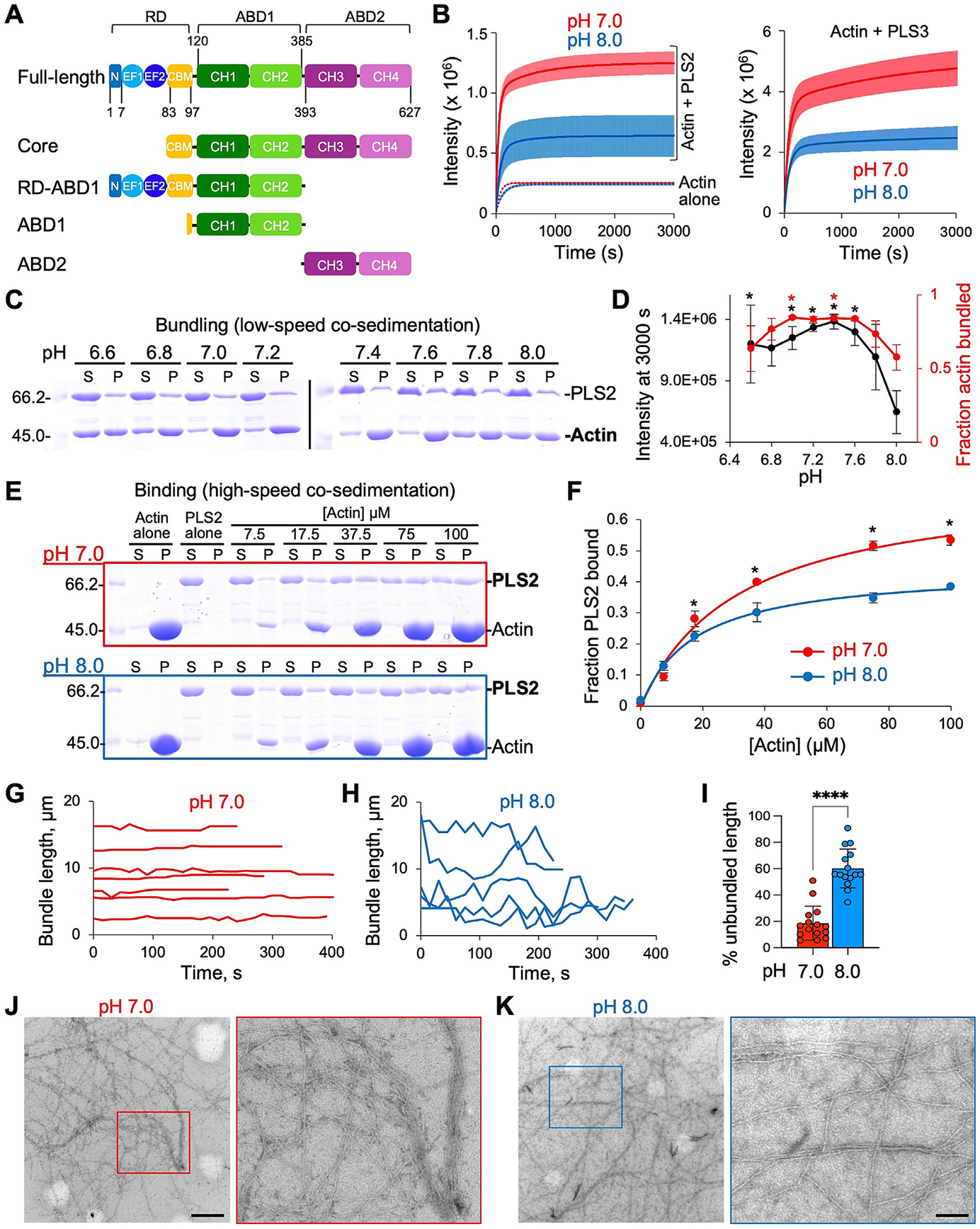
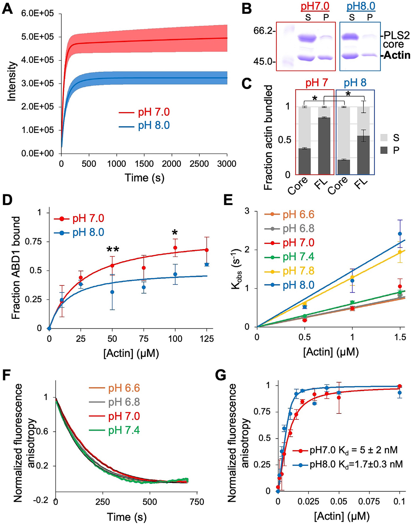
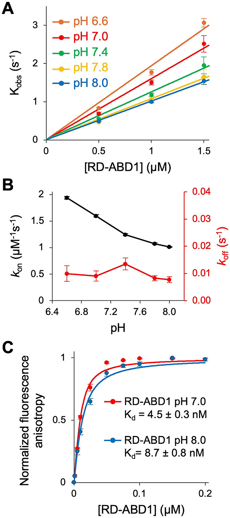
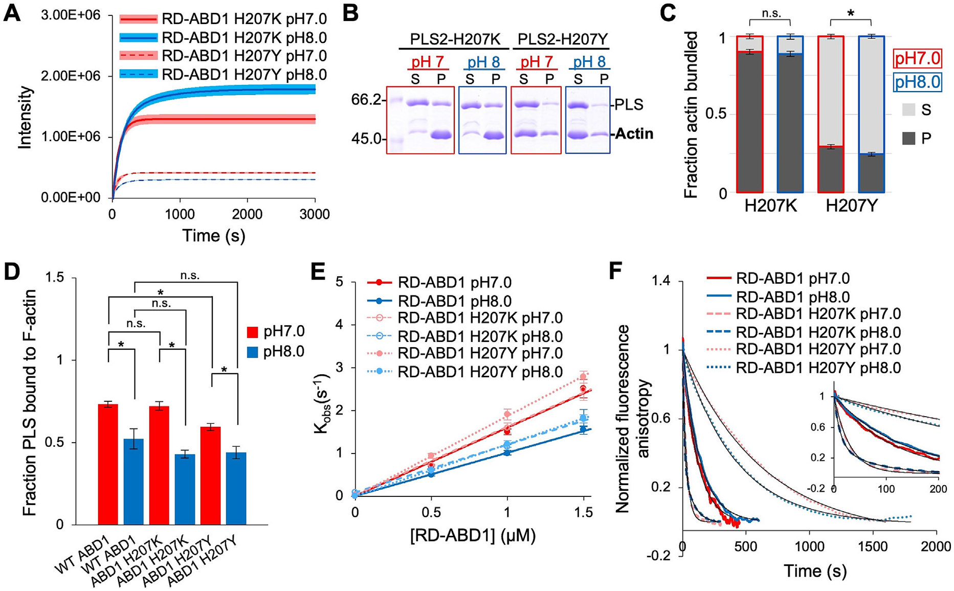
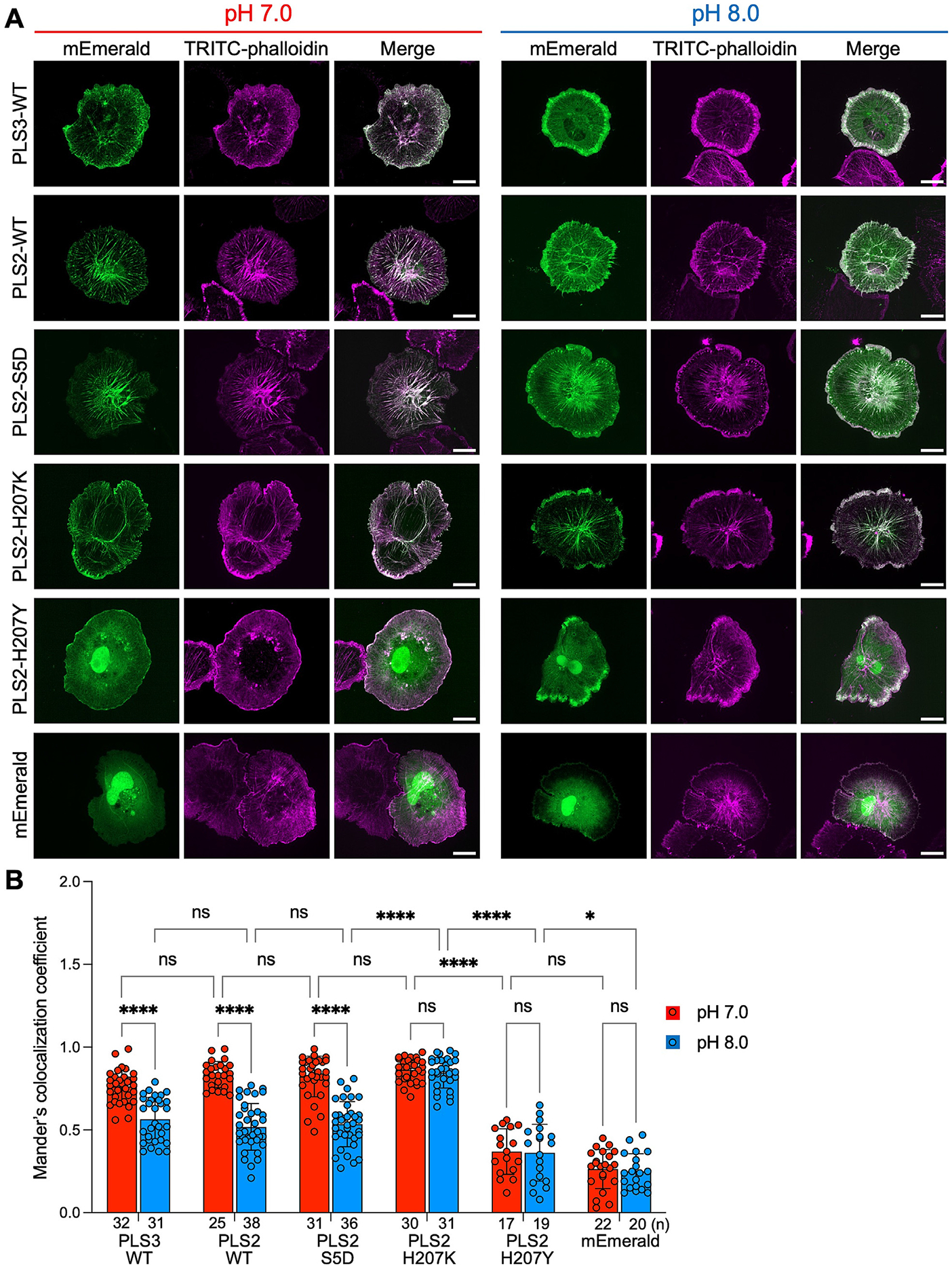
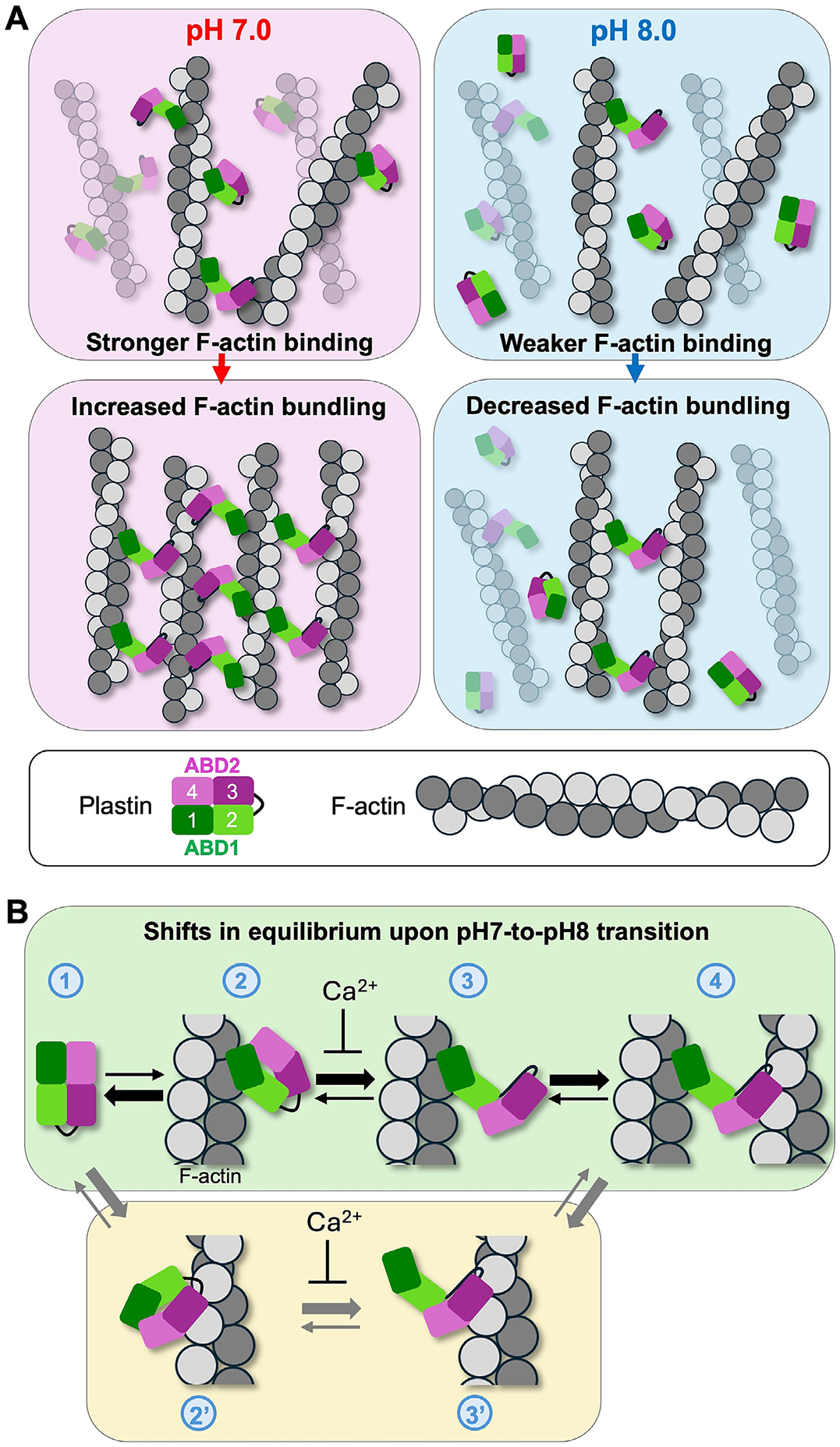
Verification
Claims the paper makes
The claims and statistics of this paper haven’t been checked yet.
Run the check — free, no account needed. It reads the open-access text and checks the sources it cites.
Self-check
Percentages, sums and reported differences re-computed from the paper’s own numbers.
The self-check needs the paper’s full text.
Citations
Cites
Highly accurate protein structure prediction with AlphaFold
Jumper, J., Evans, R., Pritzel, A., Green, T., Figurnov, M., Ronneberger, O., Tunyasuvunakool, K., Bates, R., Žídek, A., Potapenko, A., et al. (2021) · Nature · doi:10.1038/s41586-021-03819-2 · Cited by 43,764
Research works printed but not linked: 95
- 10.1038/nrm2820 · DOI · Paper page
- Spear, J. S. & White, K. A . Single-cell intracellular pH dynamics regulate the cell cycle by timing the G1 exit and G2 transition. J. Cell Sci. 136, 2021.06.04.447151 (2023). · DOI
- 2020, Cancer and pH dynamics: Transcriptional regulation, proteostasis, and the need for new molecular tools, Cancers (Basel, 12, 1
- 10.1002/jnr.23757 · DOI
- 10.1111/j.1748-1716.2006.01543.x · DOI
- 2012, The Na+ / H+ - Exchanger (NHE1) Generates pH Nanodomains at Focal Adhesions, J. Cell. Physiol, 228, 1351
- 10.1083/jcb.200208050 · DOI
- 10.1083/jcb.201302131 · DOI
- 10.1152/physrev.00018.2011 · DOI · Paper page
- 10.1038/nrc1713 · DOI · Paper page
- 10.1111/wrr.12526 · DOI
- 10.1152/ajpcell.00280.2010 · DOI
- 10.1083/jcb.200704169 · DOI
- 10.1074/jbc.M310148200 · DOI
- 10.1083/jcb.200804161 · DOI
- 10.1073/pnas.0805163105 · DOI
- 10.1083/jcb.201103045 · DOI
- 10.1074/jbc.M115.713495 · DOI
- 10.1042/BST0330891 · DOI
- Hawkins, M. , Pope, B. , Maciver, S. K. & Weeds, A. G . Human Actin Depolymerizing Factor Mediates Actin Filaments pH-Sensitive Destruction of Actin Filaments. Biochemistry 9985–9993 (1993). · DOI
- Chen, H. et al. In Vitro Activity Differences between Proteins of the ADF / Cofilin Family Define Two Distinct Subgroups. Biochemistry 7127–7142 (2004). · DOI
- 10.1021/acs.biochem.8b01001 · DOI
- 10.1242/jcs.01631 · DOI
- 10.1083/jcb.94.2.466 · DOI
- 10.1083/jcb.100.2.375 · DOI
- 10.1101/cshperspect.a022053 · DOI · Paper page
- 10.1016/j.jmb.2017.06.021 · DOI
- 10.1016/j.str.2004.04.010 · DOI
- 10.1038/s41594-022-00771-1 · DOI
- 10.1073/pnas.2110877119 · DOI
- 10.1152/physrev.00018.2013 · DOI
- Pollard, T. D. Actin and actin-binding proteins. Cold Spring Harb. Perspect. Biol. 8, (2016). · DOI
- 10.1016/S0021-9258(18)53842-4 · DOI
- 10.1155/2012/282041 · DOI · Paper page
- 10.1128/MCB.14.4.2457 · DOI
- 10.1111/cge.13626 · DOI
- 10.1155/2012/282041 · DOI · Paper page
- 10.1007/s00018-021-03843-5 · DOI
- 1994, Activation of the Leukocyte Plastin Gene Occurs in Most Human Cancer Cells, Cancer Res, 54, 1775
- 10.1002/ijc.21593 · DOI
- 10.1186/1476-4598-13-1 · DOI
- 10.1186/s12964-021-00728-9 · DOI
- 2019, Plastin 3 down-regulation augments the sensitivity of MDA-MB-231 cells to paclitaxel via the p38 MAPK signalling pathway. Artif. Cells, Nanomedicine, Biotechnol, 47, 684
- 10.3390/ijms19061551 · DOI
- 10.1002/jcp.29685 · DOI
- 10.1002/tox.23872 · DOI
- 10.1242/jcs.02874 · DOI
- 10.1038/s41413-020-0095-2 · DOI
- 10.1038/srep41926 · DOI
- 10.1016/j.cell.2010.12.001 · DOI · Paper page
- 10.1074/mcp.M111.016410 · DOI · Paper page
- 10.1038/nature18003 · DOI · Paper page
- 10.1074/mcp.M113.036392 · DOI · Paper page
- 10.1021/pr300630k · DOI · Paper page
- 10.4049/jimmunol.1103242 · DOI
- 10.1093/oxfordjournals.jbchem.a123929 · DOI
- 10.1021/bi00510a015 · DOI
- 10.1016/S0006-3495(89)82804-8 · DOI
- 10.1016/S0091-679X(08)60661-5 · DOI
- 10.1021/acs.biochem.6b01117 · DOI
- 10.1038/s41467-016-0009-6 · DOI · Paper page
- 10.7554/eLife.52760 · DOI · Paper page
- 10.1002/prot.10177 · DOI
- 10.1021/bi00578a012 · DOI
- 10.1016/B978-0-12-420138-5.00023-9 · DOI
- 10.1002/eji.201040345 · DOI
- 10.4049/jimmunol.1001424 · DOI
- Zhou, J. Y. et al. L-Plastin promotes podosome longevity and supports macrophage motility. Mol Immunol October, 79–88 (2016). · DOI
- 10.1002/cm.21717 · DOI
- Chellaiah, M. A. et al. L-Plastin deficiency produces increased trabecular bone due to attenuation of sealing ring formation and osteoclast dysfunction. Bone Res. 8, (2020). · DOI
- 10.4049/jimmunol.1101033 · DOI
- 10.1189/jlb.69.4.522 · DOI · Paper page
- 10.1371/journal.pone.0015746 · DOI
- 10.1152/ajpregu.00154.2014 · DOI
- 1989, Regulation of podosomes by intracellular pH in avian osteoclasts, Boll. Soc. Ital. Biol. Sper, 65, 597
- 10.1172/JCI114235 · DOI
- 2011, Dysregulated pH: a perfect storm for cancer progression, Nat. Publ. Gr, 11, 671
- 2009, The coordination between actin filaments and adhesion in mesenchymal migration, Cell Adhes. Migr, 3, 2
- 10.1371/journal.pone.0089680 · DOI · Paper page
- Garbett, D. et al. T-Plastin reinforces membrane protrusions to bridge matrix gaps during cell migration. Nat. Commun. 11, (2020). · DOI
- 10.5483/BMBRep.2024-0040 · DOI
- 10.1038/nsmb.1789 · DOI
- 10.1073/pnas.1106453108 · DOI
- 10.1016/j.bpj.2018.05.039 · DOI
- 10.1091/mbc.E19-06-0317 · DOI
- 10.3390/ijms23137026 · DOI
- 2006, Similarity of salt influences on the pH of buffers, polyelectrolytes, and proteins, J. Phys. Chem. B, 110, 8870, 10.1021/jp0600209 · DOI
- 10.1385/1-59259-890-0:571 · DOI
- 10.1038/nmeth.2019 · DOI · Paper page
- 10.1002/mrd.22489 · DOI
- Watanabe, N . Fluorescence single-molecule imaging of actin turnover and regulatory mechanisms. Methods in Enzymology vol. 505 (Elsevier Inc., 2012). · DOI
- Uphoff, C. C. & Drexler, H. G . Detection of Mycoplasma Contamination in Cell Cultures. Curr. Protoc. Mol. Biol. 106, (2014). · DOI · Paper page
- Rizzo, M. A. , Davidson, M. W. & Piston, D. W . Fluorescent protein tracking and detection: fluorescent protein structure and color variants. Cold Spring Harb. Protoc. 2009, pdb.top63 (2009). · DOI
- 10.1083/jcb.200908086 · DOI
- 10.1093/NAR/GKAB1061 · DOI · Paper page
Metadata
- Authors
- Runyan, L. A., Kudryashova, E., Agrawal, R., Mohamed, M., & Kudryashov, D. S.
- Affiliations
- The Ohio State University (US)
- Corresponding author
- Kudryashov, Dmitri S.
- Venue
- Journal of Molecular Biology
- Publisher
- openRxiv
- Published
- 2025-03-28
- Volume
- 437
- Issue
- 19
- Pages
- 169306-169306
- ISSN
- 0022-2836 (ISSN-L)
- PMC
- PMC12309418
- OpenAlex
- W4411632033
- Cited by
- 3
- References
- 1
- Citations by year
- 2026: 3
- FWCI
- 1.49
- Type
- journal article
- Language
- en
- Open access
- hybrid
- doi.org
- Licence
- Locations
- Journal of Molecular Biology (publishedVersion, cc-by)
- bioRxiv (Cold Spring Harbor Laboratory) (acceptedVersion, cc-by-nd)
- PubMed (publishedVersion)
- PubMed Central (acceptedVersion, cc-by)
- Europe PMC (PubMed Central) (submittedVersion, cc-by-nd)
- PubMed Central (submittedVersion, cc-by-nd)
- Funders
- National Institutes of Health
- National Cancer Institute (P30CA016058)
- National Institute of General Medical Sciences (R01GM145813)
- Topics
- Life Sciences › Biochemistry, Genetics and Molecular Biology › Cell Biology › Cellular Mechanics and Interactions
- Life Sciences › Biochemistry, Genetics and Molecular Biology › Molecular Biology › Protein Kinase Regulation and GTPase Signaling
- Life Sciences › Biochemistry, Genetics and Molecular Biology › Molecular Biology › Protein Structure and Dynamics
- Keywords
- PLS3, intracellular pH, F-actin bundling, actin-binding domain, alkaline pH, actin cytoskeleton dynamics, focal adhesions, stress fibers, tumor invasion, metastasis, cell migration, cytokinesis
About these checks
Registry, retraction, Cites and statistics results are computed, not judged, with one exception: a printed reference that no index lists is read once by a model before it counts as a citation error. Claims are extracted by a model and kept only when their quote appears verbatim in the text; each cited source is then checked, with a model’s help, for whether it says what the claim relies on. Those verdicts can be wrong — read the claim’s quote and the cited source before relying on one.
Statistics and arithmetic are recomputed, not judged.
Confidence bands are shown only for fields calibrated on our annotated test set. None is calibrated yet, so no band is shown.
What counts as a claim ›How we measure confidence ›How we check this ›
Are you an author, or do you see a problem with this page? Request a review
