

The DOI matches its Crossref record; 7 of the 7 references it cites were found in a registry or an index.
Macromolecular structure determination using X-rays, neutrons and electrons: recent developments in Phenix
Abstract
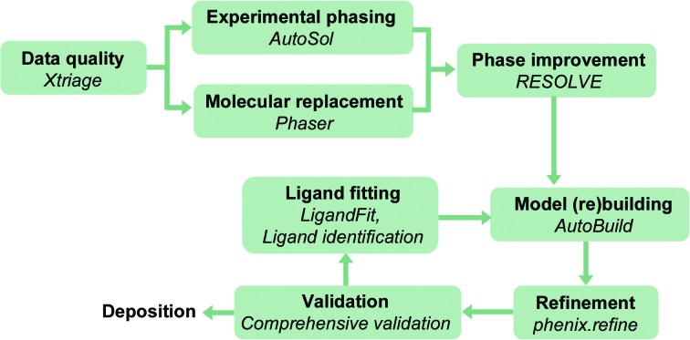
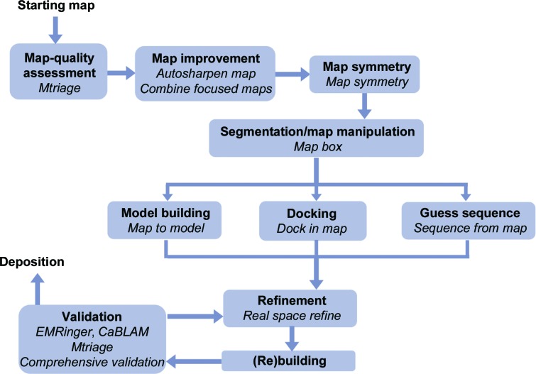
Diffraction (X-ray, neutron and electron) and electron cryo-microscopy are powerful methods to determine three-dimensional macromolecular structures, which are required to understand biological processes and to develop new therapeutics against diseases. The overall structure-solution workflow is similar for these techniques, but nuances exist because the properties of the reduced experimental data are different. Software tools for structure determination should therefore be tailored for each method. Phenix is a comprehensive software package for macromolecular structure determination that handles data from any of these techniques. Tasks performed with Phenix include data-quality assessment, map improvement, model building, the validation/rebuilding/refinement cycle and deposition. Each tool caters to the type of experimental data. The design of Phenix emphasizes the automation of procedures, where possible, to minimize repetitive and time-consuming manual tasks, while default parameters are chosen to encourage best practice. A graphical user interface provides access to many command-line features of Phenix and streamlines the transition between programs, project tracking and re-running of previous tasks.
Overview
The claims and statistics of this paper haven’t been checked yet.
Run the check — free, no account needed. It reads the open-access text and checks the sources it cites.
Integrity signals
Registry
Cites
Statistics
Figures
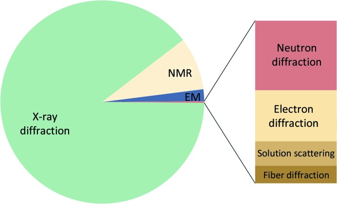
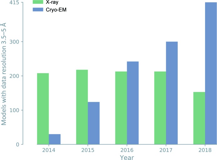
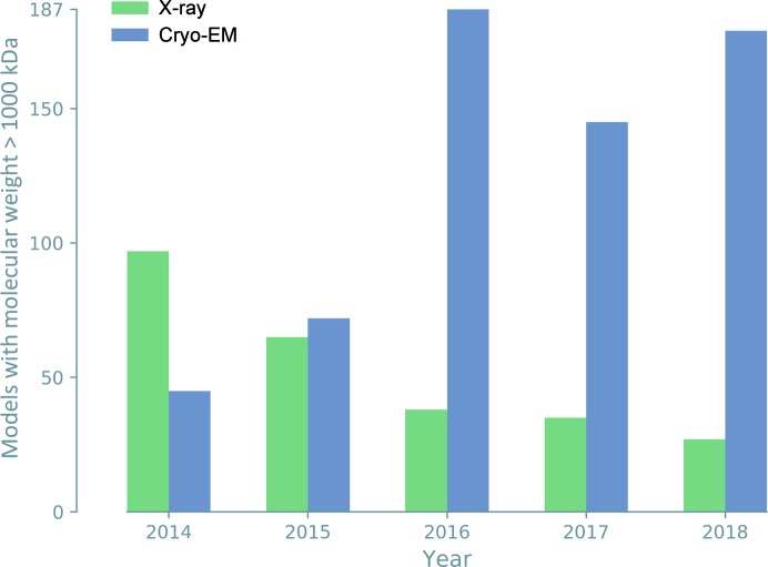
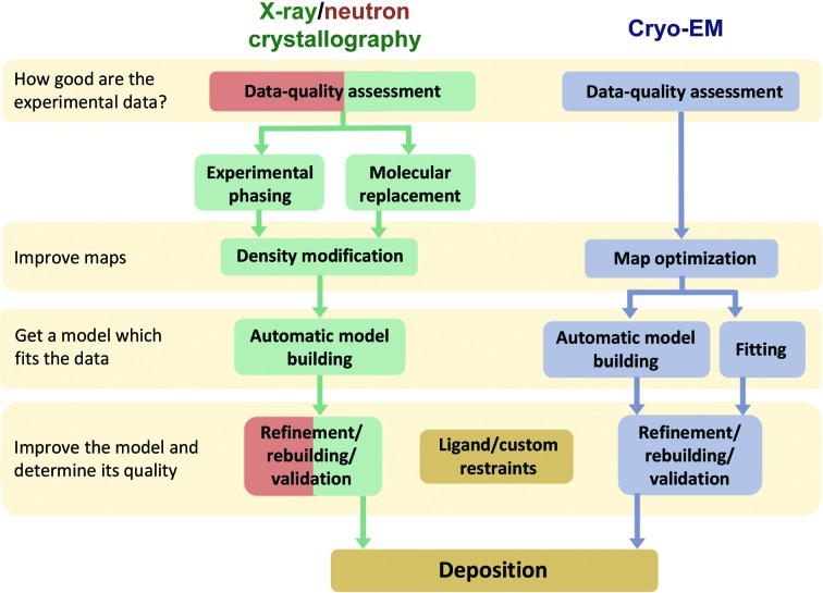
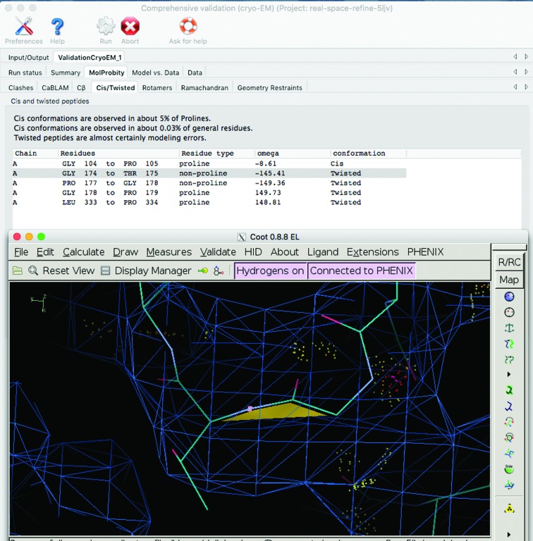
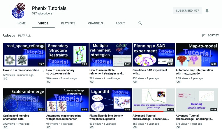
Verification
Claims the paper makes
The claims and statistics of this paper haven’t been checked yet.
Run the check — free, no account needed. It reads the open-access text and checks the sources it cites.
Claims other papers make about this one
Each is checked against this paper’s own text: does it say what the citing paper says it does?
The cited source says what the claim attributes to it.
In the citing paper
“Model geometries were assessed using the MolProbity module in Phenix, the Molprobity server68 (http://molprobity.biochem.duke.edu/), and the PDB Validation server69 (www.wwpdb.org).”
From Structural organization of the retriever–CCC endosomal recycling complex (2024) · cites this paper as [69]
Self-check
Percentages, sums and reported differences re-computed from the paper’s own numbers.
The self-check needs the paper’s full text.
Citations
Cites
Matplotlib: a 2D graphics environment
Hunter, J. D. (2007) · Computing in Science & Engineering · doi:10.1109/mcse.2007.55 · Cited by 39,249
PHENIX : a comprehensive Python-based system for macromolecular structure solution
Adams, P. D., Afonine, P. V., Bunkóczi, G., Chen, V. B., Davis, I. W., Echols, N., Headd, J. J., Hung, L.-W., Kapral, G. J., Grosse-Kunstleve, R. W., et al. (2010) · Acta Crystallographica Section D Biological Crystallography · doi:10.1107/s0907444909052925 · Cited by 23,716
Self-citationPhaser crystallographic software
McCoy, A. J., Grosse-Kunstleve, R. W., Adams, P. D., Winn, M. D., Storoni, L. C., & Read, R. J. (2007) · Journal of Applied Crystallography · doi:10.1107/s0021889807021206 · Cited by 20,612
Self-citationTowards automated crystallographic structure refinement with phenix.refine
Afonine, P. V., Grosse-Kunstleve, R. W., Echols, N., Headd, J. J., Moriarty, N. W., Mustyakimov, M., Terwilliger, T. C., Urzhumtsev, A., Zwart, P. H., & Adams, P. D. (2012) · Acta Crystallographica Section D Biological Crystallography · doi:10.1107/s0907444912001308 · Cited by 5,511
Self-citationTerwilliger, T. C., Grosse-Kunstleve, R. W., Afonine, P. V., Moriarty, N. W., Zwart, P. H., Hung, L.-W., Read, R. J., & Adams, P. D. (2008) · Acta Crystallographica Section D Biological Crystallography · doi:10.1107/s090744490705024x · Cited by 1,462
Self-citationKühlbrandt, W. (2014) · Science · doi:10.1126/science.1251652 · Cited by 1,325
De novo modeling in cryo-EM density maps with Pathwalking
Chen, M., Baldwin, P. R., Ludtke, S. J., & Baker, M. L. (2016) · Journal of Structural Biology · doi:10.1016/j.jsb.2016.06.004 · Cited by 82
Research works printed but not linked: 149
- Abrahams, 2003, C/C++ Users J., 21, 29
- Adams, 2019, Acta Cryst. D, 75, 451, 10.1107/S2059798319004522 · DOI
- Adams, 2009, Curr. Opin. Struct. Biol., 19, 566, 10.1016/j.sbi.2009.07.014 · DOI
- Adams, 2002, Acta Cryst. D, 58, 1948, 10.1107/S0907444902016657 · DOI · Paper page
- Adams, 2009, Acta Cryst. D, 65, 567, 10.1107/S0907444909011548 · DOI
- Afonine, 2018, Acta Cryst. D, 74, 621, 10.1107/S2059798318005764 · DOI
- Afonine, 2005, Acta Cryst. D, 61, 850, 10.1107/S0907444905007894 · DOI
- Afonine, 2007, Acta Cryst. D, 63, 1194, 10.1107/S0907444907046148 · DOI
- Afonine, 2013, Acta Cryst. D, 69, 625, 10.1107/S0907444913000462 · DOI
- Afonine, 2009, J. Appl. Cryst., 42, 607, 10.1107/S0021889809023528 · DOI
- Afonine, 2018, Acta Cryst. D, 74, 814, 10.1107/S2059798318009324 · DOI
- Afonine, 2015, Acta Cryst. D, 71, 646, 10.1107/S1399004714028132 · DOI
- Afonine, 2010, Acta Cryst. D, 66, 1153, 10.1107/S0907444910026582 · DOI
- Afonine, 2018, Acta Cryst. D, 74, 531, 10.1107/S2059798318006551 · DOI
- Bai, 2015, Trends Biochem. Sci., 40, 49, 10.1016/j.tibs.2014.10.005 · DOI
- Baker, 2012, Structure, 20, 450, 10.1016/j.str.2012.01.008 · DOI
- Barad, 2015, Nature Methods, 12, 943, 10.1038/nmeth.3541 · DOI · Paper page
- Berkholz, 2009, Structure, 17, 1316, 10.1016/j.str.2009.08.012 · DOI
- Bernstein, 1977, J. Mol. Biol., 112, 535, 10.1016/S0022-2836(77)80200-3 · DOI
- Blow, D. M., Navaza, J., Tong, L., Arnold, E. & Rossmann, M. G. (2012). International Tables for Crystallography, Vol. F, edited by E. Arnold, D. M. Himmel & M. G. Rossmann, pp. 333-366. Chichester: John Wiley & Sons.
- Borbulevych, 2014, Comput. Crystallogr. Newsl., 5, 26
- Brändén, 1990, Nature (London), 343, 687, 10.1038/343687a0 · DOI
- Britton, 1972, Acta Cryst. A, 28, 296, 10.1107/S0567739472000786 · DOI
- Brünger, 1992, Nature (London), 355, 472, 10.1038/355472a0 · DOI
- Brünger, 1998, Acta Cryst. D, 54, 905, 10.1107/S0907444998003254 · DOI
- Brunger, 2012, Acta Cryst. D, 68, 391, 10.1107/S090744491104978X · DOI
- Bryan, 2013, Acta Cryst. F, 69, 1015, 10.1107/S1744309113021386 · DOI
- Bunkóczi, 2013, Acta Cryst. D, 69, 2276, 10.1107/S0907444913022750 · DOI
- Bunkóczi, 2011, Acta Cryst. D, 67, 303, 10.1107/S0907444910051218 · DOI
- Burley, 2019, Nucleic Acids Res., 47, D464, 10.1093/nar/gky1004 · DOI
- Burnley, 2012, Elife, 1, e00311, 10.7554/eLife.00311 · DOI
- Busing, 1964, Acta Cryst., 17, 142, 10.1107/S0365110X64000408 · DOI
- Cardone, 2013, J. Struct. Biol., 184, 226, 10.1016/j.jsb.2013.08.002 · DOI
- Chapman, 2011, Nature (London), 470, 73, 10.1038/nature09750 · DOI
- Chen, 2010, Acta Cryst. D, 66, 12, 10.1107/S0907444909042073 · DOI · Paper page
- Chen, 2009, Protein Sci., 18, 2403, 10.1002/pro.250 · DOI
- Chou, 2013, Nature Methods, 10, 74, 10.1038/nmeth.2262 · DOI
- Clowney, 1996, J. Am. Chem. Soc., 118, 509, 10.1021/ja952883d · DOI
- Coppens, 1967, Science, 158, 1577, 10.1126/science.158.3808.1577 · DOI
- Croll, 2015, Acta Cryst. D, 71, 706, 10.1107/S1399004715000826 · DOI
- Croll, 2018, Acta Cryst. D, 74, 519, 10.1107/S2059798318002425 · DOI
- Dauter, 2017, Methods Mol. Biol., 1607, 349, 10.1007/978-1-4939-7000-1_14 · DOI
- Deis, 2013, Comput. Crystallogr. Newsl., 4, 9
- Diederichs, 2017, Methods Mol. Biol., 1607, 239, 10.1007/978-1-4939-7000-1_10 · DOI
- DiMaio, 2013, Nature Methods, 10, 1102, 10.1038/nmeth.2648 · DOI
- DiMaio, 2011, Nature (London), 473, 540, 10.1038/nature09964 · DOI
- Emsley, 2010, Acta Cryst. D, 66, 486, 10.1107/S0907444910007493 · DOI · Paper page
- Engh, 1991, Acta Cryst. A, 47, 392, 10.1107/S0108767391001071 · DOI
- Evans, 2007, Acta Cryst. D, 63, 58, 10.1107/S090744490604604X · DOI
- Evans, 2008, Acta Cryst. D, 64, 1, 10.1107/S0907444907051554 · DOI
- Fernández, 2008, J. Struct. Biol., 164, 170, 10.1016/j.jsb.2008.05.010 · DOI
- Gelbin, 1996, J. Am. Chem. Soc., 118, 519, 10.1021/ja9528846 · DOI
- Goddard, 2018, Protein Sci., 27, 14, 10.1002/pro.3235 · DOI · Paper page
- Grosse-Kunstleve, 2003, Acta Cryst. D, 59, 1966, 10.1107/S0907444903018043 · DOI
- Grosse-Kunstleve, R. W., Moriarty, N. W. & Adams, P. D. (2009). Proceedings of the ASME 2009 International Design Engineering Technical Conferences and Computers and Information in Engineering Conference, Vol. 4, pp. 1477-1485. New York: ASME.
- Grosse-Kunstleve, 2002, J. Appl. Cryst., 35, 126, 10.1107/S0021889801017824 · DOI
- Gruene, 2018, Angew. Chem. Int. Ed., 57, 16313, 10.1002/anie.201811318 · DOI
- Hahn, T. & Klapper, H. (2006). International Tables for Crystallography, Vol. D, edited by A. Authier, pp. 393-448. Dordrecht: Kluwer.
- Headd, 2012, Acta Cryst. D, 68, 381, 10.1107/S0907444911047834 · DOI
- Headd, 2014, Acta Cryst. D, 70, 1346, 10.1107/S1399004714003277 · DOI
- Heel, 1986, Optik, 73, 119
- Hintze, 2016, Proteins, 84, 1177, 10.1002/prot.25039 · DOI
- Hoppe, 1957, Acta Cryst., 10, 750
- Howells, 1950, Acta Cryst., 3, 210, 10.1107/S0365110X50000513 · DOI
- Jain, 2015, Methods Enzymol., 558, 181, 10.1016/bs.mie.2015.01.007 · DOI
- Jensen, 2010, Methods Enzymol., 483, xv, 10.1016/S0076-6879(10)83021-6 · DOI
- Jones, 2018, ACS Cent. Sci., 4, 1587, 10.1021/acscentsci.8b00760 · DOI
- Jones, 1991, Acta Cryst. A, 47, 110, 10.1107/S0108767390010224 · DOI
- Kendrew, 1958, Nature (London), 181, 662, 10.1038/181662a0 · DOI
- Kim, D. N., Moriarty, N. W., Kirmizialtin, S., Afonine, P. V., Poon, B. K., Sobolev, O. V., Adams, P. D. & Sanbonmatsu, K. Y. (2019). In the press.
- Kirmizialtin, 2015, Methods Enzymol., 558, 497, 10.1016/bs.mie.2015.02.011 · DOI
- Klei, 2014, Acta Cryst. D, 70, 134, 10.1107/S1399004713030071 · DOI
- Kleywegt, 1996, Structure, 4, 897, 10.1016/S0969-2126(96)00097-4 · DOI
- Kleywegt, 1995, Structure, 3, 535, 10.1016/S0969-2126(01)00187-3 · DOI
- Knihtila, 2015, J. Biol. Chem., 290, 31025, 10.1074/jbc.M115.679860 · DOI
- Kucukelbir, 2014, Nature Methods, 11, 63, 10.1038/nmeth.2727 · DOI
- Li, 2013, Nature Methods, 10, 584, 10.1038/nmeth.2472 · DOI
- Liebschner, 2018, Acta Cryst. D, 74, 800, 10.1107/S2059798318004588 · DOI
- Liebschner, 2017, Acta Cryst. D, 73, 148, 10.1107/S2059798316018210 · DOI
- Liu, 2017, Protein Sci., 26, 8, 10.1002/pro.2989 · DOI
- Loeffelholz, 2017, Curr. Opin. Struct. Biol., 46, 140, 10.1016/j.sbi.2017.07.007 · DOI
- Lovell, 2003, Proteins, 50, 437, 10.1002/prot.10286 · DOI · Paper page
- Lutz, M. & Ascher, D. (1999). Learning Python. Sebastopol: O'Reilly Media.
- McPherson, A. (2009). Introduction to Macromolecular Crystallography. Hoboken: Wiley-Blackwell. · DOI
- Moriarty, 2019, Acta Cryst. D, 75, 16, 10.1107/S205979831801519X · DOI
- Moriarty, 2017, Acta Cryst. D, 73, 123, 10.1107/S2059798316016570 · DOI
- Moriarty, 2009, Acta Cryst. D, 65, 1074, 10.1107/S0907444909029436 · DOI
- Moriarty, 2018, Protein Sci., 27, 182, 10.1002/pro.3296 · DOI
- Moriarty, 2014, FEBS J., 281, 4061, 10.1111/febs.12860 · DOI
- Moriarty, 2016, Acta Cryst. D, 72, 176, 10.1107/S2059798315022408 · DOI
- Nannenga, 2018, Emerg. Top. Life Sci., 2, 1, 10.1042/ETLS20170082 · DOI
- Natchiar, 2017, Nature (London), 551, 472, 10.1038/nature24482 · DOI
- Oeffner, 2018, Acta Cryst. D, 74, 245, 10.1107/S2059798318004357 · DOI
- Oldfield, 2001, Acta Cryst. D, 57, 82, 10.1107/S0907444900014098 · DOI
- Orpen, 1978, Acta Cryst. B, 34, 2466, 10.1107/S0567740878014697 · DOI
- Padilla, 2003, Acta Cryst. D, 59, 1124, 10.1107/S0907444903007947 · DOI
- Penczek, 2010, Methods Enzymol., 482, 73, 10.1016/S0076-6879(10)82003-8 · DOI
- Perutz, 1960, Nature (London), 185, 416, 10.1038/185416a0 · DOI
- Podjarny, 1996, Methods Mol. Biol., 56, 205
- Popov, 2003, Acta Cryst. D, 59, 1145, 10.1107/S0907444903008163 · DOI
- Read, 2011, Structure, 19, 1395, 10.1016/j.str.2011.08.006 · DOI
- Richardson, 2015, Comput. Crystallogr. Newsl., 6, 47
- Richardson J S Schneider B Murray L W Kapral G J Immormino R M Headd J J Richardson D C Ham D Hershkovits E Williams L D Keating K S Pyle A M Micallef D Westbrook J Berman H M & RNA Ontology Consortium, 2008, RNA, 14, 465, 10.1261/rna.657708 · DOI
- Richardson, 2018, Acta Cryst. D, 74, 132, 10.1107/S2059798317009834 · DOI
- Richardson, 2018, J. Struct. Biol., 204, 301, 10.1016/j.jsb.2018.08.007 · DOI
- Roseman, 2000, Acta Cryst. D, 56, 1332, 10.1107/S0907444900010908 · DOI
- Rosenthal, 2003, J. Mol. Biol., 333, 721, 10.1016/j.jmb.2003.07.013 · DOI · Paper page
- Rossmann, 2014, IUCrJ, 1, 84, 10.1107/S2052252514000499 · DOI
- Rotkiewicz, 2008, J. Comput. Chem., 29, 1460, 10.1002/jcc.20906 · DOI
- Schlichting, 2015, IUCrJ, 2, 246, 10.1107/S205225251402702X · DOI
- Schomaker, 1968, Acta Cryst. B, 24, 63, 10.1107/S0567740868001718 · DOI
- Sheldrick, 2008, Acta Cryst. A, 64, 112, 10.1107/S0108767307043930 · DOI
- Sheldrick, 1997, Methods Enzymol., 277, 319, 10.1016/S0076-6879(97)77018-6 · DOI
- Sobolev, 2015, J. Appl. Cryst., 48, 1130, 10.1107/S1600576715010432 · DOI
- Standfuss, 2017, IUCrJ, 4, 100, 10.1107/S2052252517001877 · DOI
- Taylor, 2010, Acta Cryst. D, 66, 325, 10.1107/S0907444910006694 · DOI
- Terwilliger, 2000, Acta Cryst. D, 56, 965, 10.1107/S0907444900005072 · DOI
- Terwilliger, 2002, Acta Cryst. D, 58, 1937, 10.1107/S0907444902016438 · DOI
- Terwilliger, 2018, Comput. Crystallogr. Newsl., 9, 51
- Terwilliger, 2018, Nature Methods, 15, 905, 10.1038/s41592-018-0173-1 · DOI
- Terwilliger, 2007, Acta Cryst. D, 63, 101, 10.1107/S0907444906046233 · DOI
- Terwilliger, 2009, Acta Cryst. D, 65, 582, 10.1107/S0907444909012098 · DOI
- Terwilliger, 1999, Acta Cryst. D, 55, 849, 10.1107/S0907444999000839 · DOI
- Terwilliger, 2016, Acta Cryst. D, 72, 346, 10.1107/S2059798315019269 · DOI
- Terwilliger, 2016, Acta Cryst. D, 72, 359, 10.1107/S2059798315019403 · DOI
- Terwilliger, 2012, J. Struct. Funct. Genomics, 13, 81, 10.1007/s10969-012-9129-3 · DOI
- Terwilliger, 2006, Acta Cryst. D, 62, 915, 10.1107/S0907444906017161 · DOI
- Terwilliger, 2013, Acta Cryst. D, 69, 2244, 10.1107/S0907444913017770 · DOI
- Terwilliger, 2018, Acta Cryst. D, 74, 545, 10.1107/S2059798318004655 · DOI
- Urzhumtsev, 2014, Acta Cryst. D, 70, 2593, 10.1107/S1399004714016289 · DOI
- Urzhumtsev, 2016, Acta Cryst. D, 72, 1073, 10.1107/S2059798316013048 · DOI
- Urzhumtseva, 2009, Acta Cryst. D, 65, 297, 10.1107/S0907444908044296 · DOI · Paper page
- Vagin, 2004, IUCr Comput. Comm. Newsl., 4, 59
- Vagin, 2004, Acta Cryst. D, 60, 2184, 10.1107/S0907444904023510 · DOI
- Wall, 2014, Structure, 22, 182, 10.1016/j.str.2014.01.002 · DOI
- Wall, 2018, Curr. Opin. Struct. Biol., 50, 109, 10.1016/j.sbi.2018.01.009 · DOI
- Wang, 2017, Protein Sci., 26, 122, 10.1002/pro.3060 · DOI
- Williams, 2018, Protein Sci., 27, 293, 10.1002/pro.3330 · DOI · Paper page
- Williams, 2018, BioRxiv, 324517
- Wlodawer, 1980, Acta Cryst. B, 36, 1826, 10.1107/S0567740880007315 · DOI
- Wlodawer, 1982, Acta Cryst. A, 38, 239, 10.1107/S0567739482000527 · DOI
- Wlodawer, 2013, FEBS J., 280, 5705, 10.1111/febs.12495 · DOI
- Word, 1999, J. Mol. Biol., 285, 1711, 10.1006/jmbi.1998.2400 · DOI
- Word, 1999, J. Mol. Biol., 285, 1735, 10.1006/jmbi.1998.2401 · DOI
- Yamaguchi, 2009, Proc. Natl. Acad. Sci. USA, 106, 440, 10.1073/pnas.0811882106 · DOI
- Yeates, 1997, Methods Enzymol., 276, 344, 10.1016/S0076-6879(97)76068-3 · DOI
- Zheng, 2017, Acta Cryst. D, 73, 1020, 10.1107/S2059798317016746 · DOI
- Zheng, 2017, Acta Cryst. D, 73, 45, 10.1107/S2059798316019847 · DOI
- Zwart, 2005, Acta Cryst. D, 61, 1437, 10.1107/S0907444905023589 · DOI
Cited by
Sorted by citations
Items 101–113 of 113
Phosphorylation-mediated conformational change regulates human SLFN11
Kugler, M., Metzner, F. J., Witte, G., Hopfner, K.-P., & Lammens, K. (2024) · Nature Communications · doi:10.1038/s41467-024-54833-7 · Cited by 13
Nanobody inhibitors of Plexin-B1 identify allostery in plexin–semaphorin interactions and signaling
Cowan, R., Trokter, M., Oleksy, A., Fedorova, M., Sawmynaden, K., Worzfeld, T., Offermanns, S., Matthews, D., Carr, M. D., & Hall, G. (2023) · Journal of Biological Chemistry · doi:10.1016/j.jbc.2023.104740 · Cited by 11
Chen, Z., Chi, G., Balo, T., Chen, X., Montes, B. R., Clifford, S. C., D’Angiolella, V., Szabo, T., Kiss, A., Novak, T., et al. (2025) · Nature Communications · doi:10.1038/s41467-025-58350-z · Cited by 10
Structural evolution of the ancient enzyme, dissimilatory sulfite reductase
Colman, D. R., Labesse, G., Swapna, G. V. T., Stefanakis, J., Montelione, G. T., Boyd, E. S., & Royer, C. A. (2022) · Proteins: Structure, Function, and Bioinformatics · doi:10.1002/prot.26315 · Cited by 10
Körner, C., Schäfer, J.-H., Esch, B. M., Parey, K., Walter, S., Teis, D., Januliene, D., Schmidt, O., Moeller, A., & Fröhlich, F. (2024) · Cell Reports · doi:10.1016/j.celrep.2024.114627 · Cited by 10
Furlanetto, V., Kalyani, D. C., Kostelac, A., Puc, J., Haltrich, D., Hällberg, B. M., & Divne, C. (2024) · Journal of Molecular Biology · doi:10.1016/j.jmb.2024.168547 · Cited by 9
Production and characterisation of stabilised PV-3 virus-like particles using Pichia pastoris
Sherry, L., Grehan, K., Swanson, J. J., Bahar, M. W., Porta, C., Fry, E. E., Stuart, D. I., Rowlands, D. J., & Stonehouse, N. J. (2022) · doi:10.1101/2022.09.16.508282 · Cited by 18
Cereblon induces G3BP2 neosubstrate degradation using molecular surface mimicry
Annunziato, S., Quan, C., Donckele, E. J., Lamberto, I., Bunker, R. D., Zlotosch, M., Schwander, L., Murthy, A., Wiedmer, L., Staehly, C., et al. (2026) · Nature Structural & Molecular Biology · doi:10.1038/s41594-025-01738-8 · Cited by 9
Cryo-EM structure of native honey bee vitellogenin
Montserrat-Canals, M., Schnelle, K., Leipart, V., Halskau, Ø., Amdam, G. V., Moeller, A., Cunha, E. S., & Luecke, H. (2025) · Nature Communications · doi:10.1038/s41467-025-58575-y · Cited by 7
Piccoli, L., Park, Y.-J., Tortorici, M. A., Czudnochowski, N., Walls, A. C., Beltramello, M., Silacci-Fregni, C., Pinto, D., Rosen, L. E., Bowen, J. E., et al. (2020) · Cell · doi:10.1016/j.cell.2020.09.037 · Cited by 1,395
Urem, M., Friggen, A. H., Musch, N., Silverman, M. H., Swain, C. J., Barbachyn, M. R., Mortin, L. I., Yu, X., DeLuccia, R. J., Lamers, M. H., et al. (2025) · Nature Communications · doi:10.1038/s41467-025-65324-8 · Cited by 2
Structural mechanism of mRNA decoding by mammalian GTPase GTPBP1
Susorov, D., Miścicka, A., Golovenko, D., Loveland, A. B., Zinoviev, A., Pestova, T. V., & Korostelev, A. A. (2025) · Nature Communications · doi:10.1038/s41467-025-66833-2 · Cited by 1
Structural and mechanistic insights into the constitutive Themis–Grb2 complex in T cell signalling
Clancy, D. M., Sanz-Sanjuan, A., Gilis, E., Tougaard, P., Velghe, I., Van Droogenbroeck, Y., Felix, J., Bloch, Y., Cuadrado, Á. F., Merceron, R., et al. (2026) · Nature Communications · doi:10.1038/s41467-026-73359-8
Metadata
- Authors
- Liebschner, D., Afonine, P. V., Baker, M. L., Bunkóczi, G., Chen, V. B., Croll, T. I., Hintze, B., Hung, L.-W., Jain, S., McCoy, A. J., Moriarty, N. W., Oeffner, R. D., Poon, B. K., Prisant, M. G., Read, R. J., Richardson, J. S., Richardson, D. C., Sammito, M. D., Sobolev, O. V., Stockwell, D. H., Terwilliger, T. C., Urzhumtsev, A. G., Videau, L. L., Williams, C. J., & Adams, P. D.
- Venue
- Acta Crystallographica Section D Structural Biology
- Published
- 2019-10-01
- Volume
- 75
- Issue
- 10
- Pages
- 861-877
- ISSN
- 2059-7983
- PMC
- PMC6778852
- Cited by
- 7,953
- References
- 7
- Licence
- Funders
- National Institutes of Health (P01GM063210, 1R01GM071939)
- US Department of Energy (DE-AC02-05CH1123.)
- Wellcome Trust (209407/Z/17/Z)
- Horizon 2020 (790122)
- French Infrastructure for Integrated Structural Biology (ANR-10-INBS-05)
About these checks
Registry, retraction, Cites and statistics results are computed, not judged, with one exception: a printed reference that no index lists is read once by a model before it counts as a citation error. Claims are extracted by a model and kept only when their quote appears verbatim in the text; each cited source is then checked, with a model’s help, for whether it says what the claim relies on. Those verdicts can be wrong — read the claim’s quote and the cited source before relying on one.
Statistics and arithmetic are recomputed, not judged.
Confidence bands are shown only for fields calibrated on our annotated test set. None is calibrated yet, so no band is shown.
What counts as a claim ›How we measure confidence ›How we check this ›
Are you an author, or do you see a problem with this page? Request a review
