

The DOI matches its Crossref record; 78 of the 104 references it cites were found in a registry or an index; none of the 71 sources its claims cite is checked for support yet.
Deep Convolutional Neural Networks for Computer-Aided Detection: CNN Architectures, Dataset Characteristics and Transfer Learning
Open accessAbstract
Remarkable progress has been made in image recognition, primarily due to the availability of large-scale annotated datasets and deep convolutional neural networks (CNNs). CNNs enable learning data-driven, highly representative, hierarchical image features from sufficient training data. However, obtaining datasets as comprehensively annotated as ImageNet in the medical imaging domain remains a challenge. There are currently three major techniques that successfully employ CNNs to medical image classification: training the CNN from scratch, using off-the-shelf pre-trained CNN features, and conducting unsupervised CNN pre-training with supervised fine-tuning. Another effective method is transfer learning, i.e., fine-tuning CNN models pre-trained from natural image dataset to medical image tasks. In this paper, we exploit three important, but previously understudied factors of employing deep convolutional neural networks to computer-aided detection problems. We first explore and evaluate different CNN architectures. The studied models contain 5 thousand to 160 million parameters, and vary in numbers of layers. We then evaluate the influence of dataset scale and spatial image context on performance. Finally, we examine when and why transfer learning from pre-trained ImageNet (via fine-tuning) can be useful. We study two specific computer-aided detection (CADe) problems, namely thoraco-abdominal lymph node (LN) detection and interstitial lung disease (ILD) classification. We achieve the state-of-the-art performance on the mediastinal LN detection, and report the first five-fold cross-validation classification results on predicting axial CT slices with ILD categories. Our extensive empirical evaluation, CNN model analysis and valuable insights can be extended to the design of high performance CAD systems for other medical imaging tasks.
Overview
AI snapshot
Written from the paper’s full text.
TL;DR
This paper empirically evaluates CNN architectures, dataset characteristics, and ImageNet transfer learning for two computer-aided detection tasks: thoraco-abdominal lymph node (LN) detection and interstitial lung disease (ILD) classification. It reports state-of-the-art mediastinal LN detection and the first five-fold cross-validation ILD slice classification results, finding that transfer learning and end-to-end training generally outperform off-the-shelf CNN features.
Key findings
- For mediastinal LN detection, GoogLeNet-RI-L achieved AUC = 0.95 and 0.85 TPR/3FP, surpassing prior results of AUC = 0.92 and 0.70 TPR/3FP [22] and 0.78 TPR/3FP [36].
- Transfer learning from ImageNet consistently improved performance: GoogLeNet-TL-H versus GoogLeNet-RI-H gave 0.81 versus 0.61 TPR/3FP in mediastinum and 0.70 versus 0.48 TPR/3FP in abdomen; AlexNet-TL-H versus AlexNet-RI-H gave 0.81 versus 0.79 TPR/3FP in mediastinum and 0.69 versus 0.67 TPR/3FP in abdomen.
- For ILD slice-level five-fold CV, GoogLeNet-TL reached 0.57 accuracy versus GoogLeNet-RI 0.41, and AlexNet-TL reached 0.46 versus AlexNet-RI 0.44; patch-level ILD classification was less challenging, with five deep models in the range [0.74, 0.76] and an averaged model at 0.79.
- CNNs trained from scratch or fine-tuned from ImageNet consistently outperformed CNNs using only off-the-shelf CNN features in both LN and ILD problems.
Methods
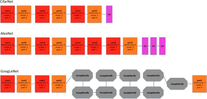
- The study evaluated CifarNet, AlexNet, and GoogLeNet architectures (later also OverFeat and VGGNet) on two CADe tasks: thoraco-abdominal LN detection using 388 mediastinal LNs in 90 patient CT scans and 595 abdominal LNs in 86 patient CT scans, and ILD classification using 905 annotated image slices from 120 patients with six ILD labels.
- LN detection used randomly sampled 2.5D views with fields-of-view spanning 35–128 mm, rescaled to 64×64 pixels, and three-fold cross-validation with FROC AUC and TPR/3FP metrics; ILD classification used slice-level and patch-level tasks under patient-level five-fold cross-validation, with CT windowing encoded into RGB channels and data augmentation.
- Training used the Caffe framework and NVidia K40 GPUs; models trained from scratch used random Gaussian initialization for 30 epochs with mini-batch size 50, while transfer learning fine-tuned all layers except the last at a learning rate 10 times smaller than default, with the last fully connected layer randomly initialized and trained at 0.01.
Limitations
- The paper states that the ILD dataset is partially labeled, with very few CT slices labeled as normal or healthy, and that this is one of its main limitations; it also states that the dataset’s limited patient population of 120 patients may not adequately represent ILD disease CT lung imaging patterns.
- The paper states that slice-level CNN learning is restricted to only 905 CT image slices, that no existing work has reached performance requirements for a realistic clinical setting, and that the most critical bottlenecks for ILD classification are cross-dataset learning and limited patient population size.
- Evident from the reported results: ILD slice-level CV5 accuracies were low (0.41–0.57), and LOO validation produced much higher accuracies than CV5 (e.g., AlexNet-TL 0.867 versus 0.46; GoogLeNet-TL 0.902 versus 0.57), indicating sensitivity to evaluation protocol; also, for abdominal LN detection the best reported CNN result (0.70 TPR/3FP) did not surpass the prior [22] result of 0.83 TPR/3FP.
Model-written summary. Check it against the paper before relying on it.
Integrity signals
Registry
Cites
Statistics
Figures
Verification
Claims the paper makes
49 Not checked yet · 31 Own result · 25 Own interpretation · 14 No basis given · 5 Sources not found · 2 Own method
A result the paper reports from its own data — it cites no other source for it, so there is none to check. The quote shows it is in the paper.
In the paper
“We achieve the state-of-the-art performance on the mediastinal LN detection, and report the first five-fold cross-validation classification results on predicting axial CT slices with ILD categories.”
- Also stated in: § Introduction · plain
“For the first time, ILD classification results under the patient-level five-fold cross-validation protocol (CV5) are investigated and reported.”
§ Deep Convolutional Neural Networks for Computer-Aided Detection: CNN Architectures, Dataset Characteristics and Transfer Learning > Abstract
plainn = 120- Asserted: passes — The authors put it forward as true.
- Checkable: passes — It can be checked against evidence.
- Load-bearing: passes — The paper’s conclusions depend on it.
- Based: passes — The paper gives a reason why it holds.
- Also stated in: § Introduction · plain
A result the paper reports from its own data — it cites no other source for it, so there is none to check. The quote shows it is in the paper.
In the paper
“Our experimental results suggest that different CNN architectures and dataset re-sampling protocols are critical for the LN detection tasks where the amount of labeled training data is sufficient and spatial contexts are local.”
§ Deep Convolutional Neural Networks for Computer-Aided Detection: CNN Architectures, Dataset Characteristics and Transfer Learning > Introduction
hedgeddirect- Asserted: passes — The authors put it forward as true.
- Checkable: passes — It can be checked against evidence.
- Load-bearing: passes — The paper’s conclusions depend on it.
- Based: passes — The paper gives a reason why it holds.
The paper states this without a source or data behind it here, so there is nothing to check it against. The quote shows it is in the paper.
In the paper
“As a result, LN datasets contain more training and testing data instances (due to data auugmentation) than ILD datasets.”
§ Deep Convolutional Neural Networks for Computer-Aided Detection: CNN Architectures, Dataset Characteristics and Transfer Learning > Introduction
plaindirect- Asserted: passes — The authors put it forward as true.
- Checkable: passes — It can be checked against evidence.
- Load-bearing: passes — The paper’s conclusions depend on it.
- Based: passes — The paper gives a reason why it holds.
A result the paper reports from its own data — it cites no other source for it, so there is none to check. The quote shows it is in the paper.
In the paper
“In the publicly available ILD dataset, very few CT slices are labeled as normal or healthy.”
§ Deep Convolutional Neural Networks for Computer-Aided Detection: CNN Architectures, Dataset Characteristics and Transfer Learning > Datasets and Related Work > Interstitial Lung Disease Dataset
plain- Asserted: passes — The authors put it forward as true.
- Checkable: passes — It can be checked against evidence.
- Load-bearing: unclear — It is unclear whether the paper’s conclusions depend on it.
- Based: passes — The paper gives a reason why it holds.
None of the sources cited for this claim have been checked yet.
In the paper
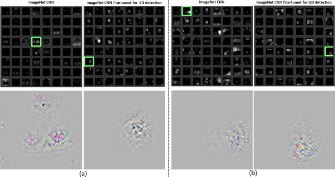
“CNN training learns to ignore them by setting very small filter weights around the corresponding regions (Fig. 13).”
§ Deep Convolutional Neural Networks for Computer-Aided Detection: CNN Architectures, Dataset Characteristics and Transfer Learning > Datasets and Related Work > Interstitial Lung Disease Dataset
plaindirect- Asserted: passes — The authors put it forward as true.
- Checkable: passes — It can be checked against evidence.
- Load-bearing: unclear — It is unclear whether the paper’s conclusions depend on it.
- Based: passes — The paper gives a reason why it holds.
- Rests on an assumption: Some areas outside the lung appear in both healthy or diseased images.
A result the paper reports from its own data — it cites no other source for it, so there is none to check. The quote shows it is in the paper.
In the paper
“There is no significant difference between two setups.”
§ Deep Convolutional Neural Networks for Computer-Aided Detection: CNN Architectures, Dataset Characteristics and Transfer Learning > Datasets and Related Work > Interstitial Lung Disease Dataset
plain- Asserted: passes — The authors put it forward as true.
- Checkable: passes — It can be checked against evidence.
- Load-bearing: passes — The paper’s conclusions depend on it.
- Based: passes — The paper gives a reason why it holds.
A result the paper reports from its own data — it cites no other source for it, so there is none to check. The quote shows it is in the paper.
In the paper
“The average number of CT slices (training instances) per fold is small, as shown in Table I.”
§ Deep Convolutional Neural Networks for Computer-Aided Detection: CNN Architectures, Dataset Characteristics and Transfer Learning > Datasets and Related Work > Interstitial Lung Disease Dataset
plainn = 5- Asserted: passes — The authors put it forward as true.
- Checkable: passes — It can be checked against evidence.
- Load-bearing: passes — The paper’s conclusions depend on it.
- Based: passes — The paper gives a reason why it holds.
A result the paper reports from its own data — it cites no other source for it, so there is none to check. The quote shows it is in the paper.
In the paper
“We found that the transfer learning strategy yields the best performance results.”
§ Deep Convolutional Neural Networks for Computer-Aided Detection: CNN Architectures, Dataset Characteristics and Transfer Learning > Methods > Training Protocols and Transfer Learning
plaindirect- Asserted: passes — The authors put it forward as true.
- Checkable: passes — It can be checked against evidence.
- Load-bearing: passes — The paper’s conclusions depend on it.
- Based: passes — The paper gives a reason why it holds.
The paper states this without a source or data behind it here, so there is nothing to check it against. The quote shows it is in the paper.
In the paper
“Another significant finding is that CNNs trained from scratch or fine-tuned from ImageNet models consistently outperform CNNs that merely use off-the-shelf CNN features, in both the LN and ILD classification problems.”
§ Deep Convolutional Neural Networks for Computer-Aided Detection: CNN Architectures, Dataset Characteristics and Transfer Learning > Introduction
boosteddirect- Asserted: passes — The authors put it forward as true.
- Checkable: passes — It can be checked against evidence.
- Load-bearing: passes — The paper’s conclusions depend on it.
- Based: passes — The paper gives a reason why it holds.
In the paper
“ILD [37] is a partially labeled database; this is one of its main limitations.”
§ Deep Convolutional Neural Networks for Computer-Aided Detection: CNN Architectures, Dataset Characteristics and Transfer Learning > Datasets and Related Work > Interstitial Lung Disease Dataset
What the sources say
- Not checked yetcites [37] Depeursinge A., “ Building a reference multimedia database for interstitial lung diseases,” Comput. Med. Imaging and. Grap., vol. 36, no. 3, pp. 227– 238, 2012. 21803548
plain- Asserted: passes — The authors put it forward as true.
- Checkable: passes — It can be checked against evidence.
- Load-bearing: unclear — It is unclear whether the paper’s conclusions depend on it.
- Based: passes — The paper gives a reason why it holds.
- It may state two separate claims as one. 32
- Probably not a substantial claim. 28
- The quote only partly states this claim. 3
- Borderline: one of the tests for a substantial claim is unclear. 1
None of the sources cited for this claim have been checked yet.
In the paper
“We obtain 86% sensitivity on 3 false positives (FP) per patient, versus the prior state-of-art sensitivities of 78% [36] (stacked shallow learning) and 70% [22] (CNN), as prior state-of-the-art.”
- Also stated in: § Abstract · plain
“We achieve the state-of-the-art performance on the mediastinal LN detection”
§ Deep Convolutional Neural Networks for Computer-Aided Detection: CNN Architectures, Dataset Characteristics and Transfer Learning > Introduction
What the sources say
- Not checked yetcites [36] Seff A., Lu L., Barbu A., Roth H., Shin H.-C., and Summers R. M., “ Leveraging mid-level semantic boundary cues for computer-aided lymph node detection,” Proc. MICCAI, 2015. source
- Not checked yetcites [22] Roth H., “ Improving computer-aided detection using convolutional neural networks and random view aggregation,” IEEE Trans. Med. Imag.. PMC7340334 26441412 source
plain- Asserted: passes — The authors put it forward as true.
- Checkable: passes — It can be checked against evidence.
- Load-bearing: passes — The paper’s conclusions depend on it.
- Based: passes — The paper gives a reason why it holds.
It may state two separate claims as one.
- Also stated in: § Abstract · plain
The paper states this without a source or data behind it here, so there is nothing to check it against. The quote shows it is in the paper.
In the paper
“They nonetheless remain less comprehensive than natural image datasets, such as ImageNet.”
§ Deep Convolutional Neural Networks for Computer-Aided Detection: CNN Architectures, Dataset Characteristics and Transfer Learning > Introduction
plain- Asserted: passes — The authors put it forward as true.
- Checkable: passes — It can be checked against evidence.
- Load-bearing: fails — The paper’s conclusions may not depend on it.
- Based: passes — The paper gives a reason why it holds.
Probably not a substantial claim. The paper’s conclusions may not depend on it.
The paper states this without a source or data behind it here, so there is nothing to check it against. The quote shows it is in the paper.
In the paper
“Fine-tuning ImageNet-trained models for ILD classification is clearly advantageous and yields early promising results, when the amount of labeled training data is highly insufficient and multi-class categorization is used, as opposed to the LN dataset's binary class categorization.”
§ Deep Convolutional Neural Networks for Computer-Aided Detection: CNN Architectures, Dataset Characteristics and Transfer Learning > Introduction
boosteddirect- Asserted: passes — The authors put it forward as true.
- Checkable: passes — It can be checked against evidence.
- Load-bearing: passes — The paper’s conclusions depend on it.
- Based: passes — The paper gives a reason why it holds.
It may state two separate claims as one.
The paper states this without a source or data behind it here, so there is nothing to check it against. The quote shows it is in the paper.
In the paper
“Hence, the purely random image resampling (with respect to scale, displacement and orientation) and reformatting (the axial, coronal, and sagittal views are in any system randomly resampled coordinates) is a natural choice, which also happens to yield high CNN performance.”
§ Deep Convolutional Neural Networks for Computer-Aided Detection: CNN Architectures, Dataset Characteristics and Transfer Learning > Datasets and Related Work > Thoracoabdominal Lymph Node Datasets
plaindirect- Asserted: passes — The authors put it forward as true.
- Checkable: passes — It can be checked against evidence.
- Load-bearing: passes — The paper’s conclusions depend on it.
- Based: passes — The paper gives a reason why it holds.
It may state two separate claims as one.
A result the paper reports from its own data — it cites no other source for it, so there is none to check. The quote shows it is in the paper.
In the paper
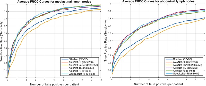
“GoogLeNet-TL-H | 0.94 | 0.81 | 0.92 | 0.70”
§ Deep Convolutional Neural Networks for Computer-Aided Detection: CNN Architectures, Dataset Characteristics and Transfer Learning > Evaluations and Discussions > Thoracoabdominal Lymph Node Detection
plain- Asserted: passes — The authors put it forward as true.
- Checkable: passes — It can be checked against evidence.
- Load-bearing: passes — The paper’s conclusions depend on it.
- Based: passes — The paper gives a reason why it holds.
It may state two separate claims as one. The quote only partly states this claim.
The paper states this without a source or data behind it here, so there is nothing to check it against. The quote shows it is in the paper.
In the paper
“GoogLeNet-TL-H yields results similar to AlexNet-TL-H's for the mediastinal LN detection, and slightly outperforms Alex-Net-H for abdominal LN detection.”
§ Deep Convolutional Neural Networks for Computer-Aided Detection: CNN Architectures, Dataset Characteristics and Transfer Learning > Evaluations and Discussions > Thoracoabdominal Lymph Node Detection
plain- Asserted: passes — The authors put it forward as true.
- Checkable: passes — It can be checked against evidence.
- Load-bearing: passes — The paper’s conclusions depend on it.
- Based: passes — The paper gives a reason why it holds.
It may state two separate claims as one.
The paper states this without a source or data behind it here, so there is nothing to check it against. The quote shows it is in the paper.
In the paper
“Likewise, AlexNet-TL-H consistently outperforms AlexNet-RI-H, though by smaller margins (0.81 versus 0.79 TPR/3FP in mediastinum; 0.69 versus 0.67 TPR/3FP in abdomen).”
§ Deep Convolutional Neural Networks for Computer-Aided Detection: CNN Architectures, Dataset Characteristics and Transfer Learning > Evaluations and Discussions > Thoracoabdominal Lymph Node Detection
plaindirect- Asserted: passes — The authors put it forward as true.
- Checkable: passes — It can be checked against evidence.
- Load-bearing: passes — The paper’s conclusions depend on it.
- Based: passes — The paper gives a reason why it holds.
It may state two separate claims as one.
A result the paper reports from its own data — it cites no other source for it, so there is none to check. The quote shows it is in the paper.
In the paper
“However, transfer learning from ImageNet pre-trained model is consistently beneficial, as evidenced by AlexNet-TL (0.46) versus AlexNet-RI (0.44), and GoogLeNet-TL (0.57) versus GoogLeNet-RI (0.41).”
§ Deep Convolutional Neural Networks for Computer-Aided Detection: CNN Architectures, Dataset Characteristics and Transfer Learning > Evaluations and Discussions > Interstitial Lung Disease Classification
boosteddirect- Asserted: passes — The authors put it forward as true.
- Checkable: passes — It can be checked against evidence.
- Load-bearing: passes — The paper’s conclusions depend on it.
- Based: passes — The paper gives a reason why it holds.
It may state two separate claims as one.
The paper states this without a source or data behind it here, so there is nothing to check it against. The quote shows it is in the paper.
In the paper
“AlexNet-RI-H exhibits less severe over-fitting than GoogLeNet-RI-H.”
§ Deep Convolutional Neural Networks for Computer-Aided Detection: CNN Architectures, Dataset Characteristics and Transfer Learning > Evaluations and Discussions > Thoracoabdominal Lymph Node Detection
plain- Asserted: passes — The authors put it forward as true.
- Checkable: passes — It can be checked against evidence.
- Load-bearing: unclear — It is unclear whether the paper’s conclusions depend on it.
- Based: unclear — It is unclear why the paper says it holds.
Borderline substantial claim. It is unclear whether the paper’s conclusions depend on it. It is unclear why the paper says it holds.
None of the sources cited for this claim have been checked yet.
In the paper
“GoogLeNet-RI-L | 0.95 | 0.85 | 0.91 | 0.69”
§ Deep Convolutional Neural Networks for Computer-Aided Detection: CNN Architectures, Dataset Characteristics and Transfer Learning > Evaluations and Discussions > Thoracoabdominal Lymph Node Detection
What the sources say
- Not checked yetcites [36] Seff A., Lu L., Barbu A., Roth H., Shin H.-C., and Summers R. M., “ Leveraging mid-level semantic boundary cues for computer-aided lymph node detection,” Proc. MICCAI, 2015. source
- Not checked yetcites [22] Roth H., “ Improving computer-aided detection using convolutional neural networks and random view aggregation,” IEEE Trans. Med. Imag.. PMC7340334 26441412 source
plain- Asserted: passes — The authors put it forward as true.
- Checkable: passes — It can be checked against evidence.
- Load-bearing: passes — The paper’s conclusions depend on it.
- Based: passes — The paper gives a reason why it holds.
It may state two separate claims as one. The quote only partly states this claim.
A result the paper reports from its own data — it cites no other source for it, so there is none to check. The quote shows it is in the paper.
In the paper
“Patch-CV5 | 0.76 | 0.74 | 0.76 | 0.75 | 0.76 | 0.79”
§ Deep Convolutional Neural Networks for Computer-Aided Detection: CNN Architectures, Dataset Characteristics and Transfer Learning > Evaluations and Discussions > Thoracoabdominal Lymph Node Detection
plain- Asserted: fails — The authors may not put it forward as true.
- Checkable: passes — It can be checked against evidence.
- Load-bearing: fails — The paper’s conclusions may not depend on it.
- Based: passes — The paper gives a reason why it holds.
Probably not a substantial claim. The authors may not put it forward as true. The paper’s conclusions may not depend on it. The quote only partly states this claim.
In the paper
“Patch-CV5 yields higher F-scores than [54] and classifies the extra consolidation (CD) class.”
§ Deep Convolutional Neural Networks for Computer-Aided Detection: CNN Architectures, Dataset Characteristics and Transfer Learning > Evaluations and Discussions > Interstitial Lung Disease Classification
What the sources say
- Not checked yetcites [54] Li Q., “ Medical image classification with convolutional neural network,” Proc. 13th Int. Conf. Control Automat. Robot. Vis., pp. 844– 848, 2014.
plainn = 120- Asserted: passes — The authors put it forward as true.
- Checkable: passes — It can be checked against evidence.
- Load-bearing: passes — The paper’s conclusions depend on it.
- Based: passes — The paper gives a reason why it holds.
It may state two separate claims as one.
A result the paper reports from its own data — it cites no other source for it, so there is none to check. The quote shows it is in the paper.
In the paper
“The quantitative results in Table III indicate that AlexNet-TL and GoogLeNet-TL have consistently better classification accuracies than AlexNet-RI and GoogLeNet-RI, respectively.”
§ Deep Convolutional Neural Networks for Computer-Aided Detection: CNN Architectures, Dataset Characteristics and Transfer Learning > Analysis Via CNN Learning Traces and Lulvisualization > ILD Classification
hedgedcorrelational- Asserted: passes — The authors put it forward as true.
- Checkable: passes — It can be checked against evidence.
- Load-bearing: passes — The paper’s conclusions depend on it.
- Based: passes — The paper gives a reason why it holds.
It may state two separate claims as one.
A result the paper reports from its own data — it cites no other source for it, so there is none to check. The quote shows it is in the paper.
In the paper
“For example, in ILD slice-level classification, the accuracy level drastically increases from 0.46 to 0.867 using AlexNet-TL, and from 0.57 to 0.902 for GoogLeNet-TL.”
§ Deep Convolutional Neural Networks for Computer-Aided Detection: CNN Architectures, Dataset Characteristics and Transfer Learning > Evaluations and Discussions > Evaluation of Five CNN Models Using ILD Classification
plaindirect- Asserted: passes — The authors put it forward as true.
- Checkable: passes — It can be checked against evidence.
- Load-bearing: passes — The paper’s conclusions depend on it.
- Based: passes — The paper gives a reason why it holds.
It may state two separate claims as one.
A result the paper reports from its own data — it cites no other source for it, so there is none to check. The quote shows it is in the paper.
In the paper
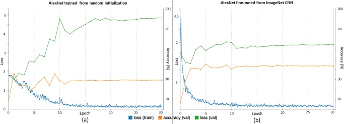
“For AlexNet-RI in Fig. 11(a), the training loss significantly decreases as the number of training epochs increases, while the validation loss notably increases and the validation accuracy does not improve much before reaching a plateau.”
§ Deep Convolutional Neural Networks for Computer-Aided Detection: CNN Architectures, Dataset Characteristics and Transfer Learning > Analysis Via CNN Learning Traces and Lulvisualization > ILD Classification
plaindirect- Asserted: passes — The authors put it forward as true.
- Checkable: passes — It can be checked against evidence.
- Load-bearing: passes — The paper’s conclusions depend on it.
- Based: passes — The paper gives a reason why it holds.
It may state two separate claims as one.
A result the paper reports from its own data — it cites no other source for it, so there is none to check. The quote shows it is in the paper.
In the paper
“During the process, the validation loss, having remained at lower values throughout, achieves higher final accuracy levels than the validation loss on a similar problem with random initialization.”
§ Deep Convolutional Neural Networks for Computer-Aided Detection: CNN Architectures, Dataset Characteristics and Transfer Learning > Analysis Via CNN Learning Traces and Lulvisualization > ILD Classification
plaindirect- Asserted: passes — The authors put it forward as true.
- Checkable: passes — It can be checked against evidence.
- Load-bearing: passes — The paper’s conclusions depend on it.
- Based: passes — The paper gives a reason why it holds.
It may state two separate claims as one.
A conclusion the paper draws from its own results — it cites no other source for it, so there is none to check. The quote shows it is in the paper.
In the paper
“Since LN images are more flexible than ILD images with respect to resampling and reformatting, LN datasets may be more readily augmented by such image transformations.”
§ Deep Convolutional Neural Networks for Computer-Aided Detection: CNN Architectures, Dataset Characteristics and Transfer Learning > Introduction
hedgedconditional- Asserted: passes — The authors put it forward as true.
- Checkable: passes — It can be checked against evidence.
- Load-bearing: passes — The paper’s conclusions depend on it.
- Based: fails — The paper may give no reason why it holds.
Probably not a substantial claim. The paper may give no reason why it holds. It may state two separate claims as one.
None of the sources cited for this claim have been checked yet.
In the paper
“There are currently three major techniques that successfully employ CNNs to medical image classification: 1) training the “CNN from scratch” [13]–[17]; 2) using “off-the-shelf CNN” features (without retraining the CNN) as complementary information channels to existing hand-crafted image features, for chest X-rays [10] and CT lung nodule identification [9], [12]; and 3) performing unsupervised pre-”
§ Deep Convolutional Neural Networks for Computer-Aided Detection: CNN Architectures, Dataset Characteristics and Transfer Learning > Introduction
What the sources say
- Not checked yetcites [13]–[17]
- Not checked yetcites [9], [12]
- Not checked yetcites [18]–[21]
plain- Asserted: passes — The authors put it forward as true.
- Checkable: passes — It can be checked against evidence.
- Load-bearing: unclear — It is unclear whether the paper’s conclusions depend on it.
- Based: passes — The paper gives a reason why it holds.
It may state two separate claims as one.
In the paper
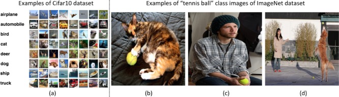
“ImageNet is currently the largest image dataset among other standard datasets for visual recognition.”
§ Deep Convolutional Neural Networks for Computer-Aided Detection: CNN Architectures, Dataset Characteristics and Transfer Learning > Methods > Imagenet: Large Scale Annotated Natural Image Dataset
What the sources say
- Not matchedcites [1] Deng J., “ ImageNet: A large-scale hierarchical image database,” in Proc. IEEE Conf. Comput. Vis. Pattern Recognit., 2009, pp. 248– 255.
Couldn’t be matched to a paper in Crossref, so it can’t be checked.
plain- Asserted: passes — The authors put it forward as true.
- Checkable: passes — It can be checked against evidence.
- Load-bearing: fails — The paper’s conclusions may not depend on it.
- Based: passes — The paper gives a reason why it holds.
Probably not a substantial claim. The paper’s conclusions may not depend on it.
- Not matched
A conclusion the paper draws from its own results — it cites no other source for it, so there is none to check. The quote shows it is in the paper.
In the paper
“This significant intra-class variation poses greater challenges to any data-driven learning system that builds a classifier to fit given data and generalize to unseen data.”
§ Deep Convolutional Neural Networks for Computer-Aided Detection: CNN Architectures, Dataset Characteristics and Transfer Learning > Methods > Imagenet: Large Scale Annotated Natural Image Dataset
plaindirect- Asserted: passes — The authors put it forward as true.
- Checkable: passes — It can be checked against evidence.
- Load-bearing: fails — The paper’s conclusions may not depend on it.
- Based: passes — The paper gives a reason why it holds.
Probably not a substantial claim. The paper’s conclusions may not depend on it.
A conclusion the paper draws from its own results — it cites no other source for it, so there is none to check. The quote shows it is in the paper.
In the paper
“Transforming axial, coronal, and sagittal representations to RGB also facilitates transfer learning from CNN models trained on ImageNet.”
§ Deep Convolutional Neural Networks for Computer-Aided Detection: CNN Architectures, Dataset Characteristics and Transfer Learning > Datasets and Related Work > Thoracoabdominal Lymph Node Datasets
plaindirect- Asserted: passes — The authors put it forward as true.
- Checkable: passes — It can be checked against evidence.
- Load-bearing: fails — The paper’s conclusions may not depend on it.
- Based: fails — The paper may give no reason why it holds.
Probably not a substantial claim. The paper’s conclusions may not depend on it. The paper may give no reason why it holds.
In the paper
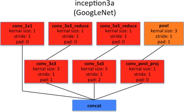
“More importantly, it also introduces a new module called “Inception”, which concatenates filters of different sizes and dimensions into a single new filter (refer to Fig. 6).”
§ Deep Convolutional Neural Networks for Computer-Aided Detection: CNN Architectures, Dataset Characteristics and Transfer Learning > Methods > Convolutional Neural Network Architectures
What the sources say
- Not checked yetcites [33] Szegedy C., “ Going deeper with convolutions,” in Proc. IEEE Comput. Soc. Conf. Comput. Vis. Pattern Recognit., 2015, pp. 1– 9. source
plaindirect- Asserted: passes — The authors put it forward as true.
- Checkable: passes — It can be checked against evidence.
- Load-bearing: fails — The paper’s conclusions may not depend on it.
- Based: passes — The paper gives a reason why it holds.
Probably not a substantial claim. The paper’s conclusions may not depend on it.
In the paper
“Evaluation protocols and details are critical to deriving significant empirical findings [34].”
§ Deep Convolutional Neural Networks for Computer-Aided Detection: CNN Architectures, Dataset Characteristics and Transfer Learning > Introduction
What the sources say
- Not checked yetcites [34] Chatfield K., Simonyan K., Vedaldi A., and Zisserman A., “ Return of the devil in the details: Delving deep into convolutional nets,” Br. Mach. Vis. Conf., 2014. source
plaindirect- Asserted: passes — The authors put it forward as true.
- Checkable: fails — It may be too vague to check against evidence.
- Load-bearing: fails — The paper’s conclusions may not depend on it.
- Based: passes — The paper gives a reason why it holds.
Probably not a substantial claim. It may be too vague to check against evidence. The paper’s conclusions may not depend on it.
In the paper
“The AlexNet architecture was published in [4], achieved significantly improved performance over the other non-deep learning methods for ImageNet Large Scale Visual Recognition Challenge (ILSVRC) 2012.”
§ Deep Convolutional Neural Networks for Computer-Aided Detection: CNN Architectures, Dataset Characteristics and Transfer Learning > Methods > Convolutional Neural Network Architectures
What the sources say
- Not checked yetcites [4] Krizhevsky A., Sutskever I., and Hinton G. E., “ ImageNet classification with deep convolutional neural networks,” Proc. NIPS, pp. 1097– 1105, 2012. source
plaindirect- Asserted: passes — The authors put it forward as true.
- Checkable: passes — It can be checked against evidence.
- Load-bearing: fails — The paper’s conclusions may not depend on it.
- Based: passes — The paper gives a reason why it holds.
Probably not a substantial claim. The paper’s conclusions may not depend on it.
A conclusion the paper draws from its own results — it cites no other source for it, so there is none to check. The quote shows it is in the paper.
In the paper
“In summary, the assumption that there are or must be pixel-wise spatial correlations among input channels does not apply to the CNN model representation.”
§ Deep Convolutional Neural Networks for Computer-Aided Detection: CNN Architectures, Dataset Characteristics and Transfer Learning > Datasets and Related Work > Thoracoabdominal Lymph Node Datasets
plain- Asserted: passes — The authors put it forward as true.
- Checkable: passes — It can be checked against evidence.
- Load-bearing: fails — The paper’s conclusions may not depend on it.
- Based: fails — The paper may give no reason why it holds.
Probably not a substantial claim. The paper’s conclusions may not depend on it. The paper may give no reason why it holds.
In the paper
“The ImageNet dataset is publicly available, and the ImageNet Large Scale Visual Recognition Challenge (ILSVRC) has become the standard benchmark for large-scale object recognition.”
§ Deep Convolutional Neural Networks for Computer-Aided Detection: CNN Architectures, Dataset Characteristics and Transfer Learning > Methods > Imagenet: Large Scale Annotated Natural Image Dataset
What the sources say
- Not matchedcites [1] Deng J., “ ImageNet: A large-scale hierarchical image database,” in Proc. IEEE Conf. Comput. Vis. Pattern Recognit., 2009, pp. 248– 255.
Couldn’t be matched to a paper in Crossref, so it can’t be checked.
plain- Asserted: passes — The authors put it forward as true.
- Checkable: passes — It can be checked against evidence.
- Load-bearing: fails — The paper’s conclusions may not depend on it.
- Based: passes — The paper gives a reason why it holds.
Probably not a substantial claim. The paper’s conclusions may not depend on it. It may state two separate claims as one.
- Not matched
A conclusion the paper draws from its own results — it cites no other source for it, so there is none to check. The quote shows it is in the paper.
In the paper
“Characterizing an arbitrary CT slice against any possible ILD type, without any manual ROI (in contrast to [38], [39]), can be useful for large-scale patient screening.”
§ Deep Convolutional Neural Networks for Computer-Aided Detection: CNN Architectures, Dataset Characteristics and Transfer Learning > Datasets and Related Work > Interstitial Lung Disease Dataset
plainconditional- Asserted: passes — The authors put it forward as true.
- Checkable: fails — It may be too vague to check against evidence.
- Load-bearing: passes — The paper’s conclusions depend on it.
- Based: fails — The paper may give no reason why it holds.
Probably not a substantial claim. It may be too vague to check against evidence. The paper may give no reason why it holds.
In the paper
“AlexNet can be fine-tuned on the PASCAL dataset to surpass the performance of the ImageNet pre-trained AlexNet, although the difference is not as significant as that between the CNN and non-CNN methods.”
§ Deep Convolutional Neural Networks for Computer-Aided Detection: CNN Architectures, Dataset Characteristics and Transfer Learning > Methods > Training Protocols and Transfer Learning
What the sources say
- Not checked yetcites [6] Girshick R., Donahue J., Darrell T., and Malik J., “ Region-based convolutional networks for accurate object detection and semantic segmentation,” IEEE Trans. Pattern Anal. Mach. Intell., vol. 38, no. 1, pp. 142– 158, Jan. 2016. 26656583 source
plaindirect- Asserted: passes — The authors put it forward as true.
- Checkable: passes — It can be checked against evidence.
- Load-bearing: unclear — It is unclear whether the paper’s conclusions depend on it.
- Based: passes — The paper gives a reason why it holds.
It may state two separate claims as one.
A conclusion the paper draws from its own results — it cites no other source for it, so there is none to check. The quote shows it is in the paper.
In the paper
“Slice-level ILD classification is a very challenging task where CNN models need to learn from very small numbers of training examples and predict ILD labels on unseen patients.”
§ Deep Convolutional Neural Networks for Computer-Aided Detection: CNN Architectures, Dataset Characteristics and Transfer Learning > Datasets and Related Work > Interstitial Lung Disease Dataset
plainn = 120- Asserted: passes — The authors put it forward as true.
- Checkable: passes — It can be checked against evidence.
- Load-bearing: passes — The paper’s conclusions depend on it.
- Based: passes — The paper gives a reason why it holds.
It may state two separate claims as one.
In the paper
“GoogLeNet is the current state-of-the-art CNN architecture for the ILSVRC challenge, where it achieved 5.5% top-5 classification error on the ImageNet challenge, compared to AlexNet's 15.3% top-5 classification error.”
§ Deep Convolutional Neural Networks for Computer-Aided Detection: CNN Architectures, Dataset Characteristics and Transfer Learning > Methods > Convolutional Neural Network Architectures
What the sources say
- Not checked yetcites [33] Szegedy C., “ Going deeper with convolutions,” in Proc. IEEE Comput. Soc. Conf. Comput. Vis. Pattern Recognit., 2015, pp. 1– 9. source
plain- Asserted: passes — The authors put it forward as true.
- Checkable: passes — It can be checked against evidence.
- Load-bearing: fails — The paper’s conclusions may not depend on it.
- Based: passes — The paper gives a reason why it holds.
Probably not a substantial claim. The paper’s conclusions may not depend on it. It may state two separate claims as one.
The paper states this without a source or data behind it here, so there is nothing to check it against. The quote shows it is in the paper.
In the paper
“On the other hand, the mainstream deep CNN architectures (e.g., AlexNet and GoogLeNet) contain tens of millions of free parameters to train, and thus require sufficiently large numbers of labeled medical images.”
§ Deep Convolutional Neural Networks for Computer-Aided Detection: CNN Architectures, Dataset Characteristics and Transfer Learning > Methods > Training Protocols and Transfer Learning
plaindirect- Asserted: passes — The authors put it forward as true.
- Checkable: passes — It can be checked against evidence.
- Load-bearing: passes — The paper’s conclusions depend on it.
- Based: unclear — It is unclear why the paper says it holds.
It may state two separate claims as one.
- Rests on an assumption: Collecting and annotating large numbers of medical images still poses significant challenges.
In the paper
“ImageNet [1] has more than 1.2 million 256×256 images categorized under 1000 object class categories.”
§ Deep Convolutional Neural Networks for Computer-Aided Detection: CNN Architectures, Dataset Characteristics and Transfer Learning > Methods > Imagenet: Large Scale Annotated Natural Image Dataset
What the sources say
- Not matchedcites [1] Deng J., “ ImageNet: A large-scale hierarchical image database,” in Proc. IEEE Conf. Comput. Vis. Pattern Recognit., 2009, pp. 248– 255.
Couldn’t be matched to a paper in Crossref, so it can’t be checked.
plain- Asserted: passes — The authors put it forward as true.
- Checkable: passes — It can be checked against evidence.
- Load-bearing: fails — The paper’s conclusions may not depend on it.
- Based: passes — The paper gives a reason why it holds.
Probably not a substantial claim. The paper’s conclusions may not depend on it.
- Not matched
A conclusion the paper draws from its own results — it cites no other source for it, so there is none to check. The quote shows it is in the paper.
In the paper
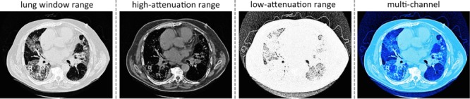
“More importantly, these CT windowing processes do not depend on the lung segmentation, which instead is directly defined in the CT HU space.”
§ Deep Convolutional Neural Networks for Computer-Aided Detection: CNN Architectures, Dataset Characteristics and Transfer Learning > Datasets and Related Work > Interstitial Lung Disease Dataset
plain- Asserted: passes — The authors put it forward as true.
- Checkable: passes — It can be checked against evidence.
- Load-bearing: fails — The paper’s conclusions may not depend on it.
- Based: passes — The paper gives a reason why it holds.
Probably not a substantial claim. The paper’s conclusions may not depend on it. It may state two separate claims as one.
What the paper did — it cites no source for this, so there is none to check. The quote shows it is in the paper.
In the paper
“We evaluate the ILD classification task with five-fold CV on patient-level split, as it is more informative for real clinical performance than LOO.”
§ Deep Convolutional Neural Networks for Computer-Aided Detection: CNN Architectures, Dataset Characteristics and Transfer Learning > Evaluations and Discussions > Interstitial Lung Disease Classification
plaindirect- Asserted: passes — The authors put it forward as true.
- Checkable: passes — It can be checked against evidence.
- Load-bearing: passes — The paper’s conclusions depend on it.
- Based: passes — The paper gives a reason why it holds.
Probably not a substantial claim. It may state two separate claims as one.
In the paper
“The experimental results in [40] are conducted with a patient-level hard split of 100 (training) and 20 (testing).”
§ Deep Convolutional Neural Networks for Computer-Aided Detection: CNN Architectures, Dataset Characteristics and Transfer Learning > Evaluations and Discussions > Interstitial Lung Disease Classification
What the sources say
- Not checked yetcites [40] Gao M., “ Holistic classification of CT attenuation patterns for interstitial lung diseases via deep convolutional neural networks,” in Proc. MICCAI First Workshop Deep Learn. Med. Image Anal., 2015. PMC5881940 29623248
plain- Asserted: passes — The authors put it forward as true.
- Checkable: passes — It can be checked against evidence.
- Load-bearing: fails — The paper’s conclusions may not depend on it.
- Based: passes — The paper gives a reason why it holds.
Probably not a substantial claim. The paper’s conclusions may not depend on it.
None of the sources cited for this claim have been checked yet.
In the paper
“This difference lies in the fact that annotated lymph node segmentation masks are required to learn a mid-level semantic boundary detector [36], whereas CNN approaches only need LN locations for training [22].”
§ Deep Convolutional Neural Networks for Computer-Aided Detection: CNN Architectures, Dataset Characteristics and Transfer Learning > Evaluations and Discussions > Thoracoabdominal Lymph Node Detection
What the sources say
- Not checked yetcites [36] Seff A., Lu L., Barbu A., Roth H., Shin H.-C., and Summers R. M., “ Leveraging mid-level semantic boundary cues for computer-aided lymph node detection,” Proc. MICCAI, 2015. source
- Not checked yetcites [22] Roth H., “ Improving computer-aided detection using convolutional neural networks and random view aggregation,” IEEE Trans. Med. Imag.. PMC7340334 26441412 source
plaindirect- Asserted: passes — The authors put it forward as true.
- Checkable: passes — It can be checked against evidence.
- Load-bearing: passes — The paper’s conclusions depend on it.
- Based: passes — The paper gives a reason why it holds.
It may state two separate claims as one.
None of the sources cited for this claim have been checked yet.
In the paper
“Both [38] and [39] use the evaluation protocol of “leave-one-patient-out” (LOO), which is arguably much easier and not directly comparable to 10-fold CV [54] or our Patch-CV5.”
§ Deep Convolutional Neural Networks for Computer-Aided Detection: CNN Architectures, Dataset Characteristics and Transfer Learning > Evaluations and Discussions > Interstitial Lung Disease Classification
What the sources say
- Not checked yetcites [38] Song Y., Cai W., Zhou Y., and Feng D. D., “ Feature-based image patch approximation for lung tissue classification,” IEEE Trans. Med. Imag., vol. 32, no. 4, pp. 797– 808, Apr. 2013. 23340591 source
- Not checked yetcites [39] Song Y., “ Large margin local estimate with applications to medical image classification,” IEEE Trans. Med. Imag., vol. 34, no. 6, pp. 1362– 1377, Jun. 2015. 25616009 source
- Not checked yetcites [54] Li Q., “ Medical image classification with convolutional neural network,” Proc. 13th Int. Conf. Control Automat. Robot. Vis., pp. 844– 848, 2014. source
hedged- Asserted: passes — The authors put it forward as true.
- Checkable: passes — It can be checked against evidence.
- Load-bearing: passes — The paper’s conclusions depend on it.
- Based: passes — The paper gives a reason why it holds.
It may state two separate claims as one.
What the paper did — it cites no source for this, so there is none to check. The quote shows it is in the paper.
In the paper
“We train and evaluate CNNs using three-fold cross-validation (folds are split into disjoint sets of patients), with the different CNN architectures described above.”
§ Deep Convolutional Neural Networks for Computer-Aided Detection: CNN Architectures, Dataset Characteristics and Transfer Learning > Evaluations and Discussions > Thoracoabdominal Lymph Node Detection
plain- Asserted: passes — The authors put it forward as true.
- Checkable: passes — It can be checked against evidence.
- Load-bearing: passes — The paper’s conclusions depend on it.
- Based: passes — The paper gives a reason why it holds.
Probably not a substantial claim.
The paper states this without a source or data behind it here, so there is nothing to check it against. The quote shows it is in the paper.
In the paper
“AlexNet is the first notably successful CNN architecture on the ImageNet challenge and has rekindled significant research interests on CNN.”
§ Deep Convolutional Neural Networks for Computer-Aided Detection: CNN Architectures, Dataset Characteristics and Transfer Learning > Evaluations and Discussions > Evaluation of Five CNN Models Using ILD Classification
plaindirect- Asserted: passes — The authors put it forward as true.
- Checkable: passes — It can be checked against evidence.
- Load-bearing: fails — The paper’s conclusions may not depend on it.
- Based: fails — The paper may give no reason why it holds.
Probably not a substantial claim. The paper’s conclusions may not depend on it. The paper may give no reason why it holds. It may state two separate claims as one.
None of the sources cited for this claim have been checked yet.
In the paper
“GoogLeNet is the state-of-the-art deep model, which has outperformed other notable models, such as AlexNet, OverFeat, and VGGNet [67], [68] in various computer vision benchmarks.”
§ Deep Convolutional Neural Networks for Computer-Aided Detection: CNN Architectures, Dataset Characteristics and Transfer Learning > Evaluations and Discussions > Evaluation of Five CNN Models Using ILD Classification
What the sources say
- Not checked yetcites [67] Sermanet P., “ Overfeat: Integrated recognition, localization and detection using convolutional networks,” Proc. ICLR, 2014. source
- Not checked yetcites [68] Simonyan K. and Zisserman A., “ Very deep convolutional networks for large-scale image recognition,” Proc. ICLR, 2014. source
plaindirect- Asserted: passes — The authors put it forward as true.
- Checkable: passes — It can be checked against evidence.
- Load-bearing: passes — The paper’s conclusions depend on it.
- Based: passes — The paper gives a reason why it holds.
It may state two separate claims as one.
In the paper
“This image sampling scheme contributes to our best reported performance results in both mediastinal LN detection (in this paper) and automated pancreas segmentation [45].”
§ Deep Convolutional Neural Networks for Computer-Aided Detection: CNN Architectures, Dataset Characteristics and Transfer Learning > Evaluations and Discussions > Thoracoabdominal Lymph Node Detection
What the sources say
- Not checked yetcites [45] Roth H., “ DeepOrgan: Multi-level deep convolutional networks for automated pancreas segmentation,” Proc. MICCAI, 2015. source
plaindirect- Asserted: passes — The authors put it forward as true.
- Checkable: passes — It can be checked against evidence.
- Load-bearing: passes — The paper’s conclusions depend on it.
- Based: passes — The paper gives a reason why it holds.
It may state two separate claims as one.
In the paper
“The overall reported LN detection results are clinically significant, as indicated in [63].”
§ Deep Convolutional Neural Networks for Computer-Aided Detection: CNN Architectures, Dataset Characteristics and Transfer Learning > Evaluations and Discussions > Thoracoabdominal Lymph Node Detection
What the sources say
- Not matchedcites [63] Kim L., “ Performance assessment of retroperitoneal lymph node computer-assisted detection using random forest and deep convolutional neural network learning algorithms in tandem,” in 102nd Annu. Meet. Radiol. Soc. N. Am., 2014.
Couldn’t be matched to a paper in Crossref, so it can’t be checked.
hedged- Asserted: passes — The authors put it forward as true.
- Checkable: passes — It can be checked against evidence.
- Load-bearing: fails — The paper’s conclusions may not depend on it.
- Based: passes — The paper gives a reason why it holds.
Probably not a substantial claim. The paper’s conclusions may not depend on it.
- Not matched
In the paper
“In general, patch-level ILD classification is less challenging than slice-level classification, as far more data samples can be sampled from the manually annotated ROIs (up to 100 image patches per ROI), available from [37].”
§ Deep Convolutional Neural Networks for Computer-Aided Detection: CNN Architectures, Dataset Characteristics and Transfer Learning > Evaluations and Discussions > Interstitial Lung Disease Classification
What the sources say
- Not checked yetcites [37] Depeursinge A., “ Building a reference multimedia database for interstitial lung diseases,” Comput. Med. Imaging and. Grap., vol. 36, no. 3, pp. 227– 238, 2012. 21803548
plaindirect- Asserted: passes — The authors put it forward as true.
- Checkable: passes — It can be checked against evidence.
- Load-bearing: unclear — It is unclear whether the paper’s conclusions depend on it.
- Based: passes — The paper gives a reason why it holds.
It may state two separate claims as one.
In the paper
“Recently, Gao et al. [40] have argued that a new CADe protocol on holistic classification of ILD diseases directly, using axial CT slice attenuation patterns and CNN, may be more realistic for clinical applications.”
§ Deep Convolutional Neural Networks for Computer-Aided Detection: CNN Architectures, Dataset Characteristics and Transfer Learning > Evaluations and Discussions > Interstitial Lung Disease Classification
What the sources say
- Not checked yetcites [40] Gao M., “ Holistic classification of CT attenuation patterns for interstitial lung diseases via deep convolutional neural networks,” in Proc. MICCAI First Workshop Deep Learn. Med. Image Anal., 2015. PMC5881940 29623248
hedgedconditional- Asserted: fails — The authors may not put it forward as true.
- Checkable: passes — It can be checked against evidence.
- Load-bearing: fails — The paper’s conclusions may not depend on it.
- Based: passes — The paper gives a reason why it holds.
Probably not a substantial claim. The authors may not put it forward as true. The paper’s conclusions may not depend on it.
In the paper
“OverFeat is described in [67] as an integrated framework for using CNN for classification, localization and detection.”
§ Deep Convolutional Neural Networks for Computer-Aided Detection: CNN Architectures, Dataset Characteristics and Transfer Learning > Evaluations and Discussions > Evaluation of Five CNN Models Using ILD Classification
What the sources say
- Not checked yetcites [67] Sermanet P., “ Overfeat: Integrated recognition, localization and detection using convolutional networks,” Proc. ICLR, 2014.
plain- Asserted: passes — The authors put it forward as true.
- Checkable: passes — It can be checked against evidence.
- Load-bearing: fails — The paper’s conclusions may not depend on it.
- Based: passes — The paper gives a reason why it holds.
Probably not a substantial claim. The paper’s conclusions may not depend on it.
In the paper
“The main difference between our dataset preparation protocol and that from [22] is a more aggressive extraction of random views within a much larger range of FOVs.”
§ Deep Convolutional Neural Networks for Computer-Aided Detection: CNN Architectures, Dataset Characteristics and Transfer Learning > Evaluations and Discussions > Thoracoabdominal Lymph Node Detection
What the sources say
- Not checked yetcites [22] Roth H., “ Improving computer-aided detection using convolutional neural networks and random view aggregation,” IEEE Trans. Med. Imag.. PMC7340334 26441412
plain- Asserted: passes — The authors put it forward as true.
- Checkable: passes — It can be checked against evidence.
- Load-bearing: passes — The paper’s conclusions depend on it.
- Based: passes — The paper gives a reason why it holds.
Probably not a substantial claim.
In the paper
“In abdominal LN detection, [22] obtains the best trade-off between its CNN model complexity and sampled data configuration.”
§ Deep Convolutional Neural Networks for Computer-Aided Detection: CNN Architectures, Dataset Characteristics and Transfer Learning > Evaluations and Discussions > Thoracoabdominal Lymph Node Detection
What the sources say
- Not checked yetcites [22] Roth H., “ Improving computer-aided detection using convolutional neural networks and random view aggregation,” IEEE Trans. Med. Imag.. PMC7340334 26441412
plaincorrelational- Asserted: passes — The authors put it forward as true.
- Checkable: passes — It can be checked against evidence.
- Load-bearing: fails — The paper’s conclusions may not depend on it.
- Based: passes — The paper gives a reason why it holds.
Probably not a substantial claim. The paper’s conclusions may not depend on it.
None of the sources cited for this claim have been checked yet.
In the paper
“Although the ILD [37] and LTRC datasets [64] (used in [19]) were generated and annotated separately, they contain many common disease labels. For instance, the ILD disease classes emphysema (EM), ground glass (GG), fibrosis (FB), and micronodules (MN) belong to both datasets, and thus can be jointly trained/tested to form a larger and unified dataset.”
§ Deep Convolutional Neural Networks for Computer-Aided Detection: CNN Architectures, Dataset Characteristics and Transfer Learning > Evaluations and Discussions > Interstitial Lung Disease Classification
What the sources say
- Not checked yetcites [37] Depeursinge A., “ Building a reference multimedia database for interstitial lung diseases,” Comput. Med. Imaging and. Grap., vol. 36, no. 3, pp. 227– 238, 2012. 21803548 source
- Not matchedcites [64] Holmes III D., Bartholmai B., Karwoski R., Zavaletta V., and Robb R., “ The lung tissue research consortium: An extensive open database containing histological, clinical, and radiological data to study chronic lung disease,” in MICCAI Open Sci. Workshop, 2006.
Couldn’t be matched to a paper in Crossref, so it can’t be checked.
plaindirect- Asserted: passes — The authors put it forward as true.
- Checkable: passes — It can be checked against evidence.
- Load-bearing: passes — The paper’s conclusions depend on it.
- Based: passes — The paper gives a reason why it holds.
It may state two separate claims as one.
A conclusion the paper draws from its own results — it cites no other source for it, so there is none to check. The quote shows it is in the paper.
In the paper
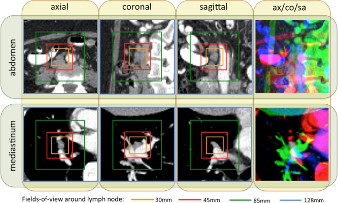
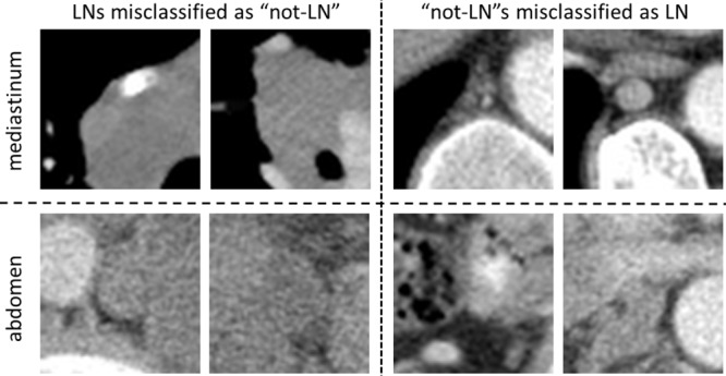
“As shown in Fig. 1, abdominal LNs are surrounded by many other similar looking objects. Meanwhile, mediastinal LNs are more easily distinguishable, due to the images' larger spatial contexts.”
§ Deep Convolutional Neural Networks for Computer-Aided Detection: CNN Architectures, Dataset Characteristics and Transfer Learning > Evaluations and Discussions > Thoracoabdominal Lymph Node Detection
plaindirect- Asserted: passes — The authors put it forward as true.
- Checkable: passes — It can be checked against evidence.
- Load-bearing: passes — The paper’s conclusions depend on it.
- Based: passes — The paper gives a reason why it holds.
It may state two separate claims as one.
A conclusion the paper draws from its own results — it cites no other source for it, so there is none to check. The quote shows it is in the paper.
In the paper
“GoogLeNet-RI-H performs poorly, as it is susceptible to over-fitting.”
§ Deep Convolutional Neural Networks for Computer-Aided Detection: CNN Architectures, Dataset Characteristics and Transfer Learning > Evaluations and Discussions > Thoracoabdominal Lymph Node Detection
plaindirect- Asserted: passes — The authors put it forward as true.
- Checkable: passes — It can be checked against evidence.
- Load-bearing: passes — The paper’s conclusions depend on it.
- Based: passes — The paper gives a reason why it holds.
It may state two separate claims as one.
In the paper
“A segmentation label propagation method was recently proposed [47] to provide full pixel-wise labeling of the ILD data images.”
§ Deep Convolutional Neural Networks for Computer-Aided Detection: CNN Architectures, Dataset Characteristics and Transfer Learning > Evaluations and Discussions > Interstitial Lung Disease Classification
What the sources say
- Not checked yetcites [47] Gao M., Lu L., Nogues I., Summers M. R., and Mollura D., “ Segmentation label propagation using deep convolutional neural networks and dense conditional random field,” Proc. IEEE Int. Conf. Comput. Vis., pp. 1377– 1385, 2015. source
plaindirect- Asserted: passes — The authors put it forward as true.
- Checkable: passes — It can be checked against evidence.
- Load-bearing: fails — The paper’s conclusions may not depend on it.
- Based: passes — The paper gives a reason why it holds.
Probably not a substantial claim. The paper’s conclusions may not depend on it.
A conclusion the paper draws from its own results — it cites no other source for it, so there is none to check. The quote shows it is in the paper.
In the paper
“For ILD classification, the most critical performance bottlenecks are the challenge of cross-dataset learning and the limited patient population size.”
§ Deep Convolutional Neural Networks for Computer-Aided Detection: CNN Architectures, Dataset Characteristics and Transfer Learning > Evaluations and Discussions > Interstitial Lung Disease Classification
plaindirect- Asserted: passes — The authors put it forward as true.
- Checkable: passes — It can be checked against evidence.
- Load-bearing: passes — The paper’s conclusions depend on it.
- Based: fails — The paper may give no reason why it holds.
Probably not a substantial claim. The paper may give no reason why it holds. It may state two separate claims as one.
In the paper
“Overfeat is the winning model of the ILSVRC 2013 in detection and classification tasks.”
§ Deep Convolutional Neural Networks for Computer-Aided Detection: CNN Architectures, Dataset Characteristics and Transfer Learning > Evaluations and Discussions > Evaluation of Five CNN Models Using ILD Classification
What the sources say
- Not checked yetcites [67] Sermanet P., “ Overfeat: Integrated recognition, localization and detection using convolutional networks,” Proc. ICLR, 2014.
plain- Asserted: passes — The authors put it forward as true.
- Checkable: passes — It can be checked against evidence.
- Load-bearing: fails — The paper’s conclusions may not depend on it.
- Based: passes — The paper gives a reason why it holds.
Probably not a substantial claim. The paper’s conclusions may not depend on it.
A conclusion the paper draws from its own results — it cites no other source for it, so there is none to check. The quote shows it is in the paper.
In the paper
“The empirical evaluation, CNN model visualization, CNN performance analysis, and conclusive insights can be generalized to the design of high performance CAD systems for other medical imaging tasks.”
§ Deep Convolutional Neural Networks for Computer-Aided Detection: CNN Architectures, Dataset Characteristics and Transfer Learning > Conclusion
plain- Asserted: passes — The authors put it forward as true.
- Checkable: fails — It may be too vague to check against evidence.
- Load-bearing: passes — The paper’s conclusions depend on it.
- Based: passes — The paper gives a reason why it holds.
Probably not a substantial claim. It may be too vague to check against evidence.
The paper relies on this without support.
Fine-tuning an ImageNet pre-trained CNN model on medical image datasets had not yet been exploited at the time of the study.
“However, the fine-tuning of an ImageNet pre-trained CNN model on medical image datasets has not yet been exploited.”
The paper relies on this without support.
Obtaining datasets as comprehensively annotated as ImageNet in the medical imaging domain remains a challenge.
“However, obtaining datasets as comprehensively annotated as ImageNet in the medical imaging domain remains a challenge.”
The paper relies on this without support.
Some areas outside the lung appear in both healthy or diseased images.
“Some areas outside of the lung appear in both healthy or diseased images.”
The paper relies on this without support.
The low-attenuation CT window is useful for visualizing certain texture patterns of lung diseases, especially emphysema.
“The low-attenuation CT window is useful for visualizing certain texture patterns of lung diseases (especially emphysema).”
The paper relies on this without support.
Collecting and annotating large numbers of medical images still poses significant challenges.
“Collecting and annotating large numbers of medical images still poses significant challenges.”
The authors state this as an assumption.
OverFeat and VGGNet may generate quantitative performance results ranked between AlexNet's and GoogLeNet's.
“Likewise, a reasonable assumption is that OverFeat and VGGNet may generate quantitative performance results ranked between AlexNet's and GoogLeNet's.”
The authors state this as an assumption.
OverFeat's and VGGNet's performance levels fall between AlexNet's and GoogLeNet's.
“The reported results validate our assumption that OverFeat's and VGGNet's performance levels fall between AlexNet's and GoogLeNet's (this observation is consistent with the computer vision findings).”
The paper relies on this without support.
Both the ILD and ImageNet datasets use full-size images.
“Indeed, both datasets use full-size images.”
The paper relies on this without support.
CifarNet is designed for images with smaller dimensions (32×32 images), and thus is not catered to classification tasks involving 256×256 images.
“CifarNet is designed for images with smaller dimensions (32×32 images), and thus is not catered to classification tasks involving 256×256 images.”
The paper relies on this without support.
Medical datasets are often biased, in that the number of healthy samples is much larger than the number of diseased instances, or that the numbers of images per class are uneven.
“Medical datasets are often “biased”, in that the number of healthy samples is much larger than the number of diseased instances, or that the numbers of images per class are uneven.”
Claims other papers make about this one
Each is checked against this paper’s own text: does it say what the citing paper says it does?
In the citing paper
“Transfer learning from natural images has been widely adopted for medical image classification Tajbakhsh et al. (2016); Shin et al. (2016); however the application to medical image segmentation has been scarce.”
From Embracing imperfect datasets: A review of deep learning solutions for medical image segmentation (2020) · cites this paper as Shin et al. (2016)
In the citing paper
“In general, shallow networks have been preferred in medical image analysis, when compared with very deep CNNs employed in computer vision applications [80] [81].”
From Medical image analysis using convolutional neural networks: a review (2018) · cites this paper as [80]
In the citing paper
“Healthcare applications of deep learning range from one-dimensional biosignal analysis [17] and the prediction of medical events, e.g. seizures [18] and cardiac arrests [19], to computer-aided detection [20] and diagnosis [21] supporting clinical decision making and survival analysis [22], to drug discovery [23] and as an aid in therapy selection and pharmacogenomics [24], to increased operational”
From An overview of deep learning in medical imaging focusing on MRI (2019) · cites this paper as [20]
In the citing paper
“After first and second fully connected layers' dropout regularization layer [45] has been used to avoid further overfitting with a neuron dropout probability of 1-p, where p is the probability of neurons kept.”
From Medical image retrieval using deep convolutional neural network (2017) · cites this paper as [45]
In the citing paper
“Instead, a specially designed small network may be more suitable to the smaller-scale training datasets that are commonly encountered in medical imaging [155].”
From Deep Learning in Medical Ultrasound Analysis: A Review (2019) · cites this paper as [155]
In the citing paper
“Shin et al. [62] reported that they obtained best performance with transfer learning from pre-trained model on ImageNet dataset and fine-tuning on lymph node and interstitial lung disease rather than training from scratch.”
From Deep learning for brain MRI segmentation: state of the art and future directions (2017) · cites this paper as [62]
In the citing paper
“The convolutional encoder-decoder network using VGG16 shares many features with the commonly used U-Net network (29) but has unique advantages, including lower computation cost, presence of batch normalization layers to improve training efficiency, and more versatility for use as both a segmentation and classification network, which allows effective transfer learning in the current setup (30,31).”
From Deep learning approach for evaluating knee MR images: achieving high diagnostic performance for cartilage lesion detection. (2018) · cites this paper as [30]
Self-check
Percentages, sums and reported differences re-computed from the paper’s own numbers.
Each contradiction between two passages listed here was confirmed by an independent check.
Rated minor: it would not touch a result — a value no result rests on, a p-value that stays on the same side of the significance level, or a conflict that is clerical, resolved elsewhere in the paper, or outside its own findings.
In the paper
§ Deep Convolutional Neural Networks for Computer-Aided Detection: CNN Architectures, Dataset Characteristics and Transfer Learning > Evaluations and Discussions > Thoracoabdominal Lymph Node Detection
“We also evaluate a simple ensemble by averaging the probability scores from five CNNs:”
§ Deep Convolutional Neural Networks for Computer-Aided Detection: CNN Architectures, Dataset Characteristics and Transfer Learning > Evaluations and Discussions > Thoracoabdominal Lymph Node Detection
“AlexNet-RI-H, AlexNet-TL-H, AlexNet-RI-H, GoogLeNet-TL-H and GoogLeNet-RI-L.”
Rated minor: it would not touch a result — a value no result rests on, a p-value that stays on the same side of the significance level, or a conflict that is clerical, resolved elsewhere in the paper, or outside its own findings.
In the paper
§ Deep Convolutional Neural Networks for Computer-Aided Detection: CNN Architectures, Dataset Characteristics and Transfer Learning > Methods > Convolutional Neural Network Architectures
“Googlenet [33] contains two convolution layers, three pooling layers, and nine inception layers.”
§ Deep Convolutional Neural Networks for Computer-Aided Detection: CNN Architectures, Dataset Characteristics and Transfer Learning > Methods > Convolutional Neural Network Architectures
“Overall, GoogLeNet has two convolution layers, two pooling layers, and nine “Inception” layers.”
Rated minor: it would not touch a result — a value no result rests on, a p-value that stays on the same side of the significance level, or a conflict that is clerical, resolved elsewhere in the paper, or outside its own findings.
In the paper
§ Deep Convolutional Neural Networks for Computer-Aided Detection: CNN Architectures, Dataset Characteristics and Transfer Learning > Evaluations and Discussions > Training With “equal Prior” vs.“biased Prior”
“Table: Table VIII Classification accuracies for ILD slice and LN patch-level detection with “equal prior” and “biased prior”, using googlenet-TL”
§ Deep Convolutional Neural Networks for Computer-Aided Detection: CNN Architectures, Dataset Characteristics and Transfer Learning > Evaluations and Discussions > Training With “equal Prior” vs.“biased Prior”
“| | ILD-Slice | ILD-Patch |”
Rated minor: it would not touch a result — a value no result rests on, a p-value that stays on the same side of the significance level, or a conflict that is clerical, resolved elsewhere in the paper, or outside its own findings.
In the paper
§ Deep Convolutional Neural Networks for Computer-Aided Detection: CNN Architectures, Dataset Characteristics and Transfer Learning > Introduction
“We obtain 86% sensitivity on 3 false positives (FP) per patient”
§ Deep Convolutional Neural Networks for Computer-Aided Detection: CNN Architectures, Dataset Characteristics and Transfer Learning > Evaluations and Discussions > Thoracoabdominal Lymph Node Detection
“| Combined | 0.95 | 0.85 | 0.93 | 0.70 |”
Citations
Cites
Very Deep Convolutional Networks for Large-Scale Image Recognition
Simonyan, K., & Zisserman, A. (2014) · arXiv · doi:10.48550/arxiv.1409.1556 · Cited by 114,148 · cited 2×
“GoogLeNet is the state-of-the-art deep model, which has outperformed other notable models, such as AlexNet, OverFeat, and VGGNet [67], [68] in various computer vision benchmarks.”
BackgroundMethodImageNet classification with deep convolutional neural networks
Krizhevsky, A., Sutskever, I., & Hinton, G. E. (2017) · Communications of the ACM · doi:10.1145/3065386 · Cited by 112,155 · cited 9×
“For datadriven learning, large-scale well-annotated datasets with representative data distribution characteristics are crucial to learning more accurate or generalizable models [5], [4].”
BackgroundMethodInfluentialImageNet: A large-scale hierarchical image database
Deng, J., Dong, W., Socher, R., Li, L.-J., Li, K., & Fei-Fei, L. (2009) · 2009 IEEE Conference on Computer Vision and Pattern Recognition · doi:10.1109/cvpr.2009.5206848 · Cited by 65,333 · cited 4×
“datasets with representative data distribution characteristics are crucial to learning more accurate or generalizable models [5], [4]. Unlike previous image datasets used in computer vision, ImageNet [1] offers a very comprehensive database of more than 1.2 million categorized natural images of 1000+ classes. The CNN…”
BackgroundMethodInfluentialGradient-based learning applied to document recognition
Lecun, Y., Bottou, L., Bengio, Y., & Haffner, P. (1998) · Proceedings of the IEEE · doi:10.1109/5.726791 · Cited by 58,805 · cited 2×
“achieved significantly improved performance over the other non-deep learning methods for ImageNet Large Scale Visual Recognition Challenge (ILSVRC) 2012. This success has revived the interest in CNNs [3] in computer vision. ImageNet consists of 1.2 million 256 256 images belonging to 1000 categories. At times, the obje…”
BackgroundInfluentialDistinctive Image Features from Scale-Invariant Keypoints
Lowe, D. G. (2004) · International Journal of Computer Vision · doi:10.1023/b:visi.0000029664.99615.94 · Cited by 55,589 · cited 2×
“Although natural images and medical images differ significantly, conventional image descriptors developed for object recognition in natural images, such as the scale-invariant feature transform (SIFT) [30] and the histogram of oriented gradients (HOG) [31], have been widely used for object detection and segmentation i…”
BackgroundGoing deeper with convolutions
Szegedy, C., Liu, W., Jia, Y., Sermanet, P., Reed, S., Anguelov, D., Erhan, D., Vanhoucke, V., & Rabinovich, A. (2015) · 2015 IEEE Conference on Computer Vision and Pattern Recognition (CVPR) · doi:10.1109/cvpr.2015.7298594 · Cited by 48,562 · cited 9×
“AlexNet [4] and GoogLeNet [33] are also modified to evaluate these state-of-the-art CNN architecture from ImageNet classification task [2] to our CADe problems and datasets.”
BackgroundMethodInfluentialImageNet Large Scale Visual Recognition Challenge
Russakovsky, O., Deng, J., Su, H., Krause, J., Satheesh, S., Ma, S., Huang, Z., Karpathy, A., Khosla, A., Bernstein, M., et al. (2015) · International Journal of Computer Vision · doi:10.1007/s11263-015-0816-y · Cited by 39,380
Fully convolutional networks for semantic segmentation
Long, J., Shelhamer, E., & Darrell, T. (2015) · 2015 IEEE Conference on Computer Vision and Pattern Recognition (CVPR) · doi:10.1109/cvpr.2015.7298965 · Cited by 38,834 · cited 1×
“However, current FCNN approaches [65], [66] lack adequate spatial resolution in their directly output label space.”
BackgroundHistograms of Oriented Gradients for Human Detection
Dalal, N., & Triggs, B. (2005) · 2005 IEEE Computer Society Conference on Computer Vision and Pattern Recognition (CVPR'05) · doi:10.1109/cvpr.2005.177 · Cited by 33,584
Learning Multiple Layers of Features from Tiny Images
Krizhevsky, A. (2024) · doi:10.57702/zp44cu3g · Cited by 23,889
Research works printed but not linked: 3
- seff, 2015, Leveraging mid-level semantic boundary cues for computer-aided lymph node detection, Proc MICCAI
- kim, 2014, Performance assessment of retroperitoneal lymph node computer-assisted detection using random forest and deep convolutional neural network learning algorithms in tandem, 102nd Annu Meet Radiol Soc N Am
- sermanet, 2014, Overfeat: Integrated recognition, localization and detection using convolutional networks, Proc ICLR
Cited by
Sorted by citations
Items 201–300 of 1,020
Qian, X., Pei, J., Zheng, H., Xie, X., Lin, Y., Zhang, H., Han, C., Gao, X., Zhang, H., Zheng, W., et al. (2021) · Nature Biomedical Engineering · doi:10.1038/s41551-021-00711-2 · Cited by 257
Self-citationSelf-supervision with Superpixels: Training Few-Shot Medical Image Segmentation Without Annotation
Ouyang, C., Biffi, C., Chen, C., Kart, T., Qiu, H., & Rueckert, D. (2020) · Lecture notes in computer science · doi:10.1007/978-3-030-58526-6_45 · Cited by 293
Deep learning in mammography images segmentation and classification: Automated CNN approach
Salama, W. M., & Aly, M. H. (2021) · Alexandria Engineering Journal · doi:10.1016/j.aej.2021.03.048 · Cited by 255
Cheng, P. M., & Malhi, H. S. (2017) · Journal of Digital Imaging · doi:10.1007/s10278-016-9929-2 · Cited by 253
Yang, Q., Zhang, Y., Dai, W., & Pan, S. J. (2020) · Cambridge University Press eBooks · doi:10.1017/9781139061773 · Cited by 286
Transfer Learning for Domain Adaptation in MRI: Application in Brain Lesion Segmentation
Ghafoorian, M., Mehrtash, A., Kapur, T., Karssemeijer, N., Marchiori, E., Pesteie, M., Guttmann, C. R. G., de Leeuw, F., Tempany, C. M., van Ginneken, B., et al. (2017) · Lecture notes in computer science · doi:10.1007/978-3-319-66179-7_59 · Cited by 337
Electrocardiogram screening for aortic valve stenosis using artificial intelligence
Cohen-Shelly, M., Attia, Z. I., Friedman, P. A., Ito, S., Essayagh, B. A., Ko, W.-Y., Murphree, D. H., Michelena, H. I., Enriquez-Sarano, M., Carter, R. E., et al. (2021) · European Heart Journal · doi:10.1093/eurheartj/ehab153 · Cited by 252
Automatic defect detection and segmentation of tunnel surface using modified Mask R-CNN
Xu, Y., Li, D., Xie, Q., Wu, Q., & Wang, J. (2021) · Measurement · doi:10.1016/j.measurement.2021.109316 · Cited by 255
Artificial Intelligence in Radiotherapy Treatment Planning: Present and Future
Wang, C., Zhu, X., Hong, J. C., & Zheng, D. (2019) · Technology in Cancer Research & Treatment · doi:10.1177/1533033819873922 · Cited by 263
He, Z., Shao, H., Zhong, X. Y., & Zhao, X. (2020) · Knowledge-Based Systems · doi:10.1016/j.knosys.2020.106396 · Cited by 253
FUIQA: Fetal Ultrasound Image Quality Assessment with Deep Convolutional Networks
Wu, L., Cheng, J.-Z., Li, S., Lei, B., Wang, T., & Ni, D. (2017) · IEEE Transactions on Cybernetics · doi:10.1109/tcyb.2017.2671898 · Cited by 242
The impact of patient clinical information on automated skin cancer detection
Pacheco, A. G. C., & Krohling, R. A. (2019) · Computers in Biology and Medicine · doi:10.1016/j.compbiomed.2019.103545 · Cited by 270
Yu, L., Chen, H., Dou, Q., Qin, J., & Heng, P. (2016) · IEEE Journal of Biomedical and Health Informatics · doi:10.1109/jbhi.2016.2637004 · Cited by 247
Content-based brain tumor retrieval for MR images using transfer learning
Swati, Z. N. K., Zhao, Q., Kabir, M., Ali, F., Ali, Z., Ahmed, S., & Lu, J. (2019) · IEEE Access · doi:10.1109/access.2019.2892455 · Cited by 246
Ye, H., Gao, F., Yin, Y., Guo, D., Zhao, P., Lü, Y., Wang, X., Bai, J., Cao, K., Song, Q., et al. (2019) · European Radiology · doi:10.1007/s00330-019-06163-2 · Cited by 255
Deep semi-supervised learning for medical image segmentation: A review
Han, K., Sheng, V. S., Song, Y., Liu, Y., Qiu, C., Ma, S., & Liu, Z. (2024) · Expert Systems with Applications · doi:10.1016/j.eswa.2023.123052 · Cited by 256
Generative Adversarial Networks and Its Applications in Biomedical Informatics
Lan, L., You, L., Zhang, Z., Fan, Z., Zhao, W., Zeng, N., Chen, Y., & Zhou, X. (2020) · Frontiers in Public Health · doi:10.3389/fpubh.2020.00164 · Cited by 237
Knowledge Transfer for Rotary Machine Fault Diagnosis
Yan, R., Shen, F., Sun, C., & Chen, X. (2019) · IEEE Sensors Journal · doi:10.1109/jsen.2019.2949057 · Cited by 245
Fast implementation of real-time fruit detection in apple orchards using deep learning
Kang, H., & Chen, C. (2020) · Computers and Electronics in Agriculture · doi:10.1016/j.compag.2019.105108 · Cited by 243
van Velzen, S. G. M., Lessmann, N., Velthuis, B. K., Bank, I. E. M., van den Bongard, D. H. J. G., Leiner, T., de Jong, P. A., Veldhuis, W. B., Correa, A., Terry, J. G., et al. (2020) · Radiology · doi:10.1148/radiol.2020191621 · Cited by 244
Role of machine learning in medical research: A survey
Garg, A., & Mago, V. (2021) · Computer Science Review · doi:10.1016/j.cosrev.2021.100370 · Cited by 266
Surface defect classification of steels with a new semi-supervised learning method
Di, H., Ke, X., Peng, Z., & Dongdong, Z. (2019) · Optics and Lasers in Engineering · doi:10.1016/j.optlaseng.2019.01.011 · Cited by 241
Deep learning based classification of breast tumors with shear-wave elastography
Zhang, Q., Xiao, Y., Dai, W., Suo, J., Wang, C., Shi, J., & Zheng, H. (2016) · Ultrasonics · doi:10.1016/j.ultras.2016.08.004 · Cited by 240
Demystifying Multitask Deep Neural Networks for Quantitative Structure–Activity Relationships
Xu, Y., Ma, J., Liaw, A., Sheridan, R. P., & Svetnik, V. (2017) · Journal of Chemical Information and Modeling · doi:10.1021/acs.jcim.7b00087 · Cited by 238
Co-Learning Feature Fusion Maps From PET-CT Images of Lung Cancer
Kumar, A., FULHAM, M. J., Feng, D., & Kim, J. (2019) · IEEE Transactions on Medical Imaging · doi:10.1109/tmi.2019.2923601 · Cited by 261
Guan, Q., Huang, Y., Zhong, Z., Zheng, Z., Zheng, L., & Yang, Y. (2018) · arXiv (Cornell University) · doi:10.48550/arxiv.1801.09927 · Cited by 235
Machine learning enables design automation of microfluidic flow-focusing droplet generation
Lashkaripour, A., Rodriguez, C., Mehdipour, N., Mardian, R., McIntyre, D., Ortiz, L., Campbell, J., & Densmore, D. M. (2021) · Nature Communications · doi:10.1038/s41467-020-20284-z · Cited by 246
Continuous Learning AI in Radiology: Implementation Principles and Early Applications
Pianykh, O. S., Langs, G., Dewey, M., Enzmann, D. R., Herold, C. J., Schoenberg, S. O., & Brink, J. A. (2020) · Radiology · doi:10.1148/radiol.2020200038 · Cited by 237
Samala, R. K., Chan, H., Hadjiiski, L. M., Helvie, M. A., Kenny, H., & Richter, C. (2017) · Physics in Medicine and Biology · doi:10.1088/1361-6560/aa93d4 · Cited by 231
Westphal, E., & Seitz, H. (2021) · Additive manufacturing · doi:10.1016/j.addma.2021.101965 · Cited by 256
Chen, Y., Tong, Z., Zheng, Y., Samuelson, H. W., & Norford, L. K. (2019) · Journal of Cleaner Production · doi:10.1016/j.jclepro.2019.119866 · Cited by 232
A Deep Transfer Convolutional Neural Network Framework for EEG Signal Classification
Xu, G., Shen, X., Chen, S., Zong, Y., Zhang, C. Y., Yue, H., Liu, M., Chen, F., & Che, W. (2019) · IEEE Access · doi:10.1109/access.2019.2930958 · Cited by 233
Lee, J., Kim, D., & Jeong, S. (2019) · Oral Diseases · doi:10.1111/odi.13223 · Cited by 239
Single Model Deep Learning on Imbalanced Small Datasets for Skin Lesion Classification
Yao, P., Shen, S., Xu, M., Liu, P., Zhang, F., Xing, J., Shao, P., Kaffenberger, B. H., & Xu, R. X. (2021) · IEEE Transactions on Medical Imaging · doi:10.1109/tmi.2021.3136682 · Cited by 255
Quantitative imaging of cancer in the postgenomic era: Radio(geno)mics, deep learning, and habitats
Napel, S. A., Mu, W., Jardim‐Perassi, B. V., Aerts, H. J. W. L., & Gillies, R. J. (2018) · Cancer · doi:10.1002/cncr.31630 · Cited by 235
Hyperspectral Classification Based on Lightweight 3-D-CNN With Transfer Learning
Zhang, H., Li, Y., Jiang, Y., Wang, P., Shen, Q., & Shen, C. (2019) · IEEE Transactions on Geoscience and Remote Sensing · doi:10.1109/tgrs.2019.2902568 · Cited by 229
Unsupervised Reverse Domain Adaptation for Synthetic Medical Images via Adversarial Training
Mahmood, F., Chen, R., & Durr, N. J. (2018) · IEEE Transactions on Medical Imaging · doi:10.1109/tmi.2018.2842767 · Cited by 257
Recognition of peripheral blood cell images using convolutional neural networks
Acevedo, A., Alférez, S., Merino, A., Puigví, L., & Rodellar, J. (2019) · Computer Methods and Programs in Biomedicine · doi:10.1016/j.cmpb.2019.105020 · Cited by 238
Bishara, D., Xie, Y., Liu, W. K., & Li, S. (2022) · Archives of Computational Methods in Engineering · doi:10.1007/s11831-022-09795-8 · Cited by 240
Joint Prostate Cancer Detection and Gleason Score Prediction in mp-MRI via FocalNet
Cao, R., Bajgiran, A. M., Mirak, S. A., Shakeri, S., Zhong, X., Enzmann, D. R., Raman, S. S., & Sung, K. (2019) · IEEE Transactions on Medical Imaging · doi:10.1109/tmi.2019.2901928 · Cited by 224
Deep Facial Diagnosis: Deep Transfer Learning From Face Recognition to Facial Diagnosis
Jin, B., Cruz, L., & Gonçalves, N. (2020) · IEEE Access · doi:10.1109/access.2020.3005687 · Cited by 240
The state of the art of deep learning models in medical science and their challenges
Bhatt, C., Kumar, I., Vijayakumar, V., Singh, K. U., & Kumar, A. (2021) · Multimedia Systems · doi:10.1007/s00530-020-00694-1 · Cited by 222
A Comprehensive Survey on Transfer LearningPreprint
Zhuang, F., Qi, Z., Duan, K., et al. (2019) · arXiv (Cornell University) · doi:10.48550/arxiv.1911.02685 · Cited by 223
Automatic Colon Polyp Detection Using Region Based Deep CNN and Post Learning Approaches
Shin, Y., Qadir, H. A., Aabakken, L., Bergsland, J., & Balasingham, I. (2018) · IEEE Access · doi:10.1109/access.2018.2856402 · Cited by 222
Deep convolutional neural networks with transfer learning for automated brain image classification
Kaur, T., & Gandhi, T. K. (2020) · Machine Vision and Applications · doi:10.1007/s00138-020-01069-2 · Cited by 234
Sengupta, P. P., Shrestha, S., Berthon, B., Messas, E., Donal, E., Tison, G. H., Min, J. K., D’hooge, J., Voigt, J., Dudley, J. T., et al. (2020) · JACC. Cardiovascular imaging · doi:10.1016/j.jcmg.2020.07.015 · Cited by 230
Riasatian, A., Babaie, M., Maleki, D., Kalra, S., Valipour, M., Hemati, S., Zaveri, M., Safarpoor, A., Shafiei, S., Afshari, M., et al. (2021) · Medical Image Analysis · doi:10.1016/j.media.2021.102032 · Cited by 221
Fu, L., Majeed, Y., Zhang, X., Karkee, M., & Zhang, Q. (2020) · Biosystems Engineering · doi:10.1016/j.biosystemseng.2020.07.007 · Cited by 219
Review of the State of the Art of Deep Learning for Plant Diseases: A Broad Analysis and Discussion
Hasan, R. I., Yusuf, S. M., & Alzubaidi, L. (2020) · Plants · doi:10.3390/plants9101302 · Cited by 213
Radiological images and machine learning: Trends, perspectives, and prospects
Zhang, Z., & Sejdić, E. (2019) · Computers in Biology and Medicine · doi:10.1016/j.compbiomed.2019.02.017 · Cited by 249
Peng, J., Kang, S., Ning, Z., Deng, H., Shen, J., Xu, Y., Zhang, J., Zhao, W., Li, X., 龚五星, et al. (2019) · European Radiology · doi:10.1007/s00330-019-06318-1 · Cited by 218
D-UNet: A Dimension-Fusion U Shape Network for Chronic Stroke Lesion Segmentation
Zhou, Y., Huang, W., Dong, P., Xia, Y., & Wang, S. (2019) · IEEE Transactions on Computational Biology and Bioinformatics · doi:10.1109/tcbb.2019.2939522 · Cited by 221
Explainable COVID-19 Detection Using Chest CT Scans and Deep Learning
Alshazly, H. A., Linse, C., Barth, E., & Martinetz, T. (2021) · Sensors · doi:10.3390/s21020455 · Cited by 222
Automatic Segmentation of Acute Ischemic Stroke From DWI Using 3-D Fully Convolutional DenseNets
Zhang, R., Zhao, L., Lou, W., Abrigo, J., Mok, V., Chu, C. W., Wang, D., & Shi, L. (2018) · IEEE Transactions on Medical Imaging · doi:10.1109/tmi.2018.2821244 · Cited by 210
Unveiling COVID-19 from CHEST X-ray with deep learning: A hurdles race with small data
Tartaglione, E., Barbano, C. A., Berzovini, C., Calandri, M., & Grangetto, M. (2020) · International Journal of Environmental Research and Public Health · doi:10.3390/ijerph17186933 · Cited by 211
Motor imagery EEG classification algorithm based on CNN-LSTM feature fusion network
Li, H., Ding, M., Zhang, R., et al. (2021) · Biomedical Signal Processing and Control · doi:10.1016/j.bspc.2021.103342 · Cited by 210
Deep Learning in Data-Driven Pavement Image Analysis and Automated Distress Detection: A Review
Gopalakrishnan, K. (2018) · Data · doi:10.3390/data3030028 · Cited by 210
Cui, S., Mao, L., Jiang, J., Liu, C., & Xiong, S. (2018) · Journal of Healthcare Engineering · doi:10.1155/2018/4940593 · Cited by 206
Mochida, K., Koda, S., Inoue, K., Hirayama, T., Tanaka, S., Nishii, R., & Melgani, F. (2018) · GigaScience · doi:10.1093/gigascience/giy153 · Cited by 217
Nida, N., Irtaza, A., Javed, A., Yousaf, M. H., & Mahmood, M. T. (2019) · International Journal of Medical Informatics · doi:10.1016/j.ijmedinf.2019.01.005 · Cited by 209
Machine learning for landslides prevention: a survey
Ma, Z., Mei, G., & Piccialli, F. (2020) · Neural Computing and Applications · doi:10.1007/s00521-020-05529-8 · Cited by 220
Bladder Cancer Treatment Response Assessment in CT using Radiomics with Deep-Learning
Kenny, H., Hadjiiski, L. M., Chan, H., Weizer, A. Z., Alva, A. S., Cohan, R. H., Caoili, E. M., Paramagul, C., & Samala, R. K. (2017) · Scientific Reports · doi:10.1038/s41598-017-09315-w · Cited by 208
Application of Convolutional Neural Network in the Diagnosis of Jaw Tumors
Poedjiastoeti, W., & Suebnukarn, S. (2018) · Healthcare Informatics Research · doi:10.4258/hir.2018.24.3.236 · Cited by 208
Amin, S. U., Alsulaiman, M., Muhammad, G., Bencherif, M. A., & Hossain, M. S. (2019) · IEEE Access · doi:10.1109/access.2019.2895688 · Cited by 216
Qin, Z., Yu, F., Liu, C., & Chen, X. (2018) · Mathematical Foundations of Computing · doi:10.3934/mfc.2018008 · Cited by 281
Transfer Learning for Domain Adaptation in MRI: Application in Brain Lesion Segmentation
Ghafoorian, M., Mehrtash, A., Kapur, T., Karssemeijer, N., Marchiori, E., Pesteie, M., Guttmann, C. R. G., Leeuw, F.-E. D., Tempany, C. M., van Ginneken, B., et al. (2017) · Radboud Repository (Radboud University) · Cited by 192
Bakkouri, I., & Afdel, K. (2019) · Multimedia Tools and Applications · doi:10.1007/s11042-019-07988-1 · Cited by 212
Hu, Y., Guo, Y., Wang, Y., Yu, J., Li, J., Zhou, S., & Chang, C. (2018) · Medical Physics · doi:10.1002/mp.13268 · Cited by 205
The classification of construction waste material using a deep convolutional neural network
Davis, P., Aziz, F., Newaz, M. T., Sher, W., & Simon, L. (2021) · Automation in Construction · doi:10.1016/j.autcon.2020.103481 · Cited by 204
Rahman, M. M., Shakeri, M., Tiong, S., Khatun, F., Amin, N., Pasupuleti, J., & Hasan, M. K. (2021) · Sustainability · doi:10.3390/su13042393 · Cited by 162
Muduli, D., Dash, R., & Majhi, B. (2021) · Biomedical Signal Processing and Control · doi:10.1016/j.bspc.2021.102825 · Cited by 211
Deep Learning Based Module Defect Analysis for Large-Scale Photovoltaic Farms
Li, X., Yang, Q., Lou, Z., & Yan, W. (2018) · IEEE Transactions on Energy Conversion · doi:10.1109/tec.2018.2873358 · Cited by 202
Automated Brain Image Classification Based on VGG-16 and Transfer Learning
Kaur, T., & Gandhi, T. K. (2019) · doi:10.1109/icit48102.2019.00023 · Cited by 201
Automated Analysis of Unregistered Multi-View Mammograms With Deep Learning
Carneiro, G., Nascimento, J. C., & Bradley, A. P. (2017) · IEEE Transactions on Medical Imaging · doi:10.1109/tmi.2017.2751523 · Cited by 198
SpineNet: Automated classification and evidence visualization in spinal MRIs
Jamaludin, A., Kadir, T., & Zisserman, A. (2017) · Medical Image Analysis · doi:10.1016/j.media.2017.07.002 · Cited by 196
Argyris, Y. A., Wang, Z., Kim, Y., & Yin, Z. (2020) · Computers in Human Behavior · doi:10.1016/j.chb.2020.106443 · Cited by 260
Multi-level deep supervised networks for retinal vessel segmentation
Mo, J., & Zhang, L. (2017) · International Journal of Computer Assisted Radiology and Surgery · doi:10.1007/s11548-017-1619-0 · Cited by 193
Step-wise integration of deep class-specific learning for dermoscopic image segmentation
Bi, L., Kim, J., Ahn, E., Kumar, A., Feng, D. D., & FULHAM, M. J. (2018) · Pattern Recognition · doi:10.1016/j.patcog.2018.08.001 · Cited by 194
Li, X., Pang, T., Xiong, B., Liu, W., Liang, P., & Wang, T. (2017) · International Congress on Image and Signal Processing (CISP) · doi:10.1109/cisp-bmei.2017.8301998 · Cited by 210
Jayapandian, C. P., Chen, Y. M., Janowczyk, A. R., Palmer, M. B., Cassol, C. A., Sekulic, M., Hodgin, J. B., Zee, J., Hewitt, S. M., O’Toole, J., et al. (2020) · Kidney International · doi:10.1016/j.kint.2020.07.044 · Cited by 185
Shamrat, F. M. J. M., Azam, S., Karim, A., Ahmed, K., Bui, F. M., & Boer, F. D. (2023) · Computers in Biology and Medicine · doi:10.1016/j.compbiomed.2023.106646 · Cited by 215
“…learning approaches have been used for the detection and classification of a variety of diseases, such as lymph nodes, interstitial lung disease [27,28], cerebral microbleed detection [29], colon cancer classification [30], spinal radiological score prediction [31], automated pancreas segmentation…”
MethodQin, H., Zhang, D., Tang, Y., & Wang, Y. (2021) · Automation in Construction · doi:10.1016/j.autcon.2021.103830 · Cited by 194
Boundary-Weighted Domain Adaptive Neural Network for Prostate MR Image Segmentation
Zhu, Q., Du, B., & Yan, P. (2019) · IEEE Transactions on Medical Imaging · doi:10.1109/tmi.2019.2935018 · Cited by 217
Jin, X., Lu, J., Wang, S., Qi, H., & Li, S. (2018) · Remote Sensing · doi:10.3390/rs10030395 · Cited by 198
Transfer learning for the classification of sugar beet and volunteer potato under field conditions
Suh, H. K., IJsselmuiden, J. M. M., Hofstee, J. W., & Henten, E. J. V. (2018) · Biosystems Engineering · doi:10.1016/j.biosystemseng.2018.06.017 · Cited by 191
A deep learning based approach for automatic detection of COVID-19 cases using chest X-ray images
Bhattacharyya, A., Bhaik, D., Kumar, S., Thakur, P., Sharma, R., & Pachori, R. B. (2021) · Biomedical Signal Processing and Control · doi:10.1016/j.bspc.2021.103182 · Cited by 202
Development of machine learning model for diagnostic disease prediction based on laboratory tests
Park, D. J., Park, M. W., Lee, H., Kim, Y.-J., Kim, Y., & Park, Y. H. (2021) · Scientific Reports · doi:10.1038/s41598-021-87171-5 · Cited by 206
Li, S., Cai, T., & Li, H. (2021) · Journal of the Royal Statistical Society Series B (Statistical Methodology) · doi:10.1111/rssb.12479 · Cited by 200
Zhen, X., Chen, J., Zhong, Z., Hrycushko, B. A., Zhou, L., Jiang, S. B., Albuquerque, K., & Gu, X. (2017) · Physics in Medicine and Biology · doi:10.1088/1361-6560/aa8d09 · Cited by 191
Multi-branch convolutional neural network for multiple sclerosis lesion segmentation
Aslani, S., Dayan, M., Storelli, L., Filippi, M., Murino, V., Rocca, M. A., & Sona, D. (2019) · NeuroImage · doi:10.1016/j.neuroimage.2019.03.068 · Cited by 193
A deep learning framework for identifying children with ADHD using an EEG-based brain network
Chen, H., Song, Y., & Li, X. (2019) · Neurocomputing · doi:10.1016/j.neucom.2019.04.058 · Cited by 192
Wang, Y., Choi, E. J., Choi, Y., Zhang, H., Jin, G. Y., & Ko, S. (2020) · Ultrasound in Medicine & Biology · doi:10.1016/j.ultrasmedbio.2020.01.001 · Cited by 189
A machine learning-based framework for diagnosis of COVID-19 from chest X-ray images
Rasheed, J., Hameed, A. A., Djeddi, C., Jamıl, A., & Al-Turjman, F. M. (2021) · Interdisciplinary Sciences Computational Life Sciences · doi:10.1007/s12539-020-00403-6 · Cited by 194
A Comprehensive Review of Deep Learning-Based Crack Detection Approaches
Hamishebahar, Y., Guan, H., So, S., & Jo, J. (2022) · Applied Sciences · doi:10.3390/app12031374 · Cited by 188
“, ImageNet), which is known as transfer learning could be effective [36].”
BackgroundAutomated Interpretation of Blood Culture Gram Stains by Use of a Deep Convolutional Neural Network
Smith, K. P., Kang, A. D., & Kirby, J. E. (2017) · Journal of Clinical Microbiology · doi:10.1128/jcm.01521-17 · Cited by 204
DepHNN: A novel hybrid neural network for electroencephalogram (EEG)-based screening of depression
Sharma, G., Parashar, A., & Joshi, A. M. (2021) · Biomedical Signal Processing and Control · doi:10.1016/j.bspc.2020.102393 · Cited by 197
A review on spectral data preprocessing techniques for machine learning and quantitative analysis
Yan, C. (2025) · iScience · doi:10.1016/j.isci.2025.112759 · Cited by 231
“…impairing both traditional analyses (e.g., Beer-Lambert–based quantification) and machine learning models (principal component analysis (PCA), 14 partial least squares (PLS), 15 support vector machines (SVMs), 16 k-nearest neighbors (KNN), 17 and convolutional neural networks (CNNs) 18 ).”
Tan, B., Song, Y., Zhong, E., & Yang, Q. (2015) · ACM SIGKDD Conference on Knowledge Discovery and Data Mining (KDD) · doi:10.1145/2783258.2783295 · Cited by 200
Lee, K.-S., Jung, S., Ryu, J., Shin, S., & Choi, J. (2020) · Journal of Clinical Medicine · doi:10.3390/jcm9020392 · Cited by 198
Understanding the Mechanisms of Deep Transfer Learning for Medical Images
Ravishankar, H., Sudhakar, P., Venkataramani, R., Thiruvenkadam, S. R., Annangi, P., Babu, N., & Vaidya, V. (2016) · Lecture notes in computer science · doi:10.1007/978-3-319-46976-8_20 · Cited by 203
Metadata
- Authors
- Shin, H.-C., Roth, H. R., Gao, M., Lu, L., Xu, Z., Nogues, I., Yao, J., Mollura, D., & Summers, R. M.
- Affiliations
- National Institutes of Health (US)
- Corresponding author
- Shin, Hoo-Chang
- Venue
- IEEE Transactions on Medical Imaging
- Published
- 2016-05
- Volume
- 35
- Issue
- 5
- Pages
- 1285-1298
- ISSN
- 0278-0062 (ISSN-L)
- 1558-254X
- PMC
- PMC4890616
- OpenAlex
- W2253429366
- Cited by
- 5,759
- References
- 104
- Citations by year
- 2026: 279 · 2025: 480 · 2024: 572 · 2023: 614 · 2022: 725 · 2021: 872 · 2020: 808 · 2019: 712 · 2018: 426 · 2017: 223 · 2016: 44
- FWCI
- 385.74
- Type
- journal article
- Language
- en
- Open access
- green
- www.ncbi.nlm.nih.gov
- Locations
- IEEE Transactions on Medical Imaging (publishedVersion)
- PubMed Central (publishedVersion, cc-by)
- PubMed (publishedVersion)
- arXiv (Cornell University) (submittedVersion)
- arXiv (Cornell University)
- Funders
- National Institutes of Health
- NIH Clinical Center (Z01CL040004)
- Topics
- Physical Sciences › Computer Science › Artificial Intelligence › AI in cancer detection
- Physical Sciences › Computer Science › Computer Vision and Pattern Recognition › Advanced Neural Network Applications
- Health Sciences › Medicine › Radiology, Nuclear Medicine and Imaging › Radiomics and Machine Learning in Medical Imaging
- Keywords
- computer-aided detection, deep convolutional neural networks, transfer learning, CNN architecture comparison, medical image classification, ImageNet pre-training, fine-tuning, lymph node detection, interstitial lung disease classification, computed tomography
- MeSH terms
- Diagnosis, Computer-Assisted, Humans, Image Interpretation, Computer-Assisted, Lymph Nodes, Reproducibility of Results, Databases, Factual, Neural Networks, Computer, Lung Diseases, Interstitial
About these checks
Registry, retraction, Cites and statistics results are computed, not judged, with one exception: a printed reference that no index lists is read once by a model before it counts as a citation error. Claims are extracted by a model and kept only when their quote appears verbatim in the text; each cited source is then checked, with a model’s help, for whether it says what the claim relies on. Those verdicts can be wrong — read the claim’s quote and the cited source before relying on one.
A language model drew the claims from the paper on 2026-10-09, under version 3 of the claim definition (model deepseek-v4.1-flash, prompt extract-v3).
A second model labelled each claim on 2026-10-09 (model jev-1.13-20260917, definition revision gold-guideline-v0.2).
Statistics and arithmetic are recomputed, not judged.
Confidence bands are shown only for fields calibrated on our annotated test set. None is calibrated yet, so no band is shown.
What counts as a claim ›How we measure confidence ›How we check this ›
Are you an author, or do you see a problem with this page? Request a review
