

The DOI matches its Crossref record; 35 of the 35 references it cites were found in a registry or an index.
SonoNet: Real-Time Detection and Localisation of Fetal Standard Scan Planes in Freehand Ultrasound
Open accessAbstract
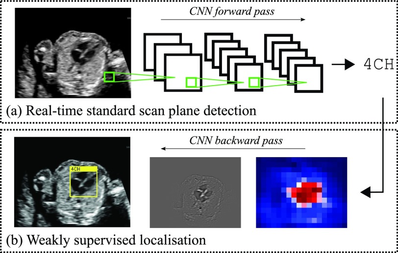
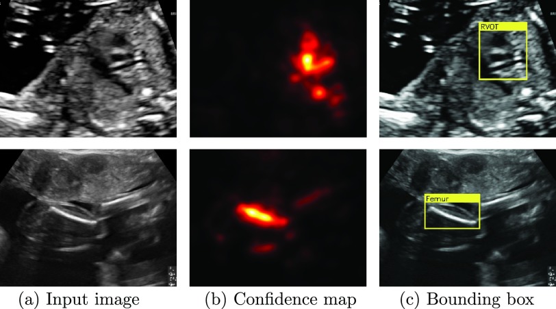
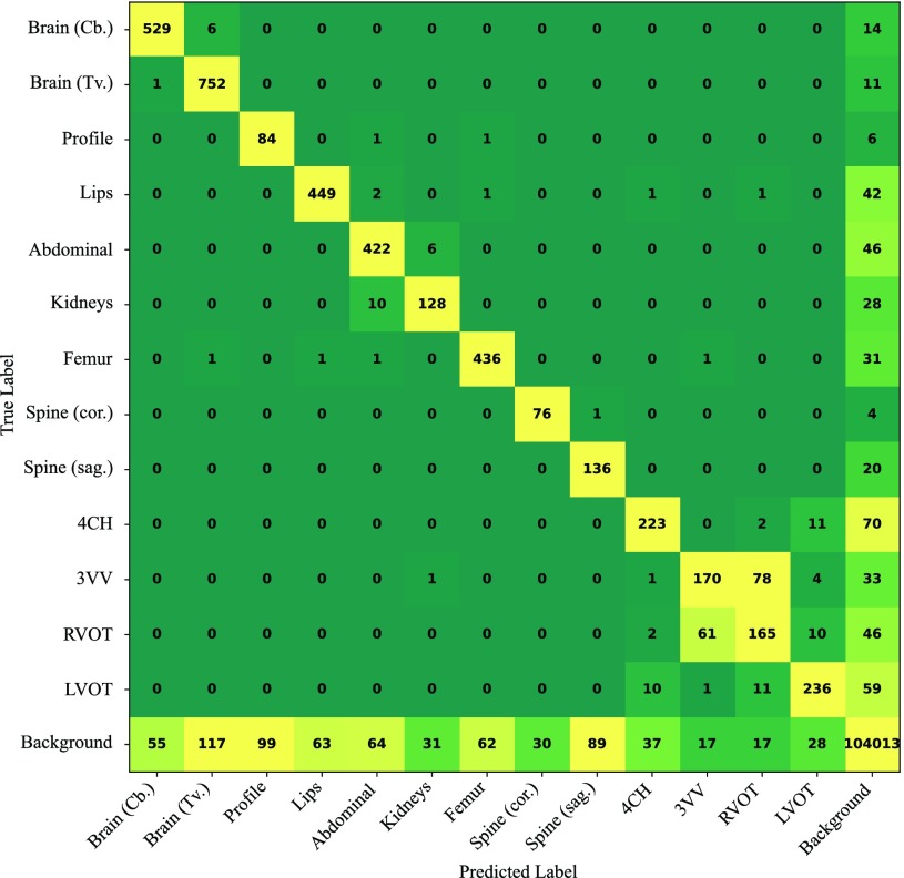
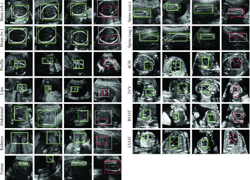
Identifying and interpreting fetal standard scan planes during 2-D ultrasound mid-pregnancy examinations are highly complex tasks, which require years of training. Apart from guiding the probe to the correct location, it can be equally difficult for a non-expert to identify relevant structures within the image. Automatic image processing can provide tools to help experienced as well as inexperienced operators with these tasks. In this paper, we propose a novel method based on convolutional neural networks, which can automatically detect 13 fetal standard views in freehand 2-D ultrasound data as well as provide a localization of the fetal structures via a bounding box. An important contribution is that the network learns to localize the target anatomy using weak supervision based on image-level labels only. The network architecture is designed to operate in real-time while providing optimal output for the localization task. We present results for real-time annotation, retrospective frame retrieval from saved videos, and localization on a very large and challenging dataset consisting of images and video recordings of full clinical anomaly screenings. We found that the proposed method achieved an average F1-score of 0.798 in a realistic classification experiment modeling real-time detection, and obtained a 90.09% accuracy for retrospective frame retrieval. Moreover, an accuracy of 77.8% was achieved on the localization task.
Overview
The claims and statistics of this paper haven’t been checked yet.
Run the check — free, no account needed. It reads the open-access text and checks the sources it cites.
Integrity signals
Registry
Cites
Statistics
Figures
Verification
Claims the paper makes
The claims and statistics of this paper haven’t been checked yet.
Run the check — free, no account needed. It reads the open-access text and checks the sources it cites.
Claims other papers make about this one
Each is checked against this paper’s own text: does it say what the citing paper says it does?
The cited source says what the claim attributes to it.
In the citing paper
“the possibility of real-time applicability [15,25,32,33]”
The check wasn’t confident either way, so it gives no verdict — never disputed.
In the citing paper
“Conventional assessments of fetal lung maturity rely on invasive amniocentesis and surface-level ultrasound scoring, achieving moderate predictive performance (AUC ≈ 0.82) but lacking dynamic integration of placental function (23, 24).”
From Placenta-pulmonary coupling-guided multimodal AI for fetal lung maturity staging and individualized glucocorticoid therapy (2026) · cites this paper as [23]
Experimental — accuracy not yet measuredIn the citing paper
“Baumgartner et al. [117,118] and Chen et al. [78,119] accomplished the detection of 13 fetal standard views (e.g., kidneys, brain, abdominal, spine, femur, and cardiac plane) and the fetal abdominal (or face and four-chamber view) standard plane in 2D US images through the transferred deep models, respectively.”
From Deep Learning in Medical Ultrasound Analysis: A Review (2019) · cites this paper as [118]
Self-check
Percentages, sums and reported differences re-computed from the paper’s own numbers.
The self-check needs the paper’s full text.
Citations
Cites
Deep Residual Learning for Image Recognition
He, K., Zhang, X., Ren, S., & Sun, J. (2016) · 2016 IEEE Conference on Computer Vision and Pattern Recognition (CVPR) · doi:10.1109/cvpr.2016.90 · Cited by 235,474
Very Deep Convolutional Networks for Large-Scale Image Recognition
Simonyan, K., & Zisserman, A. (2014) · arXiv · doi:10.48550/arxiv.1409.1556 · Cited by 114,148
ImageNet classification with deep convolutional neural networks
Krizhevsky, A., Sutskever, I., & Hinton, G. E. (2017) · Communications of the ACM · doi:10.1145/3065386 · Cited by 112,155
Batch Normalization: Accelerating Deep Network Training by Reducing Internal Covariate Shift
Ioffe, S., & Szegedy, C. (2015) · arXiv · doi:10.48550/arxiv.1502.03167 · Cited by 27,818
The pascal visual object classes (VOC) challenge
Everingham, M., Van Gool, L., Williams, C. K. I., Winn, J., & Zisserman, A. (2010) · International Journal of Computer Vision · doi:10.1007/s11263-009-0275-4 · Cited by 19,599
Batch Normalization: Accelerating Deep Network Training by Reducing Internal Covariate ShiftPreprint
Ioffe, S. (2024) · arXiv (Cornell University) · doi:10.57702/o9raffed · Cited by 15,273
He, H., & Garcia, E. A. (2009) · IEEE Transactions on Knowledge and Data Engineering · doi:10.1109/tkde.2008.239 · Cited by 10,359
Fast approximate energy minimization via graph cuts
Boykov, Y., Veksler, O., & Zabih, R. (2001) · IEEE Transactions on Pattern Analysis and Machine Intelligence · doi:10.1109/34.969114 · Cited by 6,988
Deep Inside Convolutional Networks: Visualising Image Classification Models and Saliency Maps
Simonyan, K., Vedaldi, A., & Zisserman, A. (2013) · arXiv · doi:10.48550/arxiv.1312.6034 · Cited by 5,370
Deep Residual Learning for Image Recognition
He, K., Zhang, X., Ren, S., & Sun, J. (2015) · arXiv · doi:10.48550/arxiv.1512.03385 · Cited by 4,196
Research works printed but not linked: 2
- 2015, Fetal Anomalie Screen Programme Handbook NHS Screening Programmes
- springett, 2014, British Isles Network of Congenital Anomaly Registers Congenital Anomaly Statistics 2010 England and Wales
Cited by
Sorted by citations
Items 201–300 of 413
Ultrasound detection of subquadricipital recess distension
Colussi, M., Civitarese, G., Ahmetovic, D., Bettini, C., Gualtierotti, R., Peyvandi, F., & Mascetti, S. (2023) · Intelligent Systems with Applications · doi:10.1016/j.iswa.2023.200183 · Cited by 9
Early Pregnancy Fetal Facial Ultrasound Standard Plane‐Assisted Recognition Algorithm
Xue, H., Yu, W., Liu, Z., & Liu, P. (2023) · Journal of Ultrasound in Medicine · doi:10.1002/jum.16209 · Cited by 8
Artificial Intelligence in Obstetric Anomaly Scan: Heart and Brain
Enache, I.-A., Iovoaica-Rămescu, C., Ciobanu, Ș. G., Berbecaru, E. I. A., Vochin, A., Băluță, I. D., Istrate-Ofiţeru, A.-M., Comănescu, C. M., Nagy, R. D., & Iliescu, D. G. (2024) · Life · doi:10.3390/life14020166 · Cited by 12
Generating Controllable Ultrasound Images of the Fetal Head
Lee, L. H., & Noble, J. A. (2020) · IEEE International Symposium on Biomedical Imaging (ISBI) · doi:10.1109/isbi45749.2020.9098578 · Cited by 9
Chotzoglou, E., Day, T. G., Tan, J., Matthew, J., Lloyd, D. F. A., Razavi, R., Simpson, J. M., & Kainz, B. (2021) · The Journal of Machine Learning for Biomedical Imaging · doi:10.59275/j.melba.2021-g4dg · Cited by 10
Self-citationKnowledge distillation for efficient standard scanplane detection of fetal ultrasound
Dapueto, J., Zini, L., & Odone, F. (2023) · Medical & Biological Engineering & Computing · doi:10.1007/s11517-023-02881-4 · Cited by 10
Alshagathrh, F. M., Alzubaidi, M. S., Gecík, S., Alswat, K., Aldhebaib, A., Alahmadi, B., Alkubeyyer, M., Alosaimi, A., Alsadoon, A., Alkhamash, M., et al. (2024) · Diagnostics · doi:10.3390/diagnostics14232646 · Cited by 11
Wang, L., Fatemi, M., & Alizad, A. (2025) · Computers in Biology and Medicine · doi:10.1016/j.compbiomed.2025.110312 · Cited by 12
Sejer, E. P. F., Pegios, P., Lin, M., Bashir, Z., Wulff, C. B., Christensen, A. N., Nielsen, M., Feragen, A., & Tolsgaard, M. G. (2025) · American Journal of Obstetrics and Gynecology · doi:10.1016/j.ajog.2025.09.012 · Cited by 10
Automatic Shadow Detection in 2D Ultrasound Images
Meng, Q., Baumgartner, C. F., Sinclair, M. D., Housden, R. J., Rajchl, M., Gómez, A., Hou, B., Toussaint, N., Zimmer, V. A., Tan, J., et al. (2018) · Lecture notes in computer science · doi:10.1007/978-3-030-00807-9_7 · Cited by 8
Self-citationBelciug, S. (2024) · BMC Medical Informatics and Decision Making · doi:10.1186/s12911-024-02505-3 · Cited by 13
Automatic Measurement of Fetal Head Circumference from 2-Dimensional Ultrasound
Aji, C. P., Fatoni, M. H., & Sardjono, T. A. (2019) · International Conference on Computer Engineering, Network, and Intelligent Multimedia (CENIM) · doi:10.1109/cenim48368.2019.8973258 · Cited by 8
Multi-Feature Semi-Supervised Learning for COVID-19 Diagnosis from Chest X-Ray Images
Xiao, Q., Foran, D. J., Nosher, J. L., & Hacihaliloglu, I. (2021) · Lecture notes in computer science · doi:10.1007/978-3-030-87589-3_16 · Cited by 8
Offset regression networks for view plane estimation in 3D fetal ultrasound
Schmidt-Richberg, A., Schadewaldt, N., Klinder, T., Lenga, M., Trahms, R., Canfield, E., Roundhill, D. N., & Cristian Lorenz, A. (2019) · SPIE Medical Imaging: Image Processing · doi:10.1117/12.2512697 · Cited by 8
Ambroise-Grandjean, G., Hossu, G., Banasiak, C., Ciofolo, C., Raynaud, C., Rouet, L., Morel, O., & Beaumont, M. (2019) · BMJ Open · doi:10.1136/bmjopen-2019-031777 · Cited by 8
Towards Capturing Sonographic Experience: Cognition-Inspired Ultrasound Video Saliency Prediction
Droste, R., Cai, Y., Sharma, H., Chatelain, P., Papageorghiou, A. T., & Noble, J. A. (2020) · Communications in computer and information science · doi:10.1007/978-3-030-39343-4_15 · Cited by 8
Learning Cross-domain Generalizable Features by Representation DisentanglementPreprint
Meng, Q., Rueckert, D., & Kainz, B. (2020) · arXiv (Cornell University) · doi:10.48550/arxiv.2003.00321 · Cited by 9
Self-citationUnsupervised Detection of Fetal Brain Anomalies Using Denoising Diffusion Models
Olsen, M. D. S., Ambsdorf, J., Lin, M., Taksøe‐Vester, C., Svendsen, M. B. S., Christensen, A. N., Nielsen, M., Tolsgaard, M. G., Feragen, A., & Pegios, P. (2024) · Lecture notes in computer science · doi:10.1007/978-3-031-73647-6_20 · Cited by 7
MMSummary: Multimodal Summary Generation for Fetal Ultrasound Video
Guo, X., Men, Q., & Noble, J. A. (2024) · Lecture notes in computer science · doi:10.1007/978-3-031-72083-3_63 · Cited by 7
Li, B. (2021) · Complexity · doi:10.1155/2021/5539793 · Cited by 9
Adaptive 3D Localization of 2D Freehand Ultrasound Brain Images
Yeung, P.-H., Aliasi, M., Haak, M. C., the INTERGROWTH-21st Consortium, Xie, W., & Namburete, A. I. L. (2022) · Lecture notes in computer science · doi:10.1007/978-3-031-16440-8_20 · Cited by 7
Medical Image Detection Using Deep Learning
Ocaña, M. I. G., López‐Linares, K., Lete, N., Ballester, M. Á. G., & Macía, I. (2019) · Intelligent systems reference library · doi:10.1007/978-3-030-32606-7_1 · Cited by 7
Detection of Anomalies in Fetus using Convolution Neural Network
Bindiya, H. M., Chethana, H. T., & Pavan Kumar, S. P. (2018) · International Journal of Information Technology and Computer Science · doi:10.5815/ijitcs.2018.11.08 · Cited by 7
Fetal Movement Detection and Anatomical Plane Recognition using YOLOv5 Network in Ultrasound Scans
Dandıl, E., Turkan, M., Urfalı, F. E., Bıyık, İ., & Korkmaz, M. (2021) · European Journal of Science and Technology · doi:10.31590/ejosat.951786 · Cited by 8
Automatic quality assessment for 2D fetal sonographic standard plane based on multi-task learningPreprint
Luo, H., Liu, H., Li, K., & Zhang, B. (2019) · arXiv (Cornell University) · doi:10.48550/arxiv.1912.05260 · Cited by 7
PRETUS: A plug-in based platform for real-time ultrasound imaging research
Gómez, A., Zimmer, V. A., Wheeler, G., Toussaint, N., Deng, S., Wright, R., Skelton, E., Matthews, J., Kainz, B., Hajnal, J. V., et al. (2022) · SoftwareX · doi:10.1016/j.softx.2021.100959 · Cited by 7
Self-citationYu, T., Tsui, P., Leonov, D. V., Wu, S., Bin, G., & Zhou, Z. (2024) · Sensors · doi:10.3390/s24237510 · Cited by 6
Mishra, D., Saha, P., Zhao, H., Hernandez-Cruz, N., Patey, O. V., Papageorghiou, A. T., & Noble, J. A. (2025) · Medical Image Analysis · doi:10.1016/j.media.2025.103611 · Cited by 7
Role of artificial intelligence in congenital heart disease
Niyogi, S. G., Nag, D. S., Shah, M. M., Swain, A., Naskar, C., Srivastava, P., & Kant, R. (2025) · World Journal of Clinical Pediatrics · doi:10.5409/wjcp.v14.i3.105926 · Cited by 8
Yenkikar, A., Singh, V. K., Tamboli, G., Charkha, P., Bodke, S., Bidwe, R. V., & Bali, M. (2025) · MethodsX · doi:10.1016/j.mex.2025.103618 · Cited by 6
Goetz-Fu, M., Haller, M., Collins, T., BEGUSIC, N., Jochum, F., Keeza, Y., Uwineza, J., Marescaux, J., Weingertner, A. S., Sananès, N., et al. (2025) · Journal of Gynecology Obstetrics and Human Reproduction · doi:10.1016/j.jogoh.2025.103039 · Cited by 8
From light to sound: Seeing and hearing the placenta in health and disease
Lee, D., Kim, J., Heo, J., Jeon, H., Chang, S., Choi, W., Kim, C., & Yang, Z. (2026) · Science Advances · doi:10.1126/sciadv.aed2184 · Cited by 7
Towards Standard Plane Prediction of Fetal Head Ultrasound with Domain Adaption
Men, Q., Zhao, H., Drukker, L., Papageorghiou, A. T., & Noble, J. A. (2023) · IEEE International Symposium on Biomedical Imaging (ISBI) · doi:10.1109/isbi53787.2023.10230542 · Cited by 6
Visual attribution using Adversarial Latent Transformations
Zia, T., Wahab, A., Windridge, D., Tirunagari, S., & Bashir, N. (2023) · Computers in Biology and Medicine · doi:10.1016/j.compbiomed.2023.107521 · Cited by 6
Leveraging Shape and Spatial Information for Spontaneous Preterm Birth Prediction
Pegios, P., Sejer, E. P. F., Lin, M., Bashir, Z., Svendsen, M. B. S., Nielsen, M., Petersen, E., Christensen, A. N., Tolsgaard, M. G., & Feragen, A. (2023) · Lecture notes in computer science · doi:10.1007/978-3-031-44521-7_6 · Cited by 7
Ultrasound Plane Pose Regression: Assessing Generalized Pose Coordinates in the Fetal Brain
Vece, C. D., Lous, M. L., Dromey, B., Vasconcelos, F., David, A. L., Peebles, D., & Stoyanov, D. V. (2023) · IEEE Transactions on Medical Robotics and Bionics · doi:10.1109/tmrb.2023.3328638 · Cited by 7
Deslandes, A., Avery, J., Chen, H., Leonardi, M., Condous, G. S., & Hull, M. L. (2023) · Australasian Journal of Ultrasound in Medicine · doi:10.1002/ajum.12368 · Cited by 7
FetusMap: Fetal Pose Estimation in 3D Ultrasound
Yang, X., Shi, W., Dou, H., Qian, J., Wang, Y., Xue, W., Li, S., Ni, D., & Heng, P. (2019) · Lecture notes in computer science · doi:10.1007/978-3-030-32254-0_32 · Cited by 6
Heart Position Estimation based on Bone Distribution toward Autonomous Robotic Fetal Ultrasonography
Shida, Y., Tsumura, R., Watanabe, T., & Iwata, H. (2021) · Proceedings - IEEE International Conference on Robotics and Automation/Proceedings · doi:10.1109/icra48506.2021.9560839 · Cited by 6
Automatic Recognition of Fetal Heart Standard Section Based on Fast-RCNN
Wu, B., Wu, H., Du, Y., & Liu, P. (2021) · IEEE International Conference on Anti-counterfeiting, Security, and Identification (ASID) · doi:10.1109/asid52932.2021.9651487 · Cited by 6
Artificial Intelligence in Prenatal Ultrasound: Clinical Application and Prospect
Guo, M. W., Li, M. S., Yu, M. X., Wen, B. H., Yuan, M. Y., & Yang, M. X. (2023) · Advanced ultrasound in diagnosis and therapy · doi:10.37015/audt.2023.230024 · Cited by 6
Fetal Skull Reconstruction via Deep Convolutional Autoencoders
Cerrolaza, J. J., Li, Y., Biffi, C., Gómez, A., Matthew, J., Sinclair, M. D., Gupta, C., Knight, C. L., & Rueckert, D. (2018) · Annual International Conference of the IEEE Engineering in Medicine and Biology Society (EMBC) · doi:10.1109/embc.2018.8512282 · Cited by 6
Self-citationGao, Y., & Noble, J. A. (2019) · Lecture notes in computer science · doi:10.1007/978-3-030-32254-0_34 · Cited by 6
The Classification and Segmentation of Fetal Anatomies Ultrasound Image: A Survey
Song, C., Gao, T., Wang, H., Sudirman, S., Zhang, W., & Zhu, H. (2020) · Journal of Medical Imaging and Health Informatics · doi:10.1166/jmihi.2021.3616 · Cited by 6
Classification of Fetal Heart Ultrasound Images for the Detection of CHD
Sushma, T. V., Sriraam, N., Arakeri, M. P., & Suresh, S. (2021) · Lecture notes on data engineering and communications technologies · doi:10.1007/978-981-15-9651-3_41 · Cited by 6
Self-supervised Representation Learning for Ultrasound VideoPreprint
Jiao, J., Droste, R., Drukker, L., Papageorghiou, A. T., & Noble, J. A. (2020) · arXiv (Cornell University) · doi:10.48550/arxiv.2003.00105 · Cited by 6
Qiao, S., Pang, S., Luo, G., Pan, S., Wang, X., Wang, M., Zhai, X., & Chen, T. (2020) · arXiv (Cornell University) · doi:10.48550/arxiv.2011.13096 · Cited by 6
Sriraam, N., Punyaprabha, V., Sushma, T. V., & Suresh, S. (2023) · Medical & Biological Engineering & Computing · doi:10.1007/s11517-023-02814-1 · Cited by 5
Determinants of short interpregnancy intervals in high-income countries: a systematic review
Dunne, J., Foo, D., Jancey, J. M., Pereira, G. F., Kefale, B., Belay, D. G., Dhamrait, G. K., Gebremedhin, A. T., Mruts, K. B., Nyadanu, S. D., et al. (2025) · Sexual and Reproductive Health Matters · doi:10.1080/26410397.2025.2545699 · Cited by 7
Chen, L., Cao, Z. W., Zhang, W., Tang, X., Zhao, D., Zhang, D., Liao, H., & Chen, F. (2024) · IEEE Transactions on Biomedical Engineering · doi:10.1109/tbme.2024.3510275 · Cited by 5
Gearhart, A. S., Dwork, N., & Jone, P. (2022) · Intelligence-Based Medicine · doi:10.1016/j.ibmed.2022.100082 · Cited by 7
Balagalla, U. B., Jayasooriya, V., de Alwis, C., & Akila Subasinghe, A. (2023) · Computer Methods in Biomechanics and Biomedical Engineering Imaging & Visualization · doi:10.1080/21681163.2023.2179343 · Cited by 6
Pregitha, R. E., Kumar, R. S. V., & Kumar, C. E. S. (2023) · Soft Computing · doi:10.1007/s00500-023-08187-9 · Cited by 5
RetractedSatish, S., & Herald Anantha Rufus, N. (2023) · Journal of Wireless Mobile Networks Ubiquitous Computing and Dependable Applications · doi:10.58346/jowua.2024.i1.001 · Cited by 5
Hurtado, J., Sierra-Franco, C. A., Motta, T., & Raposo, A. B. (2024) · Computers in Biology and Medicine · doi:10.1016/j.compbiomed.2024.109188 · Cited by 5
AI to Assist in the Fetal Anomaly Ultrasound Scan: A Randomized Controlled Trial
Day, T. G., Matthew, J., Budd, S., Farruggia, A., Venturini, L., Wright, R., Jamshidi, B., To, M. S., Ling, H., Lai, J., et al. (2025) · NEJM AI · doi:10.1056/aioa2400747 · Cited by 5
Self-citationMDVA-GAN: multi-domain visual attribution generative adversarial networks
Nawaz, M., Al-Obeidat, F. N., Tubaishat, A., Zia, T., Maqbool, F., & Rocha, Á. (2022) · Neural Computing and Applications · doi:10.1007/s00521-022-06969-0 · Cited by 5
Nageswari, C. S., Kumar, M. N. V., Grace, N. V. A., & Thiyagarajan, J. B. (2022) · Journal of Intelligent & Fuzzy Systems · doi:10.3233/jifs-221654 · Cited by 8
Projective Latent Interventions for Understanding and Fine-Tuning Classifiers
Hinterreiter, A., Streit, M., & Kainz, B. (2020) · Lecture notes in computer science · doi:10.1007/978-3-030-61166-8_2 · Cited by 5
Self-citationKutty, S. (2020) · Journal of the American Society of Echocardiography · doi:10.1016/j.echo.2020.07.016 · Cited by 5
Reinforcement Learning for Robot Assisted Live Ultrasound Examination
Li, C., Zhang, T., Zhou, Z., Zhao, B., Zhang, P., & Qi, X. (2025) · Electronics · doi:10.3390/electronics14183709 · Cited by 4
Liu, Z., Yu, W., Wu, X., Yang, T., Lyu, G., Liu, P., & Xue, H. (2023) · International Journal of Gynecology & Obstetrics · doi:10.1002/ijgo.15145 · Cited by 5
Algorithms, Techniques, and Applications of Intelligent Diagnosis Using Dynamic Ultrasound: A Review
Liu, J., Zhang, J., Shao, Y., Xiao, X., Huang, S., & Kong, D. (2025) · IEEE Access · doi:10.1109/access.2025.3578817 · Cited by 4
Tehsin, S., Alshaya, H., Bouchelligua, W., & Nasir, I. M. (2025) · Diagnostics · doi:10.3390/diagnostics15222879 · Cited by 4
Generative Adversarial Network based Chest Disease Detection and Binary Mask Generation
Junaid, M. A., Anwar, S., Sikander, G., & Khan, M. T. (2023) · International Conference on Robotics and Automation in Industry (ICRAI) · doi:10.1109/icrai57502.2023.10089542 · Cited by 4
Artificial Intelligence for Fetal Ultrasound
Cho, H. C., Sun, S., Park, S. W., Kwon, J., & Seo, J. K. (2023) · Mathematics in industry · doi:10.1007/978-981-99-1839-3_5 · Cited by 6
Fetal Re-Identification in Multiple Pregnancy Ultrasound Images Using Deep Learning
Gabler, E., Nissen, M., Altstidl, T., Titzmann, A., Packhäuser, K., Maier, A., Fasching, P. A., Eskofier, B. M., & Leutheuser, H. (2023) · Annual International Conference of the IEEE Engineering in Medicine and Biology Society (EMBC) · doi:10.1109/embc40787.2023.10340336 · Cited by 4
Qin, X., Bui, F. M., Han, Z., & Khademi, A. (2024) · IEEE Access · doi:10.1109/access.2024.3412608 · Cited by 4
Diffusion Models for Unsupervised Anomaly Detection in Fetal Brain Ultrasound
Mykula, H., Gasser, L., Lobmaier, S. M., Schnabel, J. A., Zimmer, V. A., & Bercea, C. I. (2024) · Lecture notes in computer science · doi:10.1007/978-3-031-73647-6_21 · Cited by 4
Horky, A., Wasenitz, M., Iacovella, C., Bahlmann, F., & Naimi, A. A. (2025) · Archives of Gynecology and Obstetrics · doi:10.1007/s00404-025-08042-2 · Cited by 4
ScanAhead: Simplifying standard plane acquisition of fetal head ultrasound
Men, Q., Zhao, H., Drukker, L., Papageorghiou, A. T., & Noble, J. A. (2025) · Medical Image Analysis · doi:10.1016/j.media.2025.103614 · Cited by 7
Artificial Intelligence in Pediatrics
Kelly, C. J., Brown, A. P. Y., & Taylor, J. A. (2021) · Springer eBooks · doi:10.1007/978-3-030-58080-3_316-1 · Cited by 4
Künstliche Intelligenz in der pränatalen kardialen Diagnostik
Weichert, J., Welp, A., Scharf, J. L., Dracopoulos, C., Rody, A., & Gembicki, M. (2021) · Der Gynäkologe · doi:10.1007/s00129-021-04890-6 · Cited by 4
USG-Net: Deep Learning-based Ultrasound Scanning-Guide for an Orthopedic Sonographer
Lee, K., Yang, J., Lee, M. H., Chang, J. H., Kim, J., & Hwang, J. Y. (2022) · Lecture notes in computer science · doi:10.1007/978-3-031-16449-1_3 · Cited by 4
Congenital Heart Defect Recognition Model Based on YOLOV5
Wu, H., Wu, B., He, S., & Liu, P. (2022) · IEEE International Conference on Anti-counterfeiting, Security, and Identification (ASID) · doi:10.1109/asid56930.2022.9995989 · Cited by 4
Deep learning models for interpretation of point of care ultrasound in military working dogs
Torres, S. I. H., Holland, L., Edwards, T. H., Venn, E. C., & Snider, E. J. (2024) · Frontiers in Veterinary Science · doi:10.3389/fvets.2024.1374890 · Cited by 4
Savochkina, E., Lee, L. H., Drukker, L., Papageorghiou, A. T., & Noble, J. A. (2021) · Lecture notes in computer science · doi:10.1007/978-3-030-80432-9_28 · Cited by 4
Standard Plane Localisation in 3D Fetal Ultrasound Using Network with Geometric and Image Loss
Li, Y., Cerrolaza, J. J., Sinclair, M. D., Hou, B., Alansary, A., Khanal, B., Matthew, J., Kainz, B., & Rueckert, D. (2018) · Cited by 4
Self-citationAn Automated Framework for Large Scale Retrospective Analysis of Ultrasound Images
Sridar, P., Kumar, A., Quinton, A. E., Kennedy, N. J., Nanan, R. K. H., & Kim, J. (2019) · IEEE Journal of Translational Engineering in Health and Medicine · doi:10.1109/jtehm.2019.2952379 · Cited by 4
Ye, J., Liu, R., Zou, B., Zhang, H., Zhan, N., Han, C., Yang, Y. Z., Zhang, H., Guo, J. H., Fang, C., et al. (2020) · doi:10.22541/au.158879105.54189382 · Cited by 4
Chen, K., Huang, Y., Yang, X., Hu, X., Zhang, Y., Tan, T., Xue, W., & Ni, D. (2024) · Biomedical Signal Processing and Control · doi:10.1016/j.bspc.2024.107249 · Cited by 3
Sarker, M. M. K., Mishra, D., Alsharid, M., Hernandez-Cruz, N., Ahuja, R., Patey, O. V., Papageorghiou, A. T., & Noble, J. A. (2025) · Medical Image Analysis · doi:10.1016/j.media.2025.103758 · Cited by 3
Komatsu, R., Komatsu, M., Takeda, K., Harada, N., Teraya, N., Wakisaka, S., Natsume, T., Taniguchi, T., Aoyama, R., Kaneko, M., et al. (2026) · Bioengineering · doi:10.3390/bioengineering13010100 · Cited by 3
Bai, J., Zhou, Z., Tang, Y., Gan, J., Liang, Z., Fan, J., Mcguire, L., Clarke, J., Cai, W., Spurway, J., et al. (2026) · Medical Image Analysis · doi:10.1016/j.media.2026.104043 · Cited by 4
Lee, K., Kim, M. C., Lim, C., & Song, T. (2021) · Sensors · doi:10.3390/s21082629 · Cited by 3
Automated Maternal Fetal Ultrasound Image Identification Using a Hybrid Vision Transformer Model
Krishna, T. B., Poreddy, A. K. R., Sindhu, K. G., & Kokil, P. (2024) · Lecture notes in computer science · doi:10.1007/978-3-031-78195-7_17 · Cited by 3
Fetal Anomaly Detection in Ultrasound Images: A Review of Deep Learning-based Approaches
Mhatre, S. R., & Bakal, J. (2024) · International Conference on Automation, Computing and Renewable Systems (ICACRS) · doi:10.1109/icacrs62842.2024.10841653 · Cited by 3
Pratap, T., Dhulipalla, V. R., & Kokil, P. (2025) · Biomedical Signal Processing and Control · doi:10.1016/j.bspc.2025.107918 · Cited by 4
Testi, M., Fiorentino, M. C., Ballabio, M., Visani, G., Ciccozzi, M., Frontoni, E., Moccia, S., & Vessio, G. (2025) · Medical & Biological Engineering & Computing · doi:10.1007/s11517-025-03436-5 · Cited by 4
Automatic recognition of fetal facial ultrasound standard planes based on improved YOLOv4
Xue, H., Liu, Z., Yu, W., & Liu, P. (2022) · IEEE International Conference on Anti-counterfeiting, Security, and Identification (ASID) · doi:10.1109/asid56930.2022.9995820 · Cited by 2
Artificial intelligence in obstetrics and gynecology
Kassir, E., Kuhn, V. C., Wong, M. S., & Han, C. S. (2023) · Elsevier eBooks · doi:10.1016/b978-0-443-15688-5.00004-8 · Cited by 3
Yolact-based Approach for Real-Time Fetal Heart Segmentation
Sapitri, A. I., Nurmaini, S., Rachmatullah, M. N., Darmawahyuni, A., Firdaus, F., & Islami, A. (2023) · International Conference on Data Science and Its Applications (ICoDSA) · doi:10.1109/icodsa58501.2023.10277564 · Cited by 3
Investigation on ultrasound images for detection of fetal congenital heart defects
Satish, S., & Rufus, N. H. A. (2024) · Biomedical Physics & Engineering Express · doi:10.1088/2057-1976/ad4f91 · Cited by 4
Automatic standard plane and diagnostic usability classification in obstetric ultrasounds
Lim, A., Abdalla, M., Abolhassani, F., Law, W., Fine, B., & Sussman, D. (2024) · WFUMB Ultrasound Open · doi:10.1016/j.wfumbo.2024.100050 · Cited by 3
Review on ultrasound image processing techniques for fetal head analysis
Prasad, K., & Patnaik, P. K. (2024) · AIP conference proceedings · doi:10.1063/5.0221469 · Cited by 3
Zukünftige Bildanalyse mit Hilfe automatisierter Algorithmen
Weichert, J., Rody, A., & Gembicki, M. (2020) · Der Gynäkologe · doi:10.1007/s00129-020-04563-w · Cited by 3
Combining Bayesian And Deep Learning Methods For The Delineation Of The Fan In Ultrasound Images
Dadoun, H., Delingette, H., Rousseau, A.-L., de Kerviler, É., & Ayache, N. (2021) · IEEE International Symposium on Biomedical Imaging (ISBI) · doi:10.1109/isbi48211.2021.9434112 · Cited by 5
Artificial Intelligence and Obstetric Ultrasound
Matsuoka, R. (2021) · Donald School Journal of Ultrasound in Obstetrics & Gynecology · doi:10.5005/jp-journals-10009-1702 · Cited by 3
Born Identity Network: Multi-way Counterfactual Map Generation to Explain a Classifier's DecisionPreprint
Oh, K., Yoon, J. S., & Suk, H. (2020) · arXiv (Cornell University) · doi:10.48550/arxiv.2011.10381 · Cited by 4
Lin, M. F., He, X., Hao, C., He, M., Guo, H., Zhang, L., Xian, J., zheng, J., Xu, Q., Feng, J., et al. (2020) · doi:10.22541/au.158880224.43356313 · Cited by 2
Metadata
- Authors
- Baumgartner, C. F., Kamnitsas, K., Matthew, J., Fletcher, T. P., Smith, S., Koch, L. M., Kainz, B., & Rueckert, D.
- Affiliations
- Imperial College London (GB)
- Guy's Hospital (GB)
- King's College London (GB)
- Corresponding author
- Baumgartner, Christian F.
- ORCID
- Venue
- IEEE Transactions on Medical Imaging
- Published
- 2017-11
- Volume
- 36
- Issue
- 11
- Pages
- 2204-2215
- ISSN
- 0278-0062 (ISSN-L)
- 1558-254X
- PMC
- PMC6051487
- OpenAlex
- W2735582614
- Cited by
- 416
- References
- 34
- Citations by year
- 2026: 52 · 2025: 48 · 2024: 54 · 2023: 56 · 2022: 55 · 2021: 57 · 2020: 44 · 2019: 28 · 2018: 21 · 2017: 1
- FWCI
- 10.67
- Type
- journal article
- Language
- en
- Open access
- hybrid
- doi.org
- Locations
- IEEE Transactions on Medical Imaging (publishedVersion, cc-by)
- Europe PMC (PubMed Central) (publishedVersion, cc-by)
- PubMed Central (publishedVersion, cc-by)
- PubMed (publishedVersion)
- arXiv (Cornell University) (submittedVersion)
- Research Portal (King's College London) (submittedVersion, cc-by)
- Spiral (Imperial College London) (submittedVersion, cc-by)
- arXiv (Cornell University)
- Funders
- Wellcome Trust IEH Award (102431)
- Topics
- Physical Sciences › Computer Science › Computer Vision and Pattern Recognition › Medical Image Segmentation Techniques
- Health Sciences › Medicine › Health Informatics › Artificial Intelligence in Healthcare and Education
- Physical Sciences › Computer Science › Computer Vision and Pattern Recognition › Advanced Neural Network Applications
- Keywords
- real-time detection, weakly supervised learning, image-level labels, convolutional neural networks, bounding box localization
- MeSH terms
- Algorithms, Female, Humans, Image Processing, Computer-Assisted, Pregnancy, Video Recording, Ultrasonography, Prenatal, Neural Networks, Computer
About these checks
Registry, retraction, Cites and statistics results are computed, not judged, with one exception: a printed reference that no index lists is read once by a model before it counts as a citation error. Claims are extracted by a model and kept only when their quote appears verbatim in the text; each cited source is then checked, with a model’s help, for whether it says what the claim relies on. Those verdicts can be wrong — read the claim’s quote and the cited source before relying on one.
Statistics and arithmetic are recomputed, not judged.
Confidence bands are shown only for fields calibrated on our annotated test set. None is calibrated yet, so no band is shown.
What counts as a claim ›How we measure confidence ›How we check this ›
Are you an author, or do you see a problem with this page? Request a review
