

The DOI matches its Crossref record; 35 of the 35 references it cites were found in a registry or an index.
SonoNet: Real-Time Detection and Localisation of Fetal Standard Scan Planes in Freehand Ultrasound
Open accessAbstract
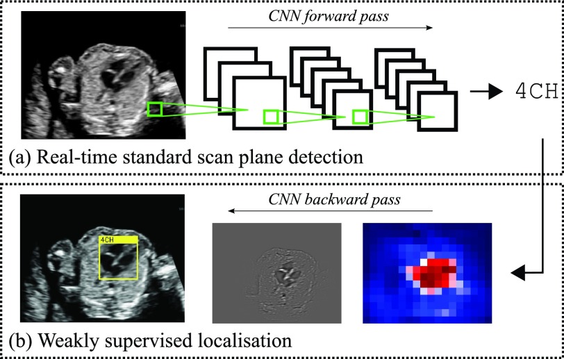
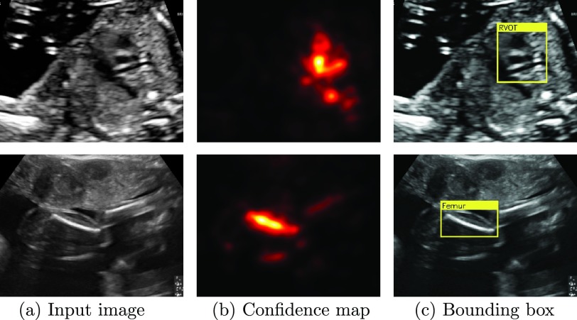
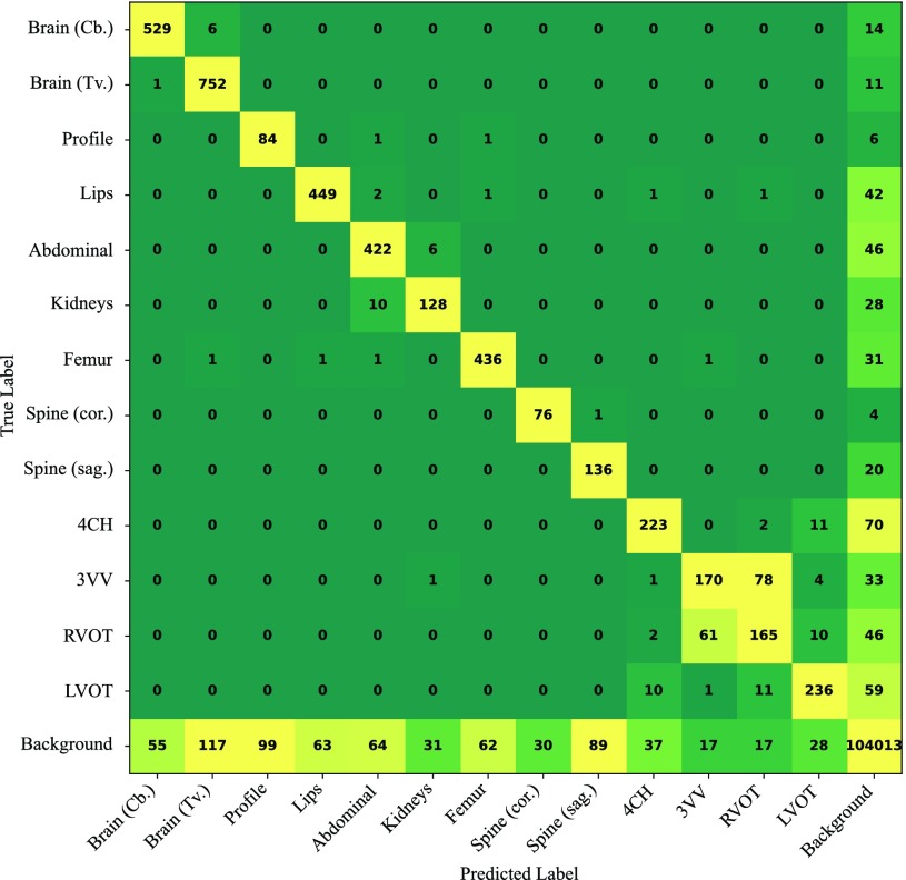
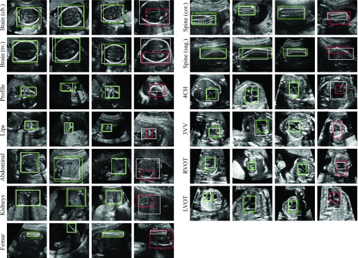
Identifying and interpreting fetal standard scan planes during 2-D ultrasound mid-pregnancy examinations are highly complex tasks, which require years of training. Apart from guiding the probe to the correct location, it can be equally difficult for a non-expert to identify relevant structures within the image. Automatic image processing can provide tools to help experienced as well as inexperienced operators with these tasks. In this paper, we propose a novel method based on convolutional neural networks, which can automatically detect 13 fetal standard views in freehand 2-D ultrasound data as well as provide a localization of the fetal structures via a bounding box. An important contribution is that the network learns to localize the target anatomy using weak supervision based on image-level labels only. The network architecture is designed to operate in real-time while providing optimal output for the localization task. We present results for real-time annotation, retrospective frame retrieval from saved videos, and localization on a very large and challenging dataset consisting of images and video recordings of full clinical anomaly screenings. We found that the proposed method achieved an average F1-score of 0.798 in a realistic classification experiment modeling real-time detection, and obtained a 90.09% accuracy for retrospective frame retrieval. Moreover, an accuracy of 77.8% was achieved on the localization task.
Overview
The claims and statistics of this paper haven’t been checked yet.
Run the check — free, no account needed. It reads the open-access text and checks the sources it cites.
Integrity signals
Registry
Cites
Statistics
Figures
Verification
Claims the paper makes
The claims and statistics of this paper haven’t been checked yet.
Run the check — free, no account needed. It reads the open-access text and checks the sources it cites.
Claims other papers make about this one
Each is checked against this paper’s own text: does it say what the citing paper says it does?
The cited source says what the claim attributes to it.
In the citing paper
“the possibility of real-time applicability [15,25,32,33]”
The check wasn’t confident either way, so it gives no verdict — never disputed.
In the citing paper
“Conventional assessments of fetal lung maturity rely on invasive amniocentesis and surface-level ultrasound scoring, achieving moderate predictive performance (AUC ≈ 0.82) but lacking dynamic integration of placental function (23, 24).”
From Placenta-pulmonary coupling-guided multimodal AI for fetal lung maturity staging and individualized glucocorticoid therapy (2026) · cites this paper as [23]
Experimental — accuracy not yet measuredIn the citing paper
“Baumgartner et al. [117,118] and Chen et al. [78,119] accomplished the detection of 13 fetal standard views (e.g., kidneys, brain, abdominal, spine, femur, and cardiac plane) and the fetal abdominal (or face and four-chamber view) standard plane in 2D US images through the transferred deep models, respectively.”
From Deep Learning in Medical Ultrasound Analysis: A Review (2019) · cites this paper as [118]
Self-check
Percentages, sums and reported differences re-computed from the paper’s own numbers.
The self-check needs the paper’s full text.
Citations
Cites
Deep Residual Learning for Image Recognition
He, K., Zhang, X., Ren, S., & Sun, J. (2016) · 2016 IEEE Conference on Computer Vision and Pattern Recognition (CVPR) · doi:10.1109/cvpr.2016.90 · Cited by 235,474
Very Deep Convolutional Networks for Large-Scale Image Recognition
Simonyan, K., & Zisserman, A. (2014) · arXiv · doi:10.48550/arxiv.1409.1556 · Cited by 114,148
ImageNet classification with deep convolutional neural networks
Krizhevsky, A., Sutskever, I., & Hinton, G. E. (2017) · Communications of the ACM · doi:10.1145/3065386 · Cited by 112,155
Batch Normalization: Accelerating Deep Network Training by Reducing Internal Covariate Shift
Ioffe, S., & Szegedy, C. (2015) · arXiv · doi:10.48550/arxiv.1502.03167 · Cited by 27,818
The pascal visual object classes (VOC) challenge
Everingham, M., Van Gool, L., Williams, C. K. I., Winn, J., & Zisserman, A. (2010) · International Journal of Computer Vision · doi:10.1007/s11263-009-0275-4 · Cited by 19,599
Batch Normalization: Accelerating Deep Network Training by Reducing Internal Covariate ShiftPreprint
Ioffe, S. (2024) · arXiv (Cornell University) · doi:10.57702/o9raffed · Cited by 15,273
He, H., & Garcia, E. A. (2009) · IEEE Transactions on Knowledge and Data Engineering · doi:10.1109/tkde.2008.239 · Cited by 10,359
Fast approximate energy minimization via graph cuts
Boykov, Y., Veksler, O., & Zabih, R. (2001) · IEEE Transactions on Pattern Analysis and Machine Intelligence · doi:10.1109/34.969114 · Cited by 6,988
Deep Inside Convolutional Networks: Visualising Image Classification Models and Saliency Maps
Simonyan, K., Vedaldi, A., & Zisserman, A. (2013) · arXiv · doi:10.48550/arxiv.1312.6034 · Cited by 5,370
Deep Residual Learning for Image Recognition
He, K., Zhang, X., Ren, S., & Sun, J. (2015) · arXiv · doi:10.48550/arxiv.1512.03385 · Cited by 4,196
Research works printed but not linked: 2
- 2015, Fetal Anomalie Screen Programme Handbook NHS Screening Programmes
- springett, 2014, British Isles Network of Congenital Anomaly Registers Congenital Anomaly Statistics 2010 England and Wales
Cited by
Sorted by citations
Items 301–400 of 413
TagGAN: A generative model for data tagging
Nawaz, M., Nasir, B., Zia, T., Hussain, Z., & Moreira, C. (2025) · Computers in Biology and Medicine · doi:10.1016/j.compbiomed.2025.111385 · Cited by 2
Sundaramoorthy, K., Ganeshbabu, M., Mithun Barath, M. R., Jayaprakash, A. N., Dhanush Kumar, B., Selvamani, S., & Dr Narayanasamy, K. (2025) · Cureus · doi:10.7759/cureus.100534 · Cited by 2
Hossain, J., Xu, P., Liu, Y., Li, J., & Kong, Z. (2026) · Expert Systems with Applications · doi:10.1016/j.eswa.2026.131996 · Cited by 3
A review of ultrasound video segmentation: From temporal modeling to clinical utility
Huang, Q., An, Z., & Li, G. (2026) · Neurocomputing · doi:10.1016/j.neucom.2026.134407 · Cited by 2
Challenges in Ultrasonography Education and Training
Lobo, M. D., & Miravent, S. (2022) · Advances in medical education, research, and ethics (AMERE) book series · doi:10.4018/978-1-7998-9578-7.ch012 · Cited by 2
Thinking Like Sonographers: A Deep CNN Model for Diagnosing Gout from Musculoskeletal Ultrasound
Cao, Z. W., Zhang, W., Chen, K., Zhao, D., Zhang, D., Liao, H., & Chen, F. (2023) · Lecture notes in computer science · doi:10.1007/978-3-031-43987-2_16 · Cited by 2
Maternal Fetal Ultrasound Planes Classification using Contrastive Language Image Pre-training Models
Bhuma, C. M. (2024) · IEEE South Asian Ultrasonics Symposium (SAUS) · doi:10.1109/saus61785.2024.10563862 · Cited by 2
Audio-visual modelling in a clinical setting
Jiao, J., Alsharid, M., Drukker, L., Papageorghiou, A. T., Zisserman, A., & Noble, J. A. (2024) · Scientific Reports · doi:10.1038/s41598-024-66160-4 · Cited by 2
Turgut, Ü. K. (2025) · Frontiers in Medicine · doi:10.3389/fmed.2025.1505450 · Cited by 2
Quarello, E., & Corno, E. (2025) · Ultrasound in Obstetrics and Gynecology · doi:10.1002/uog.29185 · Cited by 3
Initial Study of Oil Palm Crop Condition Detection Through Drone Imagery: A YOLO Approach
Nugraha, W., Ermatita, & Stiawan, D. (2024) · International Conference on Informatics, Multimedia, Cyber and Information System (ICIMCIS) · doi:10.1109/icimcis63449.2024.10957214 · Cited by 2
Diffusion-Based Iterative Counterfactual Explanations for Fetal Ultrasound Image Quality Assessment
Pegios, P., Lin, M., Weng, N., Svendsen, M. B. S., Bashir, Z., Bigdeli, S., Christensen, A. N., Tolsgaard, M. G., & Feragen, A. (2025) · Lecture notes in computer science · doi:10.1007/978-3-032-06329-8_17 · Cited by 4
Ultrasonic Image Segmentation Method based on Improved Fully Convolution Network
Hai, Y., Feng, K., Luo, L., & Xie, H. (2019) · International Conference on Mechatronics Engineering and Information Technology (ICMEIT) · doi:10.2991/icmeit-19.2019.93 · Cited by 2
Spatio-Temporal Model for EUS Video Detection of Pancreatic Anatomy Structures
Meyer, A., Fleurentin, A., Montanelli, J., Mazellier, J.-P., Swanström, L. L., Gallix, B., Exarchakis, G., Valencia, L. S., & Padoy, N. (2022) · Lecture notes in computer science · doi:10.1007/978-3-031-16902-1_2 · Cited by 2
Reconstruction of Fetal Head Surface from Few 2D Ultrasound Images Tracked in 3D Space
Marcadent, S., Hêches, J., Favre, J., Desseauve, D., & Thiran, J. (2022) · IEEE-EMBS Conference on Biomedical Engineering and Sciences · doi:10.1109/iecbes54088.2022.10079362 · Cited by 2
Prior-Guided Attribution of Deep Neural Networks for Obstetrics and Gynecology
Bonnard, J., Dapogny, A., Zsámboki, R., Braud, L. V. D., Jurkovic, D., Bailly, K., & Dhombres, F. (2023) · IEEE Journal of Biomedical and Health Informatics · doi:10.1109/jbhi.2023.3337511 · Cited by 2
Zhu, B., Yang, F., Duan, H., & Gao, Z. (2024) · Current Bioinformatics · doi:10.2174/0115748936295679240620094626 · Cited by 1
Automatic Facial Axes Standardization of 3D Fetal Ultrasound Images
Alomar, A., Rubio, R., Salort, L., Albaigés, G., Payà, A., Piella, G., & Sukno, F. M. (2024) · Lecture notes in computer science · doi:10.1007/978-3-031-73647-6_9 · Cited by 2
Fetal Ultrasound Video Representation Learning Using Contrastive Rubik’s Cube Recovery
Zhang, K., Jiao, J., & Noble, J. A. (2024) · Lecture notes in computer science · doi:10.1007/978-3-031-73647-6_18 · Cited by 1
Nuchal Translucency Assessment in Prenatal Screening: An Attention-Based U-Net Approach
Sriraam, N., Chinta, B., Seshadri, S., & Suresh, S. (2024) · International Conference on Emerging Techniques in Computational Intelligence (ICETCI) · doi:10.1109/icetci62771.2024.10704101 · Cited by 1
Comparing freehand and robotic techniques for standard scan plane localization in ultrasound imaging
Hu, Y., Huang, Y., Jones, C. K., Shepard, L. M., Ghazi, A., Basar, B., Helm, P. A., & Uneri, A. (2025) · SPIE Medical Imaging: Image-Guided Procedures, Robotic Interventions, and Modeling · doi:10.1117/12.3048812 · Cited by 1
Enhancing Fetal Cardiac Imaging With Artificial Intelligence
Martins, J. G., Horgan, R., & Sinkovskaya, E. S. (2025) · Clinical Obstetrics & Gynecology · doi:10.1097/grf.0000000000000992 · Cited by 1
Morisset, C., Logé, F., Debavelaere, V., Besson, R., Turan, Ş., Fries, N., Stirnemann, J., Oyelese, Y., & Ville, Y. G. (2026) · Obstetrics and Gynecology · doi:10.1097/aog.0000000000006164 · Cited by 1
Zhu, B., Qu, L., Zhou, L., Luo, Z., & Chen, Y. (2026) · International Journal of Medical Robotics and Computer Assisted Surgery · doi:10.1002/rcs.70140 · Cited by 1
Federated learning for prenatal detection of interrupted aortic arch using fetal ultrasound imaging
Han, J., Wang, H., Feng, Y., Yang, Q., Li, J., Zhang, H., He, Y., Liu, J., Nakamura, T., Cao, Y., et al. (2026) · Biomedical Signal Processing and Control · doi:10.1016/j.bspc.2026.109795 · Cited by 1
Deep Learning Approach for Predicting Cardiac Septal Defects Using Echocardiography
Nova, R., Sapitri, A. I., Rahayuningsih, S. E., & Putra, S. T. (2025) · International Conference on Artificial Intelligence and Technological Solutions (ICAITech) · doi:10.1109/icaitech66481.2025.11387258 · Cited by 1
Automated interpretation of fetal cardiac function evaluation from the echocardiogram
Huang, C., Zhang, L., Xie, B., Jiang, Y., Zhu, Y., Liu, X., Lei, T., He, M., Yan, Y., Wang, N., et al. (2026) · npj Digital Medicine · doi:10.1038/s41746-026-02381-3 · Cited by 1
Zhang, J., Feng, B., Cheng, Y., Zhou, F., Lu, X., Zhu, X., Wang, X., Xu, C., & Chen, L. (2026) · BMC Pregnancy and Childbirth · doi:10.1186/s12884-026-09073-6 · Cited by 3
Shaik, S., & Sunkara, K. (2026) · Engineering Research Express · doi:10.1088/2631-8695/ae68bb · Cited by 1
Fiorentino, M. C., Rosati, R., Conti, E., Tigano, C., & Mancini, A. (2026) · Neural Computing and Applications · doi:10.1007/s00521-026-12115-x · Cited by 1
Conti, E., Rosati, R., Federici, L., Mancini, A., & Fiorentino, M. C. (2026) · Neural Computing and Applications · doi:10.1007/s00521-026-12210-z · Cited by 1
Rech, L. (2026) · Medical Sciences · doi:10.3390/medsci14040434 · Cited by 1
Deteksi Otomatis Bidang Kepala Janin dari Citra Ultrasonografi 2 Dimensi
Aji, C. P., Sardjono, T. A., & Fatoni, M. H. (2020) · Jurnal Teknik ITS · doi:10.12962/j23373539.v9i1.45679 · Cited by 1
Artificial Intelligence in Pediatrics
Kelly, C. J., Brown, A. P. Y., & Taylor, J. A. (2022) · Springer eBooks · doi:10.1007/978-3-030-64573-1_316 · Cited by 1
Rojas-Azabache, C., & Romero-Olortiga, T. (2022) · Revista de investigación de Sistemas e Informática · doi:10.15381/risi.v15i2.24782 · Cited by 1
Guangzhu, X., Qian, Y., Yang, W., Rong, L., Jun, Z., & Lei, B. (2023) · Journal of Image and Graphics · doi:10.11834/jig.220347 · Cited by 1
Benjamin, J. G., Asokan, M., Alhosani, A., Alasmawi, H., Diehl, W., Bricker, L., Nandakumar, K., & Yaqub, M. K. (2023) · Lecture notes in computer science · doi:10.1007/978-3-031-44521-7_7 · Cited by 1
Pose-GuideNet: Automatic Scanning Guidance for Fetal Head Ultrasound from Pose Estimation
Men, Q., Guo, X., Papageorghiou, A. T., & Noble, J. A. (2024) · Lecture notes in computer science · doi:10.1007/978-3-031-72083-3_65 · Cited by 1
Automatic Standard Plane Detection in Fetal Ultrasound Improved by Fine-Tuning
Castellanos-Diaz, O., Pérez-González, J., Cosı́o, F. A., Camargo-Marín, L., Guzmán‐Huerta, M., Ramírez, B. E., Olveres, J., García-Ramírez, J., Medina-Bañuelos, V., & Valdés-Cristerna, R. (2025) · World Congress on Medical Physics and Biomedical Engineering, September 7 - 12, 2009, Munich, Germany · doi:10.1007/978-3-031-89514-2_31 · Cited by 1
Automatic Shadow Detection in 2D Ultrasound
Meng, Q., Baumgartner, C. F., Sinclair, M. D., Housden, R. J., Rajchl, M., Gómez, A., Hou, B., Toussaint, N., Tan, J., Matthew, J., et al. (2018) · Cited by 1
Self-citationShida, Y., Tsumura, R., Watanabe, T., FUJII, K., & Iwata, H. (2020) · Journal of the Robotics Society of Japan · doi:10.7210/jrsj.38.667 · Cited by 1
Fetal Ultrasonography: Is artificial intelligence the way forward?
Psarris, A., Syndos, M., Daskalakis, G. J., & Loutradis, D. (2020) · Hellenic Journal of Obstetrics and Gynecology · doi:10.33574/hjog.1995 · Cited by 1
Ma, B., Ran, J., Pan, T., Yan, F., Yue, Z., Yao, Y., Li, Y., & Nie, F. (2026) · Frontiers in Medicine · doi:10.3389/fmed.2026.1791481
Rawat, V., Jain, A., Shrimali, V., & Raghuvanshi, S. (2018) · Lecture notes in computer science · doi:10.1007/978-3-319-99810-7_6
Fetal Standard Plane Detection in Freehand Ultrasound Using Multi Layered Extreme Learning Machine
Sree, S. J., & Vasanthanayaki, C. (2019) · Chapman & Hall/CRC eBooks · doi:10.1201/9780429446030-11
Projective Latent Space Decluttering.Preprint
Hinterreiter, A., Streit, M., & Kainz, B. (2020) · arXiv (Cornell University)
Self-citationScreen Tracking for Clinical Translation of Live Ultrasound Image Analysis MethodsPreprint
Treivase, S., Gómez, A., Matthew, J., Skelton, E., Schnabel, J. A., & Toussaint, N. (2020) · arXiv (Cornell University) · doi:10.48550/arxiv.2007.06272
Self-citationZeng, X., Zhang, Y., & Huang, W. (2021) · Research Square · doi:10.21203/rs.3.rs-847933/v1
Detection and Classification of Pregnancy StateUsing Deep Learning Technique
Mohammed, H. A., Hussein, E. M., & Sharif, M. M. (2021) · Omdurman Islamic University Journal · doi:10.52981/oiuj.v17i2.1819
Border Sensitive Network in Weakly Supervised Thyroid Nodule Detection for Ultrasound Image
Luo, T., Xu, T., Yu, J. X., Li, X., Wei, X., Yu, M., Zhang, R., Gao, J., & Yu, R. (2021) · IEEE International Conference on Bioinformatics and Biomedicine (BIBM) · doi:10.1109/bibm52615.2021.9669500
Jiang, Y., Wu, H., Cui, C., Shi, S., Ye, X., Tian, H., Song, D., Yao, J., Wang, L., Zhang, M., et al. (2022) · SSRN Electronic Journal · doi:10.2139/ssrn.4185034
Stellenwert der KI im pränatalen sonographischen Screening
Ritgen, J. (2022) · Die Gynäkologie · doi:10.1007/s00129-022-04995-6
Wu, Y., Peng, C., Chen, X., Yao, X., & Chen, Z. (2022) · Mathematics · doi:10.3390/math10234612
Big Data and AI in Cardiac Imaging
Reddy, C. D. (2022) · Integrated science · doi:10.1007/978-3-031-11199-0_5
Foetal ultrasonographic Sparse representation evaluation of spectral trust maps
Nagarajan, P., Ramadevi, R., Lakshmi, D., Kowsalya, T., & Anita, S. J. (2023) · AIP conference proceedings · doi:10.1063/5.0110574
Automated Recognition of Congenital Heart Defects (CHD) from Ultrasonic Fetal Heart Biometry
Sushma, T. V., Sriraam, N., & Suresh, S. (2023) · Journal of Physics Conference Series · doi:10.1088/1742-6596/2571/1/012015
Review Paper on IoT Thread Detection using Deep CNN Classifier
Mr. Shirke Ganesh, S., Bhosale, P. S. B., Dere, P. K. D., & Khatri, A. A. (2023) · International Journal of Advanced Research in Science Communication and Technology · doi:10.48175/ijarsct-14235
Venous Malformation Discrimination by Phleboliths Detection with Deep Learning
Cai, Y., Gong, X., He, Q., Fan, X., & Xiong, P. (2023) · International Symposium on Artificial Intelligence for Medicine Sciences (ISAIMS) · doi:10.1145/3644116.3644164
Aop-Sam: Segment Anything Model in Intrapartum Ultrasound Image AnalysisPreprint
Zhou, Z., Lu, Y., Bai, J., Campello, V. M., Feng, F., & Lekadir, K. (2024) · SSRN Electronic Journal · doi:10.2139/ssrn.4885247
Classification of Lung Disease From Chest X-Ray Images Using Convolutional Neural Networks
Dhiman, T., & Kumar, P. (2024) · International Conference on Advances in Modern Age Technologies for Health and Engineering Science (AMATHE) · doi:10.1109/amathe61652.2024.10582137
Tang, X., Cao, Z. W., Zhang, W., Zhao, D., Liao, H., Zhang, D., & Chen, F. (2024) · Lecture notes in computer science · doi:10.1007/978-3-031-72378-0_57
An Automated Pipeline for the Identification of Liver Tissue in Ultrasound Video
Ockenden, E. S., Mpooya, S., Noble, J. A., & Chami, G. F. (2025) · Communications in computer and information science · doi:10.1007/978-3-031-79103-1_10
Automatic detection of standard anatomical planes of the fetal brain from 3D ultrasound volumes
Torres, H. R., Morais, P., Fritze, A., Birdir, C., Rüdiger, M., & Vilaça, J. L. (2025) · SPIE Medical Imaging: Image Processing · doi:10.1117/12.3047364
Real-time Landmark Guidance for Radial Head Localization in Ultrasound Imaging
Meyling, L., Großbröhmer, C., Lichtenstein, J., Heinrich, M. P., & Hansen, L. (2025) · Informatik aktuell · doi:10.1007/978-3-658-47422-5_20
Platform for Automated Assessment of Obstetric Ultrasounds, Using Machine Learning
Luna, J. S., Alanís, A., Patiñό, E., & Márquez, B. Y. (2025) · Smart innovation, systems and technologies · doi:10.1007/978-981-97-6469-3_21
Highresolution Net for Estimation of Fetal Growth Parameters from First Trimester Ultrasound Images
Sriraam, N., Chinta, B., Seshadri, S., & Suresh, S. (2024) · IEEE Region 10 Conference (TENCON) · doi:10.1109/tencon61640.2024.10902669
Künstliche (Artifizielle) Intelligenz (KI oder AI) im Ultraschall
Weichert, J., & Kollmann, C. (2025) · Springer eBooks · doi:10.1007/978-3-662-67373-7_40
Determining Fetal Orientations From Blind Sweep Ultrasound Video
Wiśniewski, J. M., Christensen, A. N., Ngo, M. L., Tolsgaard, M. G., & Wong, C. K. (2025) · Lecture notes in computer science · doi:10.1007/978-3-031-95918-9_18
Applications of Artificial Intelligence on Fetal Echocardiography
Alves, J. A., Melo, M. M., Teixeira, L. M., Bravo‐Valenzuela, N. J., & Júnior, E. A. (2025) · Congenital Heart Disease · doi:10.32604/chd.2025.066358
New Applications and Perspectives in the Assessment of Fetal Heart by 3D Ultrasound
Weichert, A., & Weichert, J. (2025) · Springer eBooks · doi:10.1007/978-3-031-87175-7_20
Liu, Y., Peng, B., Zhou, Q., Yuan, S., Yan, W., Fang, L., Jiang, J., Wang, S., Zhu, X., & Wang, W. (2025) · Computer Methods and Programs in Biomedicine · doi:10.1016/j.cmpb.2025.108957
Multi-task Weakly Supervised Intrapartum Ultrasound Measurements
AlNasi, S., Abzhanov, A., Saeed, N., & Yaqub, M. K. (2025) · Lecture notes in computer science · doi:10.1007/978-3-031-96318-6_7
Wenhao, 魏. W., Xiangling, 田. T., & Yifan, 陈. C. (2025) · Laser & Optoelectronics Progress · doi:10.3788/lop251179
Detection of Optimal Echocardiographic Video Segments Using Deep Learning
Perez-Ruiz, I., Pérez-González, J., Carrera-Pinzón, A. F., Romero, E., Ruiz, H., Benitez, Y., & Arambula Cosio, F. (2025) · International Symposium on Medical Information Processing and Analysis (SIPAIM) · doi:10.1109/sipaim67325.2025.11283345
Nhat, P. T. H., Hảo, N. V., Tho, P. V., Kerdegari, H., Pisani, L., Thu, L. N. M., Phuong, L. T., Hai, D. H. T., Thuy, D. B., McBride, A., et al. (2023) · Figshare · doi:10.6084/m9.figshare.23614177
Diff-MOCS: A Diffusion Model-Based with Multi-Object Cardiac Ultrasound Images Segmentation
Ye, S., Zhang, Y., Meng, X., Li, C., & Sun, W. (2025) · IEEE International Conference on Bioinformatics and Biomedicine (BIBM) · doi:10.1109/bibm66473.2025.11356278
SpaceTime-SonoNet: efficient classification of ultra-sound video sequences
Interlando, M., Zini, L., Guraschi, N., Noceti, N., & Odone, F. (2026) · Medical & Biological Engineering & Computing · doi:10.1007/s11517-025-03504-w
Aoyama, G., Zhou, Z., Yang, H., Li, Y., Han, B., Chapman, J. V., Asami, M., Nozaki, Y. O., Fujimoto, S., & Aoyagi, K. (2026) · Biomedical Physics & Engineering Express · doi:10.1088/2057-1976/ae4808
Application value of gastric contrast ultrasonography in gastric tumors
Liu, G., Li, J., Liang, H., Zheng, Z., Zhang, C., He, Y., Wang, Y., Gui, Y., Kang, W., & Ye, X. (2026) · Insights into Imaging · doi:10.1186/s13244-026-02242-3
Automatic detection of the fetal brain midsagittal plane on MRI using a deep learning pipeline
Zhao, Z., Grévent, D., Vaillant, S., Xu, M., Bussières, L., Salomon, L. J., & Corroënne, R. (2026) · American Journal of Obstetrics & Gynecology MFM · doi:10.1016/j.ajogmf.2026.101962
Gornale, S. S., Kamat, P., Siddalingappa, R., Li, K., & Goh, K. W. (2026) · Research Square · doi:10.21203/rs.3.rs-8461700/v1
Li, Y., Huang, Y., Liu, Z., Fu, T., He, S., Wan, Y., Fan, Y., Liu, P., Lyu, G., & Liu, S. (2026) · Scientific Reports · doi:10.1038/s41598-026-40590-8
Nagabotu, V., Prasanna, K. S. L., Bikku, T., Pujari, J. J., Kota, S. K., Shanmugasundaram, P., & Thota, S. (2026) · BMC Pediatrics · doi:10.1186/s12887-026-06924-1
Deep Learning–Based Segmentation of Fetal Anatomical Structures in the First Trimester
Hong, S., Kim, O. H., Kang, B. S., Won, S., Ko, H. S., Byun, J. H., Wie, J. H., Kwon, J. Y., Lee, K. E., Shin, J. E., et al. (2026) · Prenatal Diagnosis · doi:10.1002/pd.70156
Semi-Supervised Learning for Congenital Heart Disease Prediction in Fetal Echocardiography
Wu, H., Sun, Y., Fan, Z., & Sun, X. (2026) · IEEE International Symposium on Biomedical Imaging (ISBI) · doi:10.1109/isbi61048.2026.11515567
牛克利, Zhao, H., & Men, Q. (2026) · IEEE International Symposium on Biomedical Imaging (ISBI) · doi:10.1109/isbi61048.2026.11515627
View-Conditioned Semi-Supervised Learning for Fetal Cardiac Ultrasound Analysis
Gao, Z., Zhang, C., Liu, M., Chen, Y., Hu, X., & Tian, Q. (2026) · IEEE International Symposium on Biomedical Imaging (ISBI) · doi:10.1109/isbi61048.2026.11516031
Continual Learning for Histopathology Image Classification in Class-Incremental Learning
Wu, Y., Zhao, Y., & Ralescu, A. (2026) · Diagnostics · doi:10.3390/diagnostics16111711
Ockenden, E. S., Anguajibi, V., Mpooya, S., Ntegeka, B., Mugume, T., Nabatte, B., Kabatereine, N. B., Noble, A., & Chami, G. F. (2026) · medRxiv · doi:10.64898/2026.06.01.26354609
Hu, Y., Huang, Y., Jones, C. K., China, D., Xu, H., Basar, B., Paitel, Y., Helm, P. A., & Uneri, A. (2026) · IEEE Transactions on Medical Robotics and Bionics · doi:10.1109/tmrb.2026.3699227
Carnazzo, S. M., Balconara, D., Quatra, M. L., & Praticò, A. D. (2026) · Journal of Paediatrics and Child Health · doi:10.1111/jpc.70474
Zhang, D., & Zhang, B. (2026) · Physica Medica · doi:10.1016/j.ejmp.2026.105806
Venkatrao, K., Thanuja, K., Subhashini, V., & Naseera, S. (2026) · International Conference Inventive Material Science Applications · doi:10.1109/icima68728.2026.11564128
Ahmad, S., Chandra, K., & Ahamad, S. (2026) · Digital Medicine · doi:10.1097/dm-2026-00002
Kanakamedala, P., Deepika, B., Reddy, A. V. N., Sai, K. M. V. K., Rao, D. G. L., & Lakshman, A. S. (2026) · International Conference on Connected Intelligence for Industrial Applications (CI2A) · doi:10.1109/ci2a69097.2026.11576974
Needle tracking for free-hand ultrasound-guided percutaneous liver tumor ablations
Liu, N., Xing, S., Cool, D. W., Yuan, J. Y., Huang, L., Jiang, K., Gou, S., & Fenster, A. (2026) · Expert Systems with Applications · doi:10.1016/j.eswa.2026.133482
Lightweight Neural Models for Clinical Fetal Ultrasound Assessment
Mushtaq, G., & Veningston, K. (2026) · Procedia Computer Science · doi:10.1016/j.procs.2026.06.242
Zhao, L., Luo, B., Xu, J., & Xu, Y. (2026) · Knowledge-Based Systems · doi:10.1016/j.knosys.2026.116628
Similarity-Driven Coarse Relative Localization and Target-Driven Navigation for Robotic Ultrasound
Liu, R., Huang, J., Chen, Z., & Chong, N. Y. (2026) · International Conference on Ubiquitous Robots (UR) · doi:10.1109/ur69537.2026.11626769
Metadata
- Authors
- Baumgartner, C. F., Kamnitsas, K., Matthew, J., Fletcher, T. P., Smith, S., Koch, L. M., Kainz, B., & Rueckert, D.
- Affiliations
- Imperial College London (GB)
- Guy's Hospital (GB)
- King's College London (GB)
- Corresponding author
- Baumgartner, Christian F.
- ORCID
- Venue
- IEEE Transactions on Medical Imaging
- Published
- 2017-11
- Volume
- 36
- Issue
- 11
- Pages
- 2204-2215
- ISSN
- 0278-0062 (ISSN-L)
- 1558-254X
- PMC
- PMC6051487
- OpenAlex
- W2735582614
- Cited by
- 416
- References
- 34
- Citations by year
- 2026: 52 · 2025: 48 · 2024: 54 · 2023: 56 · 2022: 55 · 2021: 57 · 2020: 44 · 2019: 28 · 2018: 21 · 2017: 1
- FWCI
- 10.67
- Type
- journal article
- Language
- en
- Open access
- hybrid
- doi.org
- Locations
- IEEE Transactions on Medical Imaging (publishedVersion, cc-by)
- Europe PMC (PubMed Central) (publishedVersion, cc-by)
- PubMed Central (publishedVersion, cc-by)
- PubMed (publishedVersion)
- arXiv (Cornell University) (submittedVersion)
- Research Portal (King's College London) (submittedVersion, cc-by)
- Spiral (Imperial College London) (submittedVersion, cc-by)
- arXiv (Cornell University)
- Funders
- Wellcome Trust IEH Award (102431)
- Topics
- Physical Sciences › Computer Science › Computer Vision and Pattern Recognition › Medical Image Segmentation Techniques
- Health Sciences › Medicine › Health Informatics › Artificial Intelligence in Healthcare and Education
- Physical Sciences › Computer Science › Computer Vision and Pattern Recognition › Advanced Neural Network Applications
- Keywords
- real-time detection, weakly supervised learning, image-level labels, convolutional neural networks, bounding box localization
- MeSH terms
- Algorithms, Female, Humans, Image Processing, Computer-Assisted, Pregnancy, Video Recording, Ultrasonography, Prenatal, Neural Networks, Computer
About these checks
Registry, retraction, Cites and statistics results are computed, not judged, with one exception: a printed reference that no index lists is read once by a model before it counts as a citation error. Claims are extracted by a model and kept only when their quote appears verbatim in the text; each cited source is then checked, with a model’s help, for whether it says what the claim relies on. Those verdicts can be wrong — read the claim’s quote and the cited source before relying on one.
Statistics and arithmetic are recomputed, not judged.
Confidence bands are shown only for fields calibrated on our annotated test set. None is calibrated yet, so no band is shown.
What counts as a claim ›How we measure confidence ›How we check this ›
Are you an author, or do you see a problem with this page? Request a review
