

The DOI matches its Crossref record; 283 of the 284 references it cites were found in a registry or an index, and 1 are not checked yet; none of the 140 sources its claims cite is checked for support yet.
Understanding the Type 2 Immunity: An Evolving View Beyond Allergic Diseases
Open accessAbstract
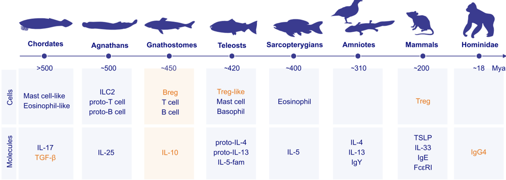
Type 2 (T2) immunity is classically associated with defense against helminths and environmental threats, from a physiological perspective, and with allergies and eosinophilic inflammatory diseases, in a pathological sense. However, growing evidence reveals that T2 pathways also support other essential homeostatic functions, including tissue protection and repair. There are major clues from the early evolution of T2 responses for protection against massive tissue damage and positive selection for survival against parasites starting over 500 million years ago. These responses likely co‐evolved stepwise with immune‐regulatory circuits, culminating in the emergence of the fully functional human‐type IgG4 in the great apes (Hominidae) within the last ~10 million years. The involvement of body barriers along with the strong influence of local epithelial cells and their interaction with the microbiome and the immune system is a common characteristic of T2 inflammation. An expanding list of new T2 diseases is being reported, characterized by epithelial cell and immune system activation, epithelial barrier damage, and microbial dysbiosis, including opportunistic pathogen colonization and loss of commensals. This evolving understanding coincides with the clinical success of biologics targeting key T2 mediators such as IL‐4, IL‐5, IL‐13, IL‐31, TSLP, and IgE, which have transformed the management of severe allergic diseases and other T2‐driven pathologies. However, the contribution of T2 immunity to homeostatic programs raises questions about the long‐term consequences of sustained T2 suppression. In this review, we examine the dual role of T2 immunity in health and disease, revisit its evolutionary origins, highlight its protective functions beyond allergy, and discuss the potential implications of prolonged T2 pathway blockade.
Checked 2026-11-12 · 1 reference not checked yet
Overview
AI snapshot
Written from the paper’s full text.
TL;DR
This review argues that type 2 (T2) immunity is not only a driver of allergic disease but an evolutionarily ancient surface-surveilling system with homeostatic roles in barrier repair, parasite defense, toxin neutralization, food aversion, cancer surveillance, cardiovascular health, aging/thymic regeneration, metabolism, and neuroimmune function. It reviews biologics targeting IL-4/IL-13, IL-5, IL-31, TSLP, and IgE, and cautions that long-term, broad T2 suppression may have understudied consequences.
Key findings
- T2 immunity evolved as a survival system at body surfaces: mast-cell-like cells date back >500 million years, ILCs arose ~500 Mya, IgE arose in early mammals 220–300 Mya, and fully functional human-type IgG4 emerged in great apes within the last ~10 million years; allergic diseases affect nearly 1 billion people.
- Beyond allergy, T2 pathways contribute to anti-helminth defense, toxin neutralization, food aversion, tissue repair, cancer immune surveillance, cardiovascular homeostasis, thymic regeneration, adipose/metabolic regulation, and neuronal function.
- New T2 diseases are characterized by epithelial cell and immune activation, epithelial barrier damage, and microbial dysbiosis; biologics targeting IL-4, IL-5, IL-13, IL-31, TSLP, and IgE have transformed management of severe allergic and other T2-driven pathologies.
- Safety signals include helminth infections with eosinophil-depleting therapies (reporting OR typically >3–4), increased lymphoma/CTCL risk with anti-IL-4Rα in real-world data, and cardiovascular event signals with omalizumab, dupilumab, and tezepelumab, though absolute risks are low and evidence remains inconclusive.
Methods
- Narrative review; no datasets were generated or analyzed (Data availability statement).
- Synthesizes evolutionary, preclinical, epidemiological, clinical trial, and pharmacovigilance literature, with figures and tables summarizing mechanisms and biologic therapies.
- No systematic search strategy, inclusion criteria, or meta-analysis is reported.
Limitations
- Stated: long-term consequences of sustained T2 suppression remain unclear, particularly for barrier integrity and repair, host defense against infections, and cancer immune surveillance; evidence linking T2-targeted therapies to infections, malignancy, and cardiovascular events remains inconclusive and requires prospective validation.
- Stated: real-world and pharmacovigilance data have inherent limitations, including potential misclassification of early CTCL as atopic dermatitis and confounding by indication.
- Evident: as a narrative review, it lacks systematic methods and primary data, and many conclusions rely on preclinical models and observational associations.
Model-written summary. Check it against the paper before relying on it.
Integrity signals
Registry
Cites
Statistics
Figures
Verification
Claims the paper makes
83 Sources not found · 36 Own interpretation · 16 No basis given · 3 Not checked yet
A conclusion the paper draws from its own results — it cites no other source for it, so there is none to check. The quote shows it is in the paper.
In the paper
“However, growing evidence reveals that T2 pathways also support other essential homeostatic functions, including tissue protection and repair.”
§ Understanding the Type 2 Immunity: An Evolving View Beyond Allergic Diseases > Abstract
plainstates a cause- Asserted: passes — The authors put it forward as true.
- Checkable: passes — It can be checked against evidence.
- Load-bearing: passes — The paper’s conclusions depend on it.
- Based: passes — The paper gives a reason why it holds.
The paper states this without a source or data behind it here, so there is nothing to check it against. The quote shows it is in the paper.
In the paper
“This evolving understanding coincides with the clinical success of biologics targeting key T2 mediators such as IL-4, IL-5, IL-13, IL-31, TSLP, and IgE, which have transformed the management of severe allergic diseases and other T2driven pathologies.”
§ Understanding the Type 2 Immunity: An Evolving View Beyond Allergic Diseases > Abstract
plainstates a cause- Asserted: passes — The authors put it forward as true.
- Checkable: passes — It can be checked against evidence.
- Load-bearing: passes — The paper’s conclusions depend on it.
- Based: unclear — It is unclear why the paper says it holds.
A conclusion the paper draws from its own results — it cites no other source for it, so there is none to check. The quote shows it is in the paper.
In the paper
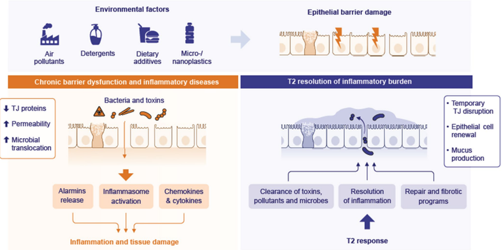
“These environmental exposures compromise epithelial surfaces of the skin, respiratory tract, and gastrointestinal system.”
§ Understanding the Type 2 Immunity: An Evolving View Beyond Allergic Diseases > 2.2.1 | Protection Against Chronic Substance Exposure-Related Danger to Body Surfaces
plainstates a cause- Asserted: passes — The authors put it forward as true.
- Checkable: passes — It can be checked against evidence.
- Load-bearing: passes — The paper’s conclusions depend on it.
- Based: passes — The paper gives a reason why it holds.
A conclusion the paper draws from its own results — it cites no other source for it, so there is none to check. The quote shows it is in the paper.
In the paper
“Persistent barrier dysfunction, sustained by microbial dysbiosis, chronic inflammation, and epigenetic alterations, leads to a self-perpetuating cycle that underlies disease chronicity.”
§ Understanding the Type 2 Immunity: An Evolving View Beyond Allergic Diseases > 2.2.1 | Protection Against Chronic Substance Exposure-Related Danger to Body Surfaces
plainstates a cause- Asserted: passes — The authors put it forward as true.
- Checkable: passes — It can be checked against evidence.
- Load-bearing: passes — The paper’s conclusions depend on it.
- Based: passes — The paper gives a reason why it holds.
None of the sources cited for this claim could be matched to a paper in Crossref, so there is nothing to check it against.
In the paper
“Comparative studies suggest that these immunoregulatory properties became further specialized during hominoid evolution [38,39].”
§ Understanding the Type 2 Immunity: An Evolving View Beyond Allergic Diseases > 2.1 | Evolutionary Origins of T2 Immunity
What the sources say
- Not matchedcites [38]
Couldn’t be matched to a paper in Crossref, so it can’t be checked.
- Not matchedcites [39]
Couldn’t be matched to a paper in Crossref, so it can’t be checked.
hedgedstates a cause- Asserted: passes — The authors put it forward as true.
- Checkable: passes — It can be checked against evidence.
- Load-bearing: unclear — It is unclear whether the paper’s conclusions depend on it.
- Based: passes — The paper gives a reason why it holds.
- Not matched
None of the sources cited for this claim could be matched to a paper in Crossref, so there is nothing to check it against.
In the paper
“In addition, air pollutants including particulate matter [58], and ozone [59], as well as micro-and nano-plastics [60][61][62], contribute significantly to epithelial barrier damage in experimental settings.”
§ Understanding the Type 2 Immunity: An Evolving View Beyond Allergic Diseases > 2.2.1 | Protection Against Chronic Substance Exposure-Related Danger to Body Surfaces
What the sources say
- Not matchedcites [58]
Couldn’t be matched to a paper in Crossref, so it can’t be checked.
- Not matchedcites [59]
Couldn’t be matched to a paper in Crossref, so it can’t be checked.
- Not matchedcites [60]
Couldn’t be matched to a paper in Crossref, so it can’t be checked.
plainstates a cause- Asserted: passes — The authors put it forward as true.
- Checkable: passes — It can be checked against evidence.
- Load-bearing: unclear — It is unclear whether the paper’s conclusions depend on it.
- Based: passes — The paper gives a reason why it holds.
- Not matched
None of the sources cited for this claim could be matched to a paper in Crossref, so there is nothing to check it against.
In the paper
“Accumulating evidence suggest that the increase in allergic and autoimmune diseases, as well as multimorbid conditions over the past six decades, parallels rising exposure to many of these agents, particularly through their detrimental effects on skin and mucosal epithelial barriers [15,[47][48][49][50][51].”
§ Understanding the Type 2 Immunity: An Evolving View Beyond Allergic Diseases > 2.2.1 | Protection Against Chronic Substance Exposure-Related Danger to Body Surfaces
What the sources say
- Not matchedcites [15]
Couldn’t be matched to a paper in Crossref, so it can’t be checked.
- Not matchedcites [47]
Couldn’t be matched to a paper in Crossref, so it can’t be checked.
- Not matchedcites [48]
Couldn’t be matched to a paper in Crossref, so it can’t be checked.
hedgedstates an association- Asserted: passes — The authors put it forward as true.
- Checkable: passes — It can be checked against evidence.
- Load-bearing: unclear — It is unclear whether the paper’s conclusions depend on it.
- Based: passes — The paper gives a reason why it holds.
- Not matched
In the paper
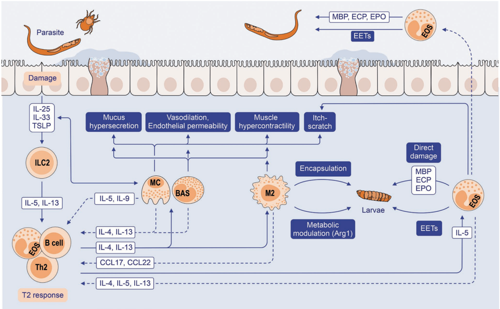
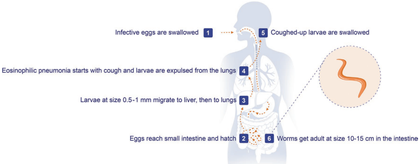
“administration of recombinant IL-33 to wild-type and Rag2 -/- mice lacking T and B cells triggered tight junction disruption, whereas Rag2 -/-Il2rg -/-and Rora sg/sg mice undergoing bone marrow transplantation that lack ILC2s did not show any barrier leakiness [75].”
§ Understanding the Type 2 Immunity: An Evolving View Beyond Allergic Diseases > FIGURE 4 | T2 response in life cycle and migration of Ascaris lumbricoides in human infection.
What the sources say
- Not matchedcites [75]
Couldn’t be matched to a paper in Crossref, so it can’t be checked.
plainstates a cause- Asserted: passes — The authors put it forward as true.
- Checkable: passes — It can be checked against evidence.
- Load-bearing: passes — The paper’s conclusions depend on it.
- Based: passes — The paper gives a reason why it holds.
- Not matched
None of the sources cited for this claim could be matched to a paper in Crossref, so there is nothing to check it against.
In the paper
“Dietary exposure to ultra-processed foods [63,64], and certain additives [65,66], emulsifiers [67], artificial sweeteners, food colorants, and flavor enhancers further exacerbate this effect [47,[68][69][70][71].”
§ Understanding the Type 2 Immunity: An Evolving View Beyond Allergic Diseases > 2.2.1 | Protection Against Chronic Substance Exposure-Related Danger to Body Surfaces
What the sources say
- Not matchedcites [63]
Couldn’t be matched to a paper in Crossref, so it can’t be checked.
- Not matchedcites [64]
Couldn’t be matched to a paper in Crossref, so it can’t be checked.
- Not matchedcites [65]
Couldn’t be matched to a paper in Crossref, so it can’t be checked.
- Not matchedcites [66]
Couldn’t be matched to a paper in Crossref, so it can’t be checked.
- Not matchedcites [67]
Couldn’t be matched to a paper in Crossref, so it can’t be checked.
plainstates a cause- Asserted: passes — The authors put it forward as true.
- Checkable: passes — It can be checked against evidence.
- Load-bearing: unclear — It is unclear whether the paper’s conclusions depend on it.
- Based: passes — The paper gives a reason why it holds.
- Not matched
A conclusion the paper draws from its own results — it cites no other source for it, so there is none to check. The quote shows it is in the paper.
In the paper
“Several conditions present with phenotypes that closely resemble those observed in allergic disease.However, the underlying immunological features distinguish them as different entities, as their central mechanism does not require allergen-specific responses (Table 1).”
§ Understanding the Type 2 Immunity: An Evolving View Beyond Allergic Diseases > 3.2 | Pathogenic Contribution of T2 Responses in Non-Allergic Disease
plain- Asserted: passes — The authors put it forward as true.
- Checkable: passes — It can be checked against evidence.
- Load-bearing: passes — The paper’s conclusions depend on it.
- Based: passes — The paper gives a reason why it holds.
- Probably not a substantial claim. 19
- It may state two separate claims as one. 67
A conclusion the paper draws from its own results — it cites no other source for it, so there is none to check. The quote shows it is in the paper.
In the paper
“These responses likely co-evolved stepwise with immune-regulatory circuits, culminating in the emergence of the fully functional human-type IgG4 in the great apes (Hominidae) within the last ~10 million years.”
§ Understanding the Type 2 Immunity: An Evolving View Beyond Allergic Diseases > Abstract
hedgedstates a cause- Asserted: passes — The authors put it forward as true.
- Checkable: passes — It can be checked against evidence.
- Load-bearing: fails — The paper’s conclusions may not depend on it.
- Based: fails — The paper may give no reason why it holds.
Probably not a substantial claim. The paper’s conclusions may not depend on it. The paper may give no reason why it holds.
In the paper
“Type 2 (T2) immunity, understood as an immunological program driven by IL-4, IL-5, IL-9, and IL-13, primarily produced by Th2 cells and group 2 innate lymphoid cells (ILC2s), enables IgE responses that help to engage effector cells such as eosinophils, basophils, and mast cells (MCs) [1].”
§ Understanding the Type 2 Immunity: An Evolving View Beyond Allergic Diseases > 1 | Introduction
What the sources say
- Not matchedcites [1]
Couldn’t be matched to a paper in Crossref, so it can’t be checked.
plainstates a cause- Asserted: passes — The authors put it forward as true.
- Checkable: passes — It can be checked against evidence.
- Load-bearing: passes — The paper’s conclusions depend on it.
- Based: passes — The paper gives a reason why it holds.
It may state two separate claims as one.
- Not matched
A conclusion the paper draws from its own results — it cites no other source for it, so there is none to check. The quote shows it is in the paper.
In the paper
“The involvement of body barriers along with the strong influence of local epithelial cells and their interaction with the microbiome and the immune system is a common characteristic of T2 inflammation.”
§ Understanding the Type 2 Immunity: An Evolving View Beyond Allergic Diseases > Abstract
plainstates an association- Asserted: passes — The authors put it forward as true.
- Checkable: passes — It can be checked against evidence.
- Load-bearing: passes — The paper’s conclusions depend on it.
- Based: passes — The paper gives a reason why it holds.
It may state two separate claims as one.
In the paper
“The outcome is bronchial eosinophilic infiltration, mucosal edema, mucus hypersecretion, and smooth muscle hyperreactivity, resulting in variable bronchial obstruction [196].”
§ Understanding the Type 2 Immunity: An Evolving View Beyond Allergic Diseases > 3.2 | Pathogenic Contribution of T2 Responses in Non-Allergic Disease
What the sources say
- Not matchedcites [196]
Couldn’t be matched to a paper in Crossref, so it can’t be checked.
plainstates a cause- Asserted: passes — The authors put it forward as true.
- Checkable: passes — It can be checked against evidence.
- Load-bearing: unclear — It is unclear whether the paper’s conclusions depend on it.
- Based: passes — The paper gives a reason why it holds.
It may state two separate claims as one.
- Not matched
A conclusion the paper draws from its own results — it cites no other source for it, so there is none to check. The quote shows it is in the paper.
In the paper
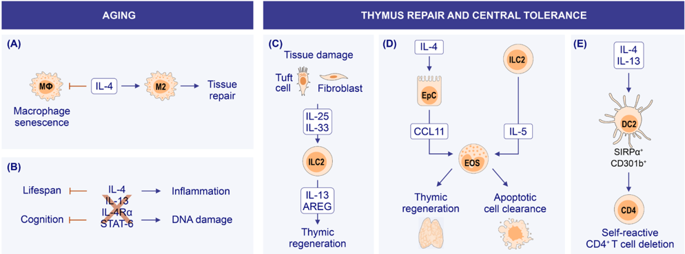
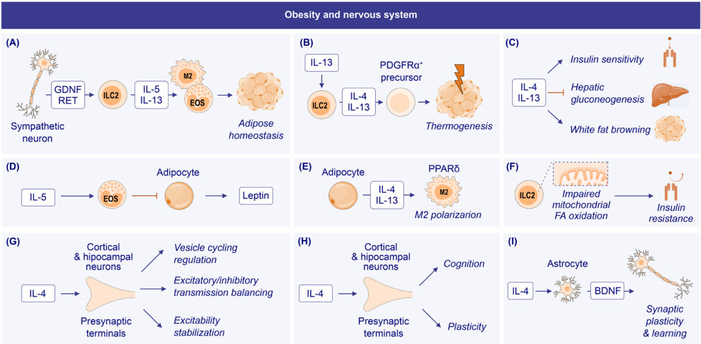
“T2 immunity and its canonical mediators have been implicated in homeostatic processes beyond classical allergy, including cancer immune surveillance, cardiovascular health, aging, thymic regeneration and tolerance, neuronal function, and metabolic regulation.”
§ Understanding the Type 2 Immunity: An Evolving View Beyond Allergic Diseases > 2.2 | T2 Immunity as a Surface-Surveilling and Survival System
plainstates an association- Asserted: passes — The authors put it forward as true.
- Checkable: passes — It can be checked against evidence.
- Load-bearing: passes — The paper’s conclusions depend on it.
- Based: unclear — It is unclear why the paper says it holds.
It may state two separate claims as one.
In the paper
“Immune regulation precedes T2 immunity, with suppressive pathways such as TGF-β signaling already present in invertebrates [33].”
§ Understanding the Type 2 Immunity: An Evolving View Beyond Allergic Diseases > 2.1 | Evolutionary Origins of T2 Immunity
What the sources say
- Not matchedcites [33]
Couldn’t be matched to a paper in Crossref, so it can’t be checked.
plainstates a cause- Asserted: passes — The authors put it forward as true.
- Checkable: passes — It can be checked against evidence.
- Load-bearing: unclear — It is unclear whether the paper’s conclusions depend on it.
- Based: passes — The paper gives a reason why it holds.
It may state two separate claims as one.
- Not matched
In the paper
“Furthermore, CD4 + T-cell responses have been reported to dynamically regulate intestinal stem cell fate, where pro-inflammatory signals (such as IL-13) promote epithelial differentiation while regulatory signals (such as IL-10) preserve stem cell renewal, thereby maintaining epithelial homeostasis [78].”
§ Understanding the Type 2 Immunity: An Evolving View Beyond Allergic Diseases > FIGURE 4 | T2 response in life cycle and migration of Ascaris lumbricoides in human infection.
What the sources say
- Not matchedcites [78]
Couldn’t be matched to a paper in Crossref, so it can’t be checked.
hedgedstates a cause- Asserted: passes — The authors put it forward as true.
- Checkable: passes — It can be checked against evidence.
- Load-bearing: unclear — It is unclear whether the paper’s conclusions depend on it.
- Based: passes — The paper gives a reason why it holds.
It may state two separate claims as one.
- Not matched
In the paper
“Also, IL-4 and IL-13 contribute to local IgE production and epithelial remodeling [201].”
§ Understanding the Type 2 Immunity: An Evolving View Beyond Allergic Diseases > 3.2 | Pathogenic Contribution of T2 Responses in Non-Allergic Disease
What the sources say
- Not matchedcites [201]
Couldn’t be matched to a paper in Crossref, so it can’t be checked.
plainstates a cause- Asserted: passes — The authors put it forward as true.
- Checkable: passes — It can be checked against evidence.
- Load-bearing: unclear — It is unclear whether the paper’s conclusions depend on it.
- Based: passes — The paper gives a reason why it holds.
It may state two separate claims as one.
- Not matched
In the paper
“Birds, once thought to lack FoxP3, were recently shown to express orthologs hidden in GC-rich genomic regions, confirming that Treg-like programs extend across amniotes [45].”
§ Understanding the Type 2 Immunity: An Evolving View Beyond Allergic Diseases > 2.1 | Evolutionary Origins of T2 Immunity
What the sources say
- Not matchedcites [45]
Couldn’t be matched to a paper in Crossref, so it can’t be checked.
boostedstates a cause- Asserted: passes — The authors put it forward as true.
- Checkable: passes — It can be checked against evidence.
- Load-bearing: unclear — It is unclear whether the paper’s conclusions depend on it.
- Based: passes — The paper gives a reason why it holds.
It may state two separate claims as one.
- Not matched
None of the sources cited for this claim have been checked yet.
In the paper
“B cells (B-cell-receptor-based adaptive immunity) and IL-10/STAT3 signaling are ancient features of jawed vertebrates.”
§ Understanding the Type 2 Immunity: An Evolving View Beyond Allergic Diseases > 2.1 | Evolutionary Origins of T2 Immunity
plain- Asserted: passes — The authors put it forward as true.
- Checkable: passes — It can be checked against evidence.
- Load-bearing: unclear — It is unclear whether the paper’s conclusions depend on it.
- Based: passes — The paper gives a reason why it holds.
It may state two separate claims as one.
None of the sources cited for this claim could be matched to a paper in Crossref, so there is nothing to check it against.
In the paper
“Most patients exhibit a local T2 environment [198] with elevated numbers of ILC2s and IL-5 driving eosinophil survival and activation [199,200].”
§ Understanding the Type 2 Immunity: An Evolving View Beyond Allergic Diseases > 3.2 | Pathogenic Contribution of T2 Responses in Non-Allergic Disease
What the sources say
- Not matchedcites [198]
Couldn’t be matched to a paper in Crossref, so it can’t be checked.
- Not matchedcites [199]
Couldn’t be matched to a paper in Crossref, so it can’t be checked.
- Not matchedcites [200]
Couldn’t be matched to a paper in Crossref, so it can’t be checked.
plainstates a cause- Asserted: passes — The authors put it forward as true.
- Checkable: passes — It can be checked against evidence.
- Load-bearing: passes — The paper’s conclusions depend on it.
- Based: passes — The paper gives a reason why it holds.
It may state two separate claims as one.
- Not matched
None of the sources cited for this claim have been checked yet.
In the paper
“IgE retained the complete IgY domain architecture (V, CH1-CH4), and co-evolved with its high-affinity receptor (FcεRI), whose α chain became fully functional in mammals, enabling the compartmentalization of humoral immunity.”
§ Understanding the Type 2 Immunity: An Evolving View Beyond Allergic Diseases > 2.1 | Evolutionary Origins of T2 Immunity
plainstates a cause- Asserted: passes — The authors put it forward as true.
- Checkable: passes — It can be checked against evidence.
- Load-bearing: unclear — It is unclear whether the paper’s conclusions depend on it.
- Based: passes — The paper gives a reason why it holds.
It may state two separate claims as one.
In the paper
“Once the epithelial barrier is compromised, commensal microorganisms and opportunistic pathogens translocate deeper into tissues leading to microbial dysbiosis, loss of biodiversity, and colonization by opportunistic pathogens [73].”
§ Understanding the Type 2 Immunity: An Evolving View Beyond Allergic Diseases > 2.2.1 | Protection Against Chronic Substance Exposure-Related Danger to Body Surfaces
What the sources say
- Not matchedcites [73]
Couldn’t be matched to a paper in Crossref, so it can’t be checked.
plainstates a cause- Asserted: passes — The authors put it forward as true.
- Checkable: passes — It can be checked against evidence.
- Load-bearing: passes — The paper’s conclusions depend on it.
- Based: passes — The paper gives a reason why it holds.
It may state two separate claims as one.
- Not matched
In the paper
“ILCs likely arose in early vertebrates (~500 Mya) alongside adaptive immune lineages [18], with signature genes already present in the most basal vertebrates, preceding the emergence of the ILC spectrum [18].”
§ Understanding the Type 2 Immunity: An Evolving View Beyond Allergic Diseases > 2.1 | Evolutionary Origins of T2 Immunity
What the sources say
- Not matchedcites [18]
Couldn’t be matched to a paper in Crossref, so it can’t be checked.
hedgedstates an association- Asserted: passes — The authors put it forward as true.
- Checkable: passes — It can be checked against evidence.
- Load-bearing: unclear — It is unclear whether the paper’s conclusions depend on it.
- Based: passes — The paper gives a reason why it holds.
It may state two separate claims as one.
- Not matched
In the paper
“Since the chemical revolution of the 1950s, around 350,000 new chemical compounds have been introduced into the environment, largely without adequate evaluation of their long-term health effects [46].”
§ Understanding the Type 2 Immunity: An Evolving View Beyond Allergic Diseases > 2.2.1 | Protection Against Chronic Substance Exposure-Related Danger to Body Surfaces
What the sources say
- Not matchedcites [46]
Couldn’t be matched to a paper in Crossref, so it can’t be checked.
plain- Asserted: passes — The authors put it forward as true.
- Checkable: passes — It can be checked against evidence.
- Load-bearing: unclear — It is unclear whether the paper’s conclusions depend on it.
- Based: passes — The paper gives a reason why it holds.
It may state two separate claims as one.
- Not matched
A conclusion the paper draws from its own results — it cites no other source for it, so there is none to check. The quote shows it is in the paper.
In the paper
“The T2 response appears to play a dual role, contributing to barrier opening and repairing while also facilitating the clearance of inflammatory burden from affected tissues (Figure 2).”
§ Understanding the Type 2 Immunity: An Evolving View Beyond Allergic Diseases > 2.2.1 | Protection Against Chronic Substance Exposure-Related Danger to Body Surfaces
hedgedstates a cause- Asserted: passes — The authors put it forward as true.
- Checkable: passes — It can be checked against evidence.
- Load-bearing: passes — The paper’s conclusions depend on it.
- Based: passes — The paper gives a reason why it holds.
It may state two separate claims as one.
In the paper
“Similarly, the eosinophil lineage is conserved across all vertebrate species, possibly with functional orthologs in invertebrates [17].”
§ Understanding the Type 2 Immunity: An Evolving View Beyond Allergic Diseases > 2.1 | Evolutionary Origins of T2 Immunity
What the sources say
- Not matchedcites [17]
Couldn’t be matched to a paper in Crossref, so it can’t be checked.
hedged- Asserted: passes — The authors put it forward as true.
- Checkable: passes — It can be checked against evidence.
- Load-bearing: unclear — It is unclear whether the paper’s conclusions depend on it.
- Based: passes — The paper gives a reason why it holds.
It may state two separate claims as one.
- Not matched
A conclusion the paper draws from its own results — it cites no other source for it, so there is none to check. The quote shows it is in the paper.
In the paper
“Consequently, local inflammation, tissue damage, and remodeling processes are triggered, contributing to disease development.”
§ Understanding the Type 2 Immunity: An Evolving View Beyond Allergic Diseases > 2.2.1 | Protection Against Chronic Substance Exposure-Related Danger to Body Surfaces
plainstates a cause- Asserted: passes — The authors put it forward as true.
- Checkable: passes — It can be checked against evidence.
- Load-bearing: passes — The paper’s conclusions depend on it.
- Based: passes — The paper gives a reason why it holds.
It may state two separate claims as one.
None of the sources cited for this claim could be matched to a paper in Crossref, so there is nothing to check it against.
In the paper
“This adaptation is thought to reflect selection by chronic antigen exposure, including parasites [40], and is now harnessed in allergen immunotherapy (AIT) [41].”
§ Understanding the Type 2 Immunity: An Evolving View Beyond Allergic Diseases > 2.1 | Evolutionary Origins of T2 Immunity
What the sources say
- Not matchedcites [40]
Couldn’t be matched to a paper in Crossref, so it can’t be checked.
- Not matchedcites [41]
Couldn’t be matched to a paper in Crossref, so it can’t be checked.
hedgedstates a possible cause- Asserted: passes — The authors put it forward as true.
- Checkable: passes — It can be checked against evidence.
- Load-bearing: unclear — It is unclear whether the paper’s conclusions depend on it.
- Based: passes — The paper gives a reason why it holds.
It may state two separate claims as one.
- Not matched
In the paper
“Beyond allergic diseases, recent studies have revealed that T2 immunity also regulates diverse biological processes, tissues, and organs [14], which conflicts with the emergence and use of several treatments targeting this pathway.”
§ Understanding the Type 2 Immunity: An Evolving View Beyond Allergic Diseases > 1 | Introduction
What the sources say
- Not matchedcites [14]
Couldn’t be matched to a paper in Crossref, so it can’t be checked.
plainstates a cause- Asserted: passes — The authors put it forward as true.
- Checkable: passes — It can be checked against evidence.
- Load-bearing: passes — The paper’s conclusions depend on it.
- Based: passes — The paper gives a reason why it holds.
It may state two separate claims as one.
- Not matched
None of the sources cited for this claim could be matched to a paper in Crossref, so there is nothing to check it against.
In the paper
“T2 cytokines and epithelial alarmins may further amplify disease activity in some patients, although their precise roles remain incompletely defined [207,208].”
§ Understanding the Type 2 Immunity: An Evolving View Beyond Allergic Diseases > 3.2 | Pathogenic Contribution of T2 Responses in Non-Allergic Disease
What the sources say
- Not matchedcites [207]
Couldn’t be matched to a paper in Crossref, so it can’t be checked.
- Not matchedcites [208]
Couldn’t be matched to a paper in Crossref, so it can’t be checked.
hedgedstates a possible cause- Asserted: passes — The authors put it forward as true.
- Checkable: passes — It can be checked against evidence.
- Load-bearing: unclear — It is unclear whether the paper’s conclusions depend on it.
- Based: passes — The paper gives a reason why it holds.
It may state two separate claims as one.
- Not matched
In the paper
“In addition to classical effector cells, IL-4/IL-13-driven alternatively activated (M2) macrophages accumulate in T2 settings, where they promote tissue remodeling and fibrosis, support eosinophilic inflammation, and reinforce Th2 responses through metabolic reprogramming and arginase-1 and IL-9-dependent pathways [8].”
§ Understanding the Type 2 Immunity: An Evolving View Beyond Allergic Diseases > 1 | Introduction
What the sources say
- Not matchedcites [8]
Couldn’t be matched to a paper in Crossref, so it can’t be checked.
plainstates a cause- Asserted: passes — The authors put it forward as true.
- Checkable: passes — It can be checked against evidence.
- Load-bearing: unclear — It is unclear whether the paper’s conclusions depend on it.
- Based: passes — The paper gives a reason why it holds.
It may state two separate claims as one.
- Not matched
A statement the paper makes itself — it cites no source for it, so there is none to check. The quote shows it is in the paper.
In the paper
“They sense environmental insults, undergo damage, and release alarmins such as IL-33, TSLP, and IL-25, which induce ILC2s, condition dendritic cells (DCs), and shape both innate and adaptive T2 responses.”
§ Understanding the Type 2 Immunity: An Evolving View Beyond Allergic Diseases > 1 | Introduction
plainstates a cause- Asserted: passes — The authors put it forward as true.
- Checkable: passes — It can be checked against evidence.
- Load-bearing: passes — The paper’s conclusions depend on it.
- Based: passes — The paper gives a reason why it holds.
It may state two separate claims as one.
None of the sources cited for this claim could be matched to a paper in Crossref, so there is nothing to check it against.
In the paper
“IL-5 promotes eosinophilia and tissue infiltration of eosinophils, which contribute to organ fibrosis and thrombosis via degranulation and the release of profibrotic mediators including TGF-β [203,204].”
§ Understanding the Type 2 Immunity: An Evolving View Beyond Allergic Diseases > 3.2 | Pathogenic Contribution of T2 Responses in Non-Allergic Disease
What the sources say
- Not matchedcites [203]
Couldn’t be matched to a paper in Crossref, so it can’t be checked.
- Not matchedcites [204]
Couldn’t be matched to a paper in Crossref, so it can’t be checked.
plainstates a cause- Asserted: passes — The authors put it forward as true.
- Checkable: passes — It can be checked against evidence.
- Load-bearing: unclear — It is unclear whether the paper’s conclusions depend on it.
- Based: passes — The paper gives a reason why it holds.
It may state two separate claims as one.
- Not matched
None of the sources cited for this claim could be matched to a paper in Crossref, so there is nothing to check it against.
In the paper
“It is thought to be triggered by stimuli that damage the bronchial epithelium and release alarmins, which directly activate ILC2s and induce IL-5 and IL-13 production [175,194,195].”
§ Understanding the Type 2 Immunity: An Evolving View Beyond Allergic Diseases > 3.2 | Pathogenic Contribution of T2 Responses in Non-Allergic Disease
What the sources say
- Not matchedcites [175]
Couldn’t be matched to a paper in Crossref, so it can’t be checked.
- Not matchedcites [194]
Couldn’t be matched to a paper in Crossref, so it can’t be checked.
- Not matchedcites [195]
Couldn’t be matched to a paper in Crossref, so it can’t be checked.
hedgedstates a cause- Asserted: passes — The authors put it forward as true.
- Checkable: passes — It can be checked against evidence.
- Load-bearing: passes — The paper’s conclusions depend on it.
- Based: passes — The paper gives a reason why it holds.
It may state two separate claims as one.
- Not matched
A conclusion the paper draws from its own results — it cites no other source for it, so there is none to check. The quote shows it is in the paper.
In the paper
“Lampreys and hagfish lack CSF2RB, suggesting that IL-5 emerged within the gnathostome lineage after divergence from agnathans, approximately 500 Mya.”
§ Understanding the Type 2 Immunity: An Evolving View Beyond Allergic Diseases > 2.1 | Evolutionary Origins of T2 Immunity
hedgedstates a cause- Asserted: passes — The authors put it forward as true.
- Checkable: passes — It can be checked against evidence.
- Load-bearing: unclear — It is unclear whether the paper’s conclusions depend on it.
- Based: passes — The paper gives a reason why it holds.
It may state two separate claims as one.
A conclusion the paper draws from its own results — it cites no other source for it, so there is none to check. The quote shows it is in the paper.
In the paper
“Indeed, T2 immunity is a finely tuned evolutionary program developed for maintaining the integrity of body surfaces, particularly in early mammals, when all core T2 components became operative for the following major functions that are overlapping (Figures 23456).”
§ Understanding the Type 2 Immunity: An Evolving View Beyond Allergic Diseases > 2.2 | T2 Immunity as a Surface-Surveilling and Survival System
plainstates a cause- Asserted: passes — The authors put it forward as true.
- Checkable: passes — It can be checked against evidence.
- Load-bearing: fails — The paper’s conclusions may not depend on it.
- Based: fails — The paper may give no reason why it holds.
Probably not a substantial claim. The paper’s conclusions may not depend on it. The paper may give no reason why it holds.
None of the sources cited for this claim could be matched to a paper in Crossref, so there is nothing to check it against.
In the paper
“Key molecular mechanisms driving this process include oxidative and endoplasmic reticulum stress, unfolded protein responses, mitochondrial dysfunction, impaired DNA repair, and dysregulated innate and adaptive immune responses [54,68,69,72].”
§ Understanding the Type 2 Immunity: An Evolving View Beyond Allergic Diseases > 2.2.1 | Protection Against Chronic Substance Exposure-Related Danger to Body Surfaces
What the sources say
- Not matchedcites [54]
Couldn’t be matched to a paper in Crossref, so it can’t be checked.
- Not matchedcites [68]
Couldn’t be matched to a paper in Crossref, so it can’t be checked.
- Not matchedcites [69]
Couldn’t be matched to a paper in Crossref, so it can’t be checked.
- Not matchedcites [72]
Couldn’t be matched to a paper in Crossref, so it can’t be checked.
plainstates a cause- Asserted: passes — The authors put it forward as true.
- Checkable: passes — It can be checked against evidence.
- Load-bearing: unclear — It is unclear whether the paper’s conclusions depend on it.
- Based: passes — The paper gives a reason why it holds.
It may state two separate claims as one.
- Not matched
In the paper
“In turn, IgE responses adapted for tissue and mucosal defense, while IgG predominated systemically [32], a division that already existed before the monotreme-therian split (~150-170 Mya).”
§ Understanding the Type 2 Immunity: An Evolving View Beyond Allergic Diseases > 2.1 | Evolutionary Origins of T2 Immunity
What the sources say
- Not matchedcites [32]
Couldn’t be matched to a paper in Crossref, so it can’t be checked.
plainstates a cause- Asserted: passes — The authors put it forward as true.
- Checkable: passes — It can be checked against evidence.
- Load-bearing: unclear — It is unclear whether the paper’s conclusions depend on it.
- Based: passes — The paper gives a reason why it holds.
It may state two separate claims as one.
- Not matched
None of the sources cited for this claim could be matched to a paper in Crossref, so there is nothing to check it against.
In the paper
“The transcription factor FoxP3 arose in early gnathostomes, with functional orthologs present in bony, but not in cartilaginous fish [42,43].”
§ Understanding the Type 2 Immunity: An Evolving View Beyond Allergic Diseases > 2.1 | Evolutionary Origins of T2 Immunity
What the sources say
- Not matchedcites [42]
Couldn’t be matched to a paper in Crossref, so it can’t be checked.
- Not matchedcites [43]
Couldn’t be matched to a paper in Crossref, so it can’t be checked.
plain- Asserted: passes — The authors put it forward as true.
- Checkable: passes — It can be checked against evidence.
- Load-bearing: unclear — It is unclear whether the paper’s conclusions depend on it.
- Based: passes — The paper gives a reason why it holds.
It may state two separate claims as one.
- Not matched
None of the sources cited for this claim could be matched to a paper in Crossref, so there is nothing to check it against.
In the paper
“Teleosts possess ancestral IL-4/IL-13 paralogues that partition innate and adaptive-like regulatory functions, whereas amniotes (birds, reptiles, mammals) evolved distinct IL-4 and IL-13 genes with conserved STAT6 pathways [19,20].”
§ Understanding the Type 2 Immunity: An Evolving View Beyond Allergic Diseases > 2.1 | Evolutionary Origins of T2 Immunity
What the sources say
- Not matchedcites [19]
Couldn’t be matched to a paper in Crossref, so it can’t be checked.
- Not matchedcites [20]
Couldn’t be matched to a paper in Crossref, so it can’t be checked.
plainstates a cause- Asserted: passes — The authors put it forward as true.
- Checkable: passes — It can be checked against evidence.
- Load-bearing: unclear — It is unclear whether the paper’s conclusions depend on it.
- Based: passes — The paper gives a reason why it holds.
It may state two separate claims as one.
- Not matched
In the paper
“However, direct functional evidence is currently strongest in bony fish, where IL-10-producing B cells modulated inflammation through conserved mechanisms, establishing an energetically favorable regulatory circuit preserved across vertebrate lineages [36].”
§ Understanding the Type 2 Immunity: An Evolving View Beyond Allergic Diseases > 2.1 | Evolutionary Origins of T2 Immunity
What the sources say
- Not matchedcites [36]
Couldn’t be matched to a paper in Crossref, so it can’t be checked.
boostedstates a cause- Asserted: passes — The authors put it forward as true.
- Checkable: passes — It can be checked against evidence.
- Load-bearing: unclear — It is unclear whether the paper’s conclusions depend on it.
- Based: passes — The paper gives a reason why it holds.
It may state two separate claims as one.
- Not matched
A conclusion the paper draws from its own results — it cites no other source for it, so there is none to check. The quote shows it is in the paper.
In the paper
“Altogether, these changes lead to goblet cell hyperplasia, mucus overproduction, and chronic mucosal edema, ultimately promoting eosinophilic polyp formation.”
§ Understanding the Type 2 Immunity: An Evolving View Beyond Allergic Diseases > 3.2 | Pathogenic Contribution of T2 Responses in Non-Allergic Disease
plainstates a cause- Asserted: passes — The authors put it forward as true.
- Checkable: passes — It can be checked against evidence.
- Load-bearing: passes — The paper’s conclusions depend on it.
- Based: passes — The paper gives a reason why it holds.
It may state two separate claims as one.
In the paper
“While these events are uncommon, with reporting OR in the range of 2-5, they suggest that pathway-specific inhibition may influence immune and tissue-regulatory processes beyond allergic inflammation [273].”
§ Understanding the Type 2 Immunity: An Evolving View Beyond Allergic Diseases > 5.2 | Helminth and Barrier Defense
What the sources say
- Not matchedcites [273]
Couldn’t be matched to a paper in Crossref, so it can’t be checked.
hedgedstates a possible cause- Asserted: passes — The authors put it forward as true.
- Checkable: passes — It can be checked against evidence.
- Load-bearing: passes — The paper’s conclusions depend on it.
- Based: passes — The paper gives a reason why it holds.
It may state two separate claims as one.
- Not matched
The paper states this without a source or data behind it here, so there is nothing to check it against. The quote shows it is in the paper.
In the paper
“Current clinical data indicate a generally favorable safety profile of T2targeted biologics in the short to medium term”
§ Understanding the Type 2 Immunity: An Evolving View Beyond Allergic Diseases > What is known
hedged- Asserted: passes — The authors put it forward as true.
- Checkable: passes — It can be checked against evidence.
- Load-bearing: fails — The paper’s conclusions may not depend on it.
- Based: fails — The paper may give no reason why it holds.
Probably not a substantial claim. The paper’s conclusions may not depend on it. The paper may give no reason why it holds.
A conclusion the paper draws from its own results — it cites no other source for it, so there is none to check. The quote shows it is in the paper.
In the paper
“While these observations do not suggest developmental harm from current therapies, they identify biological processes that fall outside the scope of conventional safety endpoints.”
§ Understanding the Type 2 Immunity: An Evolving View Beyond Allergic Diseases > 5.4 | Cardiovascular Events
hedged- Asserted: passes — The authors put it forward as true.
- Checkable: passes — It can be checked against evidence.
- Load-bearing: passes — The paper’s conclusions depend on it.
- Based: passes — The paper gives a reason why it holds.
It may state two separate claims as one.
A conclusion the paper draws from its own results — it cites no other source for it, so there is none to check. The quote shows it is in the paper.
In the paper
“The apparent clinical tolerability of T2-targeted biologics does not imply that T2 immunity is dispensable or that treatment switches the pathway entirely “off.””
§ Understanding the Type 2 Immunity: An Evolving View Beyond Allergic Diseases > 5.4 | Cardiovascular Events
plain- Asserted: passes — The authors put it forward as true.
- Checkable: passes — It can be checked against evidence.
- Load-bearing: passes — The paper’s conclusions depend on it.
- Based: passes — The paper gives a reason why it holds.
It may state two separate claims as one.
None of the sources cited for this claim could be matched to a paper in Crossref, so there is nothing to check it against.
In the paper
“For instance, in the US, dupilumab is indicated for AD from 6 months of age [290], and omalizumab for IgE-mediated food allergy from 1 year [291].”
§ Understanding the Type 2 Immunity: An Evolving View Beyond Allergic Diseases > 5.4 | Cardiovascular Events
What the sources say
- Not matchedcites [290]
Couldn’t be matched to a paper in Crossref, so it can’t be checked.
- Not matchedcites [291]
Couldn’t be matched to a paper in Crossref, so it can’t be checked.
plain- Asserted: passes — The authors put it forward as true.
- Checkable: passes — It can be checked against evidence.
- Load-bearing: unclear — It is unclear whether the paper’s conclusions depend on it.
- Based: passes — The paper gives a reason why it holds.
It may state two separate claims as one.
- Not matched
In the paper
“For example, chemosensory tuft cells detect microbial signals and release IL-25, thereby activating ILC2s to produce IL-13, which promotes goblet-cell hyperplasia and mucus secretion, facilitating pathogen clearance [250].”
§ Understanding the Type 2 Immunity: An Evolving View Beyond Allergic Diseases > 5.1 | Antiviral Response and Infection Risk
What the sources say
- Not matchedcites [250]
Couldn’t be matched to a paper in Crossref, so it can’t be checked.
plainstates a cause- Asserted: passes — The authors put it forward as true.
- Checkable: passes — It can be checked against evidence.
- Load-bearing: unclear — It is unclear whether the paper’s conclusions depend on it.
- Based: passes — The paper gives a reason why it holds.
It may state two separate claims as one.
- Not matched
In the paper
“Notably, the excess risk was most pronounced in older individuals and early after treatment initiation, supporting the hypothesis that anti-IL-4Rα may unmask or accelerate pre-existing, subclinical CTCL rather than induce de novo lymphomagenesis [277].”
§ Understanding the Type 2 Immunity: An Evolving View Beyond Allergic Diseases > 5.3 | Malignancy and Immune Surveillance
What the sources say
- Not matchedcites [277]
Couldn’t be matched to a paper in Crossref, so it can’t be checked.
hedgedstates a possible cause- Asserted: passes — The authors put it forward as true.
- Checkable: passes — It can be checked against evidence.
- Load-bearing: passes — The paper’s conclusions depend on it.
- Based: passes — The paper gives a reason why it holds.
It may state two separate claims as one.
- Not matched
The paper states this without a source or data behind it here, so there is nothing to check it against. The quote shows it is in the paper.
In the paper
“The efficacy of biologics targeting key components of T2 immunity is now well established and has transformed disease management.”
§ Understanding the Type 2 Immunity: An Evolving View Beyond Allergic Diseases > 6 | Future Prospects
boostedstates a cause- Asserted: passes — The authors put it forward as true.
- Checkable: passes — It can be checked against evidence.
- Load-bearing: passes — The paper’s conclusions depend on it.
- Based: fails — The paper may give no reason why it holds.
Probably not a substantial claim. The paper may give no reason why it holds. It may state two separate claims as one.
A conclusion the paper draws from its own results — it cites no other source for it, so there is none to check. The quote shows it is in the paper.
In the paper
“Although AIT remains limited by prolonged treatment duration, suboptimal adherence, adverse reactions, and variable long-term tolerance, its clinical efficacy provides a mechanistic rationale for the therapeutic strategy proposed here and underscores the need”
§ Understanding the Type 2 Immunity: An Evolving View Beyond Allergic Diseases > 6 | Future Prospects
plainstates a cause- Asserted: passes — The authors put it forward as true.
- Checkable: passes — It can be checked against evidence.
- Load-bearing: passes — The paper’s conclusions depend on it.
- Based: passes — The paper gives a reason why it holds.
It may state two separate claims as one.
None of the sources cited for this claim could be matched to a paper in Crossref, so there is nothing to check it against.
In the paper
“In IPF, recurrent epithelial injury and dysregulated repair lead to fibroblast expansion and matrix accumulation, with IL-4 and IL-13 promoting M2 macrophage activation and TGF-β-mediated fibrogenesis [223, 224].”
§ Understanding the Type 2 Immunity: An Evolving View Beyond Allergic Diseases > 3.2 | Pathogenic Contribution of T2 Responses in Non-Allergic Disease
What the sources say
- Not matchedcites [223]
Couldn’t be matched to a paper in Crossref, so it can’t be checked.
- Not matchedcites [224]
Couldn’t be matched to a paper in Crossref, so it can’t be checked.
plainstates a cause- Asserted: passes — The authors put it forward as true.
- Checkable: passes — It can be checked against evidence.
- Load-bearing: passes — The paper’s conclusions depend on it.
- Based: passes — The paper gives a reason why it holds.
It may state two separate claims as one.
- Not matched
The paper states this without a source or data behind it here, so there is nothing to check it against. The quote shows it is in the paper.
In the paper
“The advent of monoclonal antibody technology revolutionized therapeutic strategies across medicine.”
§ Understanding the Type 2 Immunity: An Evolving View Beyond Allergic Diseases > 6 | Future Prospects
plainstates a cause- Asserted: passes — The authors put it forward as true.
- Checkable: passes — It can be checked against evidence.
- Load-bearing: fails — The paper’s conclusions may not depend on it.
- Based: fails — The paper may give no reason why it holds.
Probably not a substantial claim. The paper’s conclusions may not depend on it. The paper may give no reason why it holds.
None of the sources cited for this claim could be matched to a paper in Crossref, so there is nothing to check it against.
In the paper
“Biological therapies have transformed the management of allergic diseases and have demonstrated efficacy in T2-mediated conditions [228][229][230].”
§ Understanding the Type 2 Immunity: An Evolving View Beyond Allergic Diseases > 4 | The Transformative Impact of Biologicals in T2 Diseases
What the sources say
- Not matchedcites [228]
Couldn’t be matched to a paper in Crossref, so it can’t be checked.
- Not matchedcites [229]
Couldn’t be matched to a paper in Crossref, so it can’t be checked.
- Not matchedcites [230]
Couldn’t be matched to a paper in Crossref, so it can’t be checked.
boostedstates a cause- Asserted: passes — The authors put it forward as true.
- Checkable: passes — It can be checked against evidence.
- Load-bearing: passes — The paper’s conclusions depend on it.
- Based: passes — The paper gives a reason why it holds.
It may state two separate claims as one.
- Not matched
In the paper
“Additional pharmacovigilance studies have identified broader safety signals associated with T2-targeted therapies, including infection clusters with anti-IL-5R [272], and noninfectious effects, such as alopecia or metabolic alterations, with anti-IL-4Rα.”
§ Understanding the Type 2 Immunity: An Evolving View Beyond Allergic Diseases > 5.2 | Helminth and Barrier Defense
What the sources say
- Not matchedcites [272]
Couldn’t be matched to a paper in Crossref, so it can’t be checked.
plainstates an association- Asserted: passes — The authors put it forward as true.
- Checkable: passes — It can be checked against evidence.
- Load-bearing: passes — The paper’s conclusions depend on it.
- Based: passes — The paper gives a reason why it holds.
It may state two separate claims as one.
- Not matched
None of the sources cited for this claim could be matched to a paper in Crossref, so there is nothing to check it against.
In the paper
“However, preliminary observational evidence suggests that a subset of patients with FPIES may harbor a T2 program involving OX40L-expressing DCs and T2 cytotoxic CD8 + T cells, with clinical responsiveness to IL-4Rα blockade [226, 227].”
§ Understanding the Type 2 Immunity: An Evolving View Beyond Allergic Diseases > 3.2 | Pathogenic Contribution of T2 Responses in Non-Allergic Disease
What the sources say
- Not matchedcites [226]
Couldn’t be matched to a paper in Crossref, so it can’t be checked.
- Not matchedcites [227]
Couldn’t be matched to a paper in Crossref, so it can’t be checked.
hedgedstates a possible cause- Asserted: passes — The authors put it forward as true.
- Checkable: passes — It can be checked against evidence.
- Load-bearing: unclear — It is unclear whether the paper’s conclusions depend on it.
- Based: passes — The paper gives a reason why it holds.
It may state two separate claims as one.
- Not matched
A conclusion the paper draws from its own results — it cites no other source for it, so there is none to check. The quote shows it is in the paper.
In the paper
“However, the homeostatic roles of T2 immunity in barrier defense, metabolism, vasculature, neuro-immune regulation, and tissue repair argue for caution regarding longterm, broad, or early-life suppression.”
§ Understanding the Type 2 Immunity: An Evolving View Beyond Allergic Diseases > 6 | Future Prospects
plainstates a possible cause- Asserted: passes — The authors put it forward as true.
- Checkable: fails — It may be too vague to check against evidence.
- Load-bearing: passes — The paper’s conclusions depend on it.
- Based: passes — The paper gives a reason why it holds.
Probably not a substantial claim. It may be too vague to check against evidence.
A statement the paper makes itself — it cites no source for it, so there is none to check. The quote shows it is in the paper.
In the paper
“Preclinical and mechanistic data implicate T2 cytokines in thymic regeneration and central tolerance, adiposetissue homeostasis, neuroglial function, and epithelial remodeling.”
§ Understanding the Type 2 Immunity: An Evolving View Beyond Allergic Diseases > 5.4 | Cardiovascular Events
hedgedstates a possible cause- Asserted: passes — The authors put it forward as true.
- Checkable: passes — It can be checked against evidence.
- Load-bearing: passes — The paper’s conclusions depend on it.
- Based: passes — The paper gives a reason why it holds.
It may state two separate claims as one.
In the paper
“Notably, eosinophil-depleting therapies have been associated with increased reporting of helminth infections, with an OR typically exceeding 3-4, although absolute case numbers remain low and exposure durations are generally limited to 6-24 months [271].”
§ Understanding the Type 2 Immunity: An Evolving View Beyond Allergic Diseases > 5.2 | Helminth and Barrier Defense
What the sources say
- Not matchedcites [271]
Couldn’t be matched to a paper in Crossref, so it can’t be checked.
plainstates an association- Asserted: passes — The authors put it forward as true.
- Checkable: passes — It can be checked against evidence.
- Load-bearing: passes — The paper’s conclusions depend on it.
- Based: passes — The paper gives a reason why it holds.
It may state two separate claims as one.
- Not matched
The paper states this without a source or data behind it here, so there is nothing to check it against. The quote shows it is in the paper.
In the paper
“For decades, aside from AIT, pharmacological treatments were broad, non-specific, and primarily symptomatic.”
§ Understanding the Type 2 Immunity: An Evolving View Beyond Allergic Diseases > 6 | Future Prospects
plain- Asserted: passes — The authors put it forward as true.
- Checkable: passes — It can be checked against evidence.
- Load-bearing: fails — The paper’s conclusions may not depend on it.
- Based: fails — The paper may give no reason why it holds.
Probably not a substantial claim. The paper’s conclusions may not depend on it. The paper may give no reason why it holds.
The paper states this without a source or data behind it here, so there is nothing to check it against. The quote shows it is in the paper.
In the paper
“Epithelial cells act as key orchestrators through alarmins (IL-33, TSLP, IL-25), linking barrier disruption to immune activation”
§ Understanding the Type 2 Immunity: An Evolving View Beyond Allergic Diseases > What is known
plainstates an association- Asserted: passes — The authors put it forward as true.
- Checkable: passes — It can be checked against evidence.
- Load-bearing: fails — The paper’s conclusions may not depend on it.
- Based: fails — The paper may give no reason why it holds.
Probably not a substantial claim. The paper’s conclusions may not depend on it. The paper may give no reason why it holds.
The paper states this without a source or data behind it here, so there is nothing to check it against. The quote shows it is in the paper.
In the paper
“Omalizumab (anti-IgE) reduces exacerbations and symptom burden in asthma and CRSwNP, remains the standard”
§ Understanding the Type 2 Immunity: An Evolving View Beyond Allergic Diseases > 4 | The Transformative Impact of Biologicals in T2 Diseases
plainstates a cause- Asserted: passes — The authors put it forward as true.
- Checkable: passes — It can be checked against evidence.
- Load-bearing: fails — The paper’s conclusions may not depend on it.
- Based: fails — The paper may give no reason why it holds.
Probably not a substantial claim. The paper’s conclusions may not depend on it. The paper may give no reason why it holds. It may state two separate claims as one.
A conclusion the paper draws from its own results — it cites no other source for it, so there is none to check. The quote shows it is in the paper.
In the paper
“In this regard, AIT merits particular emphasis as an existing precedent for immune modulation rather than pathway ablation.”
§ Understanding the Type 2 Immunity: An Evolving View Beyond Allergic Diseases > 6 | Future Prospects
plain- Asserted: passes — The authors put it forward as true.
- Checkable: fails — It may be too vague to check against evidence.
- Load-bearing: passes — The paper’s conclusions depend on it.
- Based: passes — The paper gives a reason why it holds.
Probably not a substantial claim. It may be too vague to check against evidence.
None of the sources cited for this claim could be matched to a paper in Crossref, so there is nothing to check it against.
In the paper
“However, within the SLE spectrum, accumulating evidence suggests that lupus nephritis (LN) patients present with autoreactive IgE, basophil activation, and T2 T-follicular helper responses that can enhance autoantibody production [214, 215].”
§ Understanding the Type 2 Immunity: An Evolving View Beyond Allergic Diseases > 3.2 | Pathogenic Contribution of T2 Responses in Non-Allergic Disease
What the sources say
- Not matchedcites [214]
Couldn’t be matched to a paper in Crossref, so it can’t be checked.
- Not matchedcites [215]
Couldn’t be matched to a paper in Crossref, so it can’t be checked.
hedgedstates a possible cause- Asserted: passes — The authors put it forward as true.
- Checkable: passes — It can be checked against evidence.
- Load-bearing: passes — The paper’s conclusions depend on it.
- Based: passes — The paper gives a reason why it holds.
It may state two separate claims as one.
- Not matched
In the paper
“Similarly, in a placebo-controlled trial of tezepelumab, cardiovascular events were more frequent in the active arm (1.3% vs. 0.23%), although all affected patients had ≥ 2 baseline cardiovascular risk factors [287].”
§ Understanding the Type 2 Immunity: An Evolving View Beyond Allergic Diseases > 5.4 | Cardiovascular Events
What the sources say
- Not matchedcites [287]
Couldn’t be matched to a paper in Crossref, so it can’t be checked.
plainstates an association- Asserted: passes — The authors put it forward as true.
- Checkable: passes — It can be checked against evidence.
- Load-bearing: passes — The paper’s conclusions depend on it.
- Based: passes — The paper gives a reason why it holds.
It may state two separate claims as one.
- Not matched
A conclusion the paper draws from its own results — it cites no other source for it, so there is none to check. The quote shows it is in the paper.
In the paper
“Rather, current biologics interrupt specific molecules with varying degrees of pathway interference, while substantial redundancy persists across epithelial alarmins, effector cytokines, immune and structural cells, and parallel regulatory or tissue-repair circuits.”
§ Understanding the Type 2 Immunity: An Evolving View Beyond Allergic Diseases > 5.4 | Cardiovascular Events
plainstates a cause- Asserted: passes — The authors put it forward as true.
- Checkable: passes — It can be checked against evidence.
- Load-bearing: passes — The paper’s conclusions depend on it.
- Based: passes — The paper gives a reason why it holds.
It may state two separate claims as one.
The paper states this without a source or data behind it here, so there is nothing to check it against. The quote shows it is in the paper.
In the paper
“Biologic therapies targeting T2 pathways (e.g., anti-IgE, anti-IL-5, anti-IL-4Rα, anti-TSLP) have transformed the management of severe allergic and eosinophilic diseases”
§ Understanding the Type 2 Immunity: An Evolving View Beyond Allergic Diseases > What is known
plainstates a cause- Asserted: passes — The authors put it forward as true.
- Checkable: passes — It can be checked against evidence.
- Load-bearing: fails — The paper’s conclusions may not depend on it.
- Based: fails — The paper may give no reason why it holds.
Probably not a substantial claim. The paper’s conclusions may not depend on it. The paper may give no reason why it holds.
A conclusion the paper draws from its own results — it cites no other source for it, so there is none to check. The quote shows it is in the paper.
In the paper
“Their synthesis supports a context-dependent role of T2 immunity, whereby T2-driven barrier repair and eosinophil-mediated antiviral effects may be protective in some settings, whereas unchecked T2 inflammation, barrier leakiness, and impaired early IFN responses favor higher viral loads, more frequent exacerbations of chronic airway disease, and prolonged post-viral symptoms.”
§ Understanding the Type 2 Immunity: An Evolving View Beyond Allergic Diseases > 5.1 | Antiviral Response and Infection Risk
hedgedstates a possible cause- Asserted: passes — The authors put it forward as true.
- Checkable: passes — It can be checked against evidence.
- Load-bearing: passes — The paper’s conclusions depend on it.
- Based: passes — The paper gives a reason why it holds.
It may state two separate claims as one.
In the paper
“While systemic T2 blockade maintains an excellent safety profile in non-endemic Western cohorts where environmental pathogen exposure is negligible, global datasets reveal a contrasting epidemiological reality [270].”
§ Understanding the Type 2 Immunity: An Evolving View Beyond Allergic Diseases > 5.2 | Helminth and Barrier Defense
What the sources say
- Not matchedcites [270]
Couldn’t be matched to a paper in Crossref, so it can’t be checked.
plainstates an association- Asserted: passes — The authors put it forward as true.
- Checkable: passes — It can be checked against evidence.
- Load-bearing: passes — The paper’s conclusions depend on it.
- Based: passes — The paper gives a reason why it holds.
It may state two separate claims as one.
- Not matched
A conclusion the paper draws from its own results — it cites no other source for it, so there is none to check. The quote shows it is in the paper.
In the paper
“While biologics can normalize T2 pathways, the long-term consequences of sustained pathway blockade remain incompletely defined.”
§ Understanding the Type 2 Immunity: An Evolving View Beyond Allergic Diseases > 5 | Potential Effects of Chronic T2 Pathway Suppression
plain- Asserted: passes — The authors put it forward as true.
- Checkable: passes — It can be checked against evidence.
- Load-bearing: fails — The paper’s conclusions may not depend on it.
- Based: passes — The paper gives a reason why it holds.
Probably not a substantial claim. The paper’s conclusions may not depend on it.
A conclusion the paper draws from its own results — it cites no other source for it, so there is none to check. The quote shows it is in the paper.
In the paper
“Selective blockade is not expected to be toxic per se, but tissue-specific, cumulative, or life-stage-dependent effects cannot be excluded and warrant long-term prospective study in pediatric populations, including assessments of vaccine responses, infection patterns, growth, metabolism, and organ-specific immune remodeling.”
§ Understanding the Type 2 Immunity: An Evolving View Beyond Allergic Diseases > 5.4 | Cardiovascular Events
plain- Asserted: passes — The authors put it forward as true.
- Checkable: passes — It can be checked against evidence.
- Load-bearing: passes — The paper’s conclusions depend on it.
- Based: passes — The paper gives a reason why it holds.
It may state two separate claims as one.
None of the sources cited for this claim could be matched to a paper in Crossref, so there is nothing to check it against.
In the paper
“In contrast, a pharmacovigilance study comparing anti-IL-5/ IL-5R agents to omalizumab found no overall excess cardiovascular events but suggested a potential signal for ischemic stroke with anti-IL-5/IL-5R and for hypertension and arrhythmia with omalizumab [284,285].”
§ Understanding the Type 2 Immunity: An Evolving View Beyond Allergic Diseases > 5.4 | Cardiovascular Events
What the sources say
- Not matchedcites [284]
Couldn’t be matched to a paper in Crossref, so it can’t be checked.
- Not matchedcites [285]
Couldn’t be matched to a paper in Crossref, so it can’t be checked.
hedgedstates an association- Asserted: passes — The authors put it forward as true.
- Checkable: passes — It can be checked against evidence.
- Load-bearing: unclear — It is unclear whether the paper’s conclusions depend on it.
- Based: passes — The paper gives a reason why it holds.
It may state two separate claims as one.
- Not matched
The paper states this without a source or data behind it here, so there is nothing to check it against. The quote shows it is in the paper.
In the paper
“T2 immunity plays key roles in barrier defense and repair.”
§ Understanding the Type 2 Immunity: An Evolving View Beyond Allergic Diseases > 5 | Potential Effects of Chronic T2 Pathway Suppression
plainstates a cause- Asserted: passes — The authors put it forward as true.
- Checkable: passes — It can be checked against evidence.
- Load-bearing: passes — The paper’s conclusions depend on it.
- Based: fails — The paper may give no reason why it holds.
Probably not a substantial claim. The paper may give no reason why it holds.
A conclusion the paper draws from its own results — it cites no other source for it, so there is none to check. The quote shows it is in the paper.
In the paper
“Real-world evidence from parasitic-endemic territories demonstrates a distinct, exposure-dependent risk of helminthic infections following prolonged T2 targeted biological therapy, providing concrete clinical validation for the evolutionary “surface-surveillance” hypothesis.”
§ Understanding the Type 2 Immunity: An Evolving View Beyond Allergic Diseases > 5.2 | Helminth and Barrier Defense
boostedstates a cause- Asserted: passes — The authors put it forward as true.
- Checkable: passes — It can be checked against evidence.
- Load-bearing: fails — The paper’s conclusions may not depend on it.
- Based: fails — The paper may give no reason why it holds.
Probably not a substantial claim. The paper’s conclusions may not depend on it. The paper may give no reason why it holds. It may state two separate claims as one.
None of the sources cited for this claim could be matched to a paper in Crossref, so there is nothing to check it against.
In the paper
“In SSc, endothelial injury promotes fibroblast activation and extracellular matrix deposition, with IL-4 and IL-13 directly inducing collagen synthesis and myofibroblast differentiation [219, 220].”
§ Understanding the Type 2 Immunity: An Evolving View Beyond Allergic Diseases > 3.2 | Pathogenic Contribution of T2 Responses in Non-Allergic Disease
What the sources say
- Not matchedcites [219]
Couldn’t be matched to a paper in Crossref, so it can’t be checked.
- Not matchedcites [220]
Couldn’t be matched to a paper in Crossref, so it can’t be checked.
plainstates a cause- Asserted: passes — The authors put it forward as true.
- Checkable: passes — It can be checked against evidence.
- Load-bearing: passes — The paper’s conclusions depend on it.
- Based: passes — The paper gives a reason why it holds.
It may state two separate claims as one.
- Not matched
In the paper
“Eosinophils exhibit broad antimicrobial activity, including the release of extracellular traps, reactive oxygen species, and cytotoxic granule proteins, which can damage pathogens and degrade viral RNA [257].”
§ Understanding the Type 2 Immunity: An Evolving View Beyond Allergic Diseases > 5.1 | Antiviral Response and Infection Risk
What the sources say
- Not matchedcites [257]
Couldn’t be matched to a paper in Crossref, so it can’t be checked.
plainstates a cause- Asserted: passes — The authors put it forward as true.
- Checkable: passes — It can be checked against evidence.
- Load-bearing: unclear — It is unclear whether the paper’s conclusions depend on it.
- Based: passes — The paper gives a reason why it holds.
It may state two separate claims as one.
- Not matched
A conclusion the paper draws from its own results — it cites no other source for it, so there is none to check. The quote shows it is in the paper.
In the paper
“Overall, available data support a favorable benefit-risk profile for licensed T2-targeted biologics in appropriately selected patients, with no major safety signal that would currently preclude their use.”
§ Understanding the Type 2 Immunity: An Evolving View Beyond Allergic Diseases > 6 | Future Prospects
plain- Asserted: passes — The authors put it forward as true.
- Checkable: passes — It can be checked against evidence.
- Load-bearing: passes — The paper’s conclusions depend on it.
- Based: fails — The paper may give no reason why it holds.
Probably not a substantial claim. The paper may give no reason why it holds. It may state two separate claims as one.
The paper states this without a source or data behind it here, so there is nothing to check it against. The quote shows it is in the paper.
In the paper
“In allergology, T2 immunity is considered a pathogenic program driving the diverse clinical manifestations of allergic diseases.”
§ Understanding the Type 2 Immunity: An Evolving View Beyond Allergic Diseases > 6 | Future Prospects
plainstates a cause- Asserted: passes — The authors put it forward as true.
- Checkable: passes — It can be checked against evidence.
- Load-bearing: passes — The paper’s conclusions depend on it.
- Based: fails — The paper may give no reason why it holds.
Probably not a substantial claim. The paper may give no reason why it holds.
In the paper
“MCs contribute to antimicrobial defense through the release of peptides, proteases, and extracellular traps, and through direct bacterial killing and immune cell recruitment [256].”
§ Understanding the Type 2 Immunity: An Evolving View Beyond Allergic Diseases > 5.1 | Antiviral Response and Infection Risk
What the sources say
- Not matchedcites [256]
Couldn’t be matched to a paper in Crossref, so it can’t be checked.
plainstates a cause- Asserted: passes — The authors put it forward as true.
- Checkable: passes — It can be checked against evidence.
- Load-bearing: unclear — It is unclear whether the paper’s conclusions depend on it.
- Based: passes — The paper gives a reason why it holds.
It may state two separate claims as one.
- Not matched
None of the sources cited for this claim could be matched to a paper in Crossref, so there is nothing to check it against.
In the paper
“Similar pathways operate in viral infection, where ILC2-derived factors promote epithelial repair following influenza and contribute to eosinophil recruitment [253-255].”
§ Understanding the Type 2 Immunity: An Evolving View Beyond Allergic Diseases > 5.1 | Antiviral Response and Infection Risk
What the sources say
- Not matchedcites [253]
Couldn’t be matched to a paper in Crossref, so it can’t be checked.
- Not matchedcites [254]
Couldn’t be matched to a paper in Crossref, so it can’t be checked.
- Not matchedcites [255]
Couldn’t be matched to a paper in Crossref, so it can’t be checked.
plainstates a cause- Asserted: passes — The authors put it forward as true.
- Checkable: passes — It can be checked against evidence.
- Load-bearing: unclear — It is unclear whether the paper’s conclusions depend on it.
- Based: passes — The paper gives a reason why it holds.
It may state two separate claims as one.
- Not matched
In the paper
“IL-31 further contributes to itch and correlates with eosinophil counts [213].”
§ Understanding the Type 2 Immunity: An Evolving View Beyond Allergic Diseases > 3.2 | Pathogenic Contribution of T2 Responses in Non-Allergic Disease
What the sources say
- Not matchedcites [213]
Couldn’t be matched to a paper in Crossref, so it can’t be checked.
plainstates an association- Asserted: passes — The authors put it forward as true.
- Checkable: passes — It can be checked against evidence.
- Load-bearing: unclear — It is unclear whether the paper’s conclusions depend on it.
- Based: passes — The paper gives a reason why it holds.
It may state two separate claims as one.
- Not matched
The paper states this without a source or data behind it here, so there is nothing to check it against. The quote shows it is in the paper.
In the paper
“PN involves a complex interplay between the immune and nervous systems, with evidence supporting contributions from T2 inflammation, neural sensitization, and skin barrier dysfunction.”
§ Understanding the Type 2 Immunity: An Evolving View Beyond Allergic Diseases > 3.2 | Pathogenic Contribution of T2 Responses in Non-Allergic Disease
plainstates an association- Asserted: passes — The authors put it forward as true.
- Checkable: passes — It can be checked against evidence.
- Load-bearing: passes — The paper’s conclusions depend on it.
- Based: fails — The paper may give no reason why it holds.
Probably not a substantial claim. The paper may give no reason why it holds. It may state two separate claims as one.
The paper states this without a source or data behind it here, so there is nothing to check it against. The quote shows it is in the paper.
In the paper
“T2 immunity plays a central pathogenic role in allergic diseases, including asthma, atopic dermatitis, food allergy, and eosinophilic disorders”
§ Understanding the Type 2 Immunity: An Evolving View Beyond Allergic Diseases > What is known
plainstates a cause- Asserted: passes — The authors put it forward as true.
- Checkable: passes — It can be checked against evidence.
- Load-bearing: passes — The paper’s conclusions depend on it.
- Based: fails — The paper may give no reason why it holds.
Probably not a substantial claim. The paper may give no reason why it holds.
The paper states this without a source or data behind it here, so there is nothing to check it against. The quote shows it is in the paper.
In the paper
“Beyond allergy, T2 immunity contributes to tissue homeostasis, including barrier integrity and repair:”
§ Understanding the Type 2 Immunity: An Evolving View Beyond Allergic Diseases > What is known
plainstates a cause- Asserted: passes — The authors put it forward as true.
- Checkable: passes — It can be checked against evidence.
- Load-bearing: passes — The paper’s conclusions depend on it.
- Based: unclear — It is unclear why the paper says it holds.
It may state two separate claims as one.
The paper states this without a source or data behind it here, so there is nothing to check it against. The quote shows it is in the paper.
In the paper
“Yet, T2 immunity also plays essential roles in tissue homeostasis, repair, and host defense.”
§ Understanding the Type 2 Immunity: An Evolving View Beyond Allergic Diseases > 6 | Future Prospects
plainstates a cause- Asserted: passes — The authors put it forward as true.
- Checkable: passes — It can be checked against evidence.
- Load-bearing: passes — The paper’s conclusions depend on it.
- Based: unclear — It is unclear why the paper says it holds.
It may state two separate claims as one.
None of the sources cited for this claim could be matched to a paper in Crossref, so there is nothing to check it against.
In the paper
“They also contribute to pathogen control through nitric oxide-dependent mechanisms and IFN-induced antiviral programs, and can participate in early containment of fungal and bacterial infections [258-261].”
§ Understanding the Type 2 Immunity: An Evolving View Beyond Allergic Diseases > 5.1 | Antiviral Response and Infection Risk
What the sources say
- Not matchedcites [258]
Couldn’t be matched to a paper in Crossref, so it can’t be checked.
- Not matchedcites [259]
Couldn’t be matched to a paper in Crossref, so it can’t be checked.
- Not matchedcites [260]
Couldn’t be matched to a paper in Crossref, so it can’t be checked.
- Not matchedcites [261]
Couldn’t be matched to a paper in Crossref, so it can’t be checked.
plainstates a cause- Asserted: passes — The authors put it forward as true.
- Checkable: passes — It can be checked against evidence.
- Load-bearing: passes — The paper’s conclusions depend on it.
- Based: passes — The paper gives a reason why it holds.
It may state two separate claims as one.
- Not matched
The paper states this without a source or data behind it here, so there is nothing to check it against. The quote shows it is in the paper.
In the paper
“However, emerging evidence indicates that it is also linked to tissue remodeling, sensory neuron activation, and reflex responses such as itch and cough.”
§ Understanding the Type 2 Immunity: An Evolving View Beyond Allergic Diseases > 5 | Potential Effects of Chronic T2 Pathway Suppression
hedgedstates an association- Asserted: passes — The authors put it forward as true.
- Checkable: passes — It can be checked against evidence.
- Load-bearing: passes — The paper’s conclusions depend on it.
- Based: unclear — It is unclear why the paper says it holds.
It may state two separate claims as one.
In the paper
“ILC2s can also respond to microbial cues and contribute to humoral immunity [251].”
§ Understanding the Type 2 Immunity: An Evolving View Beyond Allergic Diseases > 5.1 | Antiviral Response and Infection Risk
What the sources say
- Not matchedcites [251]
Couldn’t be matched to a paper in Crossref, so it can’t be checked.
plainstates a cause- Asserted: passes — The authors put it forward as true.
- Checkable: passes — It can be checked against evidence.
- Load-bearing: unclear — It is unclear whether the paper’s conclusions depend on it.
- Based: passes — The paper gives a reason why it holds.
It may state two separate claims as one.
- Not matched
The paper states this without a source or data behind it here, so there is nothing to check it against. The quote shows it is in the paper.
In the paper
“T2 responses evolved as a surface-surveilling system to protect against helminths, toxins, and environmental threats, promoting expulsion mechanisms, mucus production, and tissue repair”
§ Understanding the Type 2 Immunity: An Evolving View Beyond Allergic Diseases > What is known
plainstates a cause- Asserted: passes — The authors put it forward as true.
- Checkable: passes — It can be checked against evidence.
- Load-bearing: fails — The paper’s conclusions may not depend on it.
- Based: fails — The paper may give no reason why it holds.
Probably not a substantial claim. The paper’s conclusions may not depend on it. The paper may give no reason why it holds.
In the paper
“In a 5-year observational cohort, omalizumab-treated asthma patients showed a higher incidence of serious cardiovascular events compared to matched controls [283], prompting the FDA to include this risk in the US label, although it is not reflected in the European Medicines Agency (EMA) or the Agencia Española de Medicamentos y Productos Sanitarios (AEMPS) documents.”
§ Understanding the Type 2 Immunity: An Evolving View Beyond Allergic Diseases > 5.4 | Cardiovascular Events
What the sources say
- Not matchedcites [283]
Couldn’t be matched to a paper in Crossref, so it can’t be checked.
plainstates an association- Asserted: passes — The authors put it forward as true.
- Checkable: passes — It can be checked against evidence.
- Load-bearing: unclear — It is unclear whether the paper’s conclusions depend on it.
- Based: passes — The paper gives a reason why it holds.
It may state two separate claims as one.
- Not matched
The paper relies on this without support.
Findings from murine and preclinical models can be extrapolated to human T2 immunity and disease.
“Most mechanistic insights into the ontogeny of allergic disease come from murine models [163], which consistently revealed environmental cues (alarmins), usually but not exclusively of epithelial origin, that precondition the tissue and, ultimately, the immune system to sense exogenous antigens as threats, thereby promoting T2 responses [4, 164-167] (Figure 11).”
Self-check
Percentages, sums and reported differences re-computed from the paper’s own numbers.
Each contradiction between two passages listed here was confirmed by an independent check.
Rated minor: it would not touch a result — a value no result rests on, a p-value that stays on the same side of the significance level, or a conflict that is clerical, resolved elsewhere in the paper, or outside its own findings.
In the paper
§ Understanding the Type 2 Immunity: An Evolving View Beyond Allergic Diseases > 2.2.3 | Neutralization of Toxins
“However, a series of murine studies in the 2010s vindicated this idea [98,99].”
§ Understanding the Type 2 Immunity: An Evolving View Beyond Allergic Diseases > References
“98. M. Metz, A. M. Piliponsky, C. C. Chen, et al., "Mast Cells Can Enhance Resistance to Snake and Honeybee Venoms," Science 313, no. 5786 (2006): 526-530.”
Rated minor: it would not touch a result — a value no result rests on, a p-value that stays on the same side of the significance level, or a conflict that is clerical, resolved elsewhere in the paper, or outside its own findings.
In the paper
§ Understanding the Type 2 Immunity: An Evolving View Beyond Allergic Diseases > 2.6 | Adipose Tissue and the Nervous System
“sympathetic neurons stimulates adipose-resident ILC2s via mesenchymal-derived glial cell line-derived neurotrophic factor (GDNF) and rearranged during transfection (RET) signaling”
§ Understanding the Type 2 Immunity: An Evolving View Beyond Allergic Diseases > What is known
“(A) Sympathetic neurons release GDNF and RET, regulating adipose tissue homeostasis in a process mediated by ILC2s, eosinophils, and M2.”
Rated minor: it would not touch a result — a value no result rests on, a p-value that stays on the same side of the significance level, or a conflict that is clerical, resolved elsewhere in the paper, or outside its own findings.
In the paper
§ Understanding the Type 2 Immunity: An Evolving View Beyond Allergic Diseases > 3.2 | Pathogenic Contribution of T2 Responses in Non-Allergic Disease
“Hypereosinophilic syndromes (HES) comprise a heterogeneous group of disorders defined by persistent eosinophilia (> 1500/ μL) and eosinophil-mediated tissue injury in the absence of known secondary causes [202].”
§ Understanding the Type 2 Immunity: An Evolving View Beyond Allergic Diseases > 3.2 | Pathogenic Contribution of T2 Responses in Non-Allergic Disease
“Hypereosinophilic syndromes Systemic Persistent eosinophilia due to neoplastic, lymphocytic, reactive, familiar or idiopathic causes”
Citations
Cites
Tolerance, Danger and the Extended Family
Matzinger, P. (1994) · Annual Review of Immunology · doi:10.1146/annurev.iy.12.040194.005015 · Cited by 5,107
Macrophages in Tissue Repair, Regeneration, and Fibrosis
Wynn, T. A., & Vannella, K. M. (2016) · Immunity · doi:10.1016/j.immuni.2016.02.015 · Cited by 4,481
Human epithelial cells trigger dendritic cell–mediated allergic inflammation by producing TSLP
Soumelis, V., Reche, P. A., Kanzler, H., Yuan, W., Edward, G., Homey, B., Gilliet, M. F., Ho, S., Antonenko, S. V., Lauerma, A., et al. (2002) · Nature Immunology · doi:10.1038/ni805 · Cited by 2,221
Dupilumab Efficacy and Safety in Moderate-to-Severe Uncontrolled Asthma
Castro, M., Corren, J., Pavord, I. D., Maspero, J. F., Wenzel, S. E., Rabe, K. F., Busse, W. W., Ford, L., Sher, L., FitzGerald, J. M., et al. (2018) · New England Journal of Medicine · doi:10.1056/nejmoa1804092 · Cited by 2,016
Microglia Biology: One Century of Evolving Concepts
Prinz, M., Jung, S., & Priller, J. (2019) · Cell · doi:10.1016/j.cell.2019.08.053 · Cited by 1,525
Bile acid metabolites control TH17 and Treg cell differentiation
Hang, S., Paik, D., Yao, L., Kim, E., Trinath, J., Lu, J., Ha, S., Nelson, B. N., Kelly, S. P., Wu, L., et al. (2019) · Nature · doi:10.1038/s41586-019-1785-z · Cited by 1,457
Innate lymphoid cells promote lung-tissue homeostasis after infection with influenza virus
Monticelli, L. A., Sonnenberg, G. F., Abt, M. C., Alenghat, T., Ziegler, C. G. K., Doering, T. A., Angelosanto, J., Laidlaw, B. J., Yang, C. Y., Sathaliyawala, T., et al. (2011) · Nature Immunology · doi:10.1038/ni.2131 · Cited by 1,327
Zuberbier, T., Abdul Latiff, A. H., Abuzakouk, M., Aquilina, S., Asero, R., Baker, D., Ballmer‐Weber, B., Bangert, C., Ben‐Shoshan, M., Bernstein, J. A., et al. (2022) · Allergy · doi:10.1111/all.15090 · Cited by 1,263
Tuft-cell-derived IL-25 regulates an intestinal ILC2-epithelial response circuit
von Moltke, J., Ji, M., Liang, H.-E., & Locksley, R. M. (2016) · Nature · doi:10.1038/nature16161 · Cited by 1,230
Lambrecht, B. N., Hammad, H., & Fahy, J. V. (2019) · Immunity · doi:10.1016/j.immuni.2019.03.018 · Cited by 1,108
Wang, Z., Walker, G. W., Muir, D. C. G., & Nagatani-Yoshida, K. (2020) · Environmental Science & Technology · doi:10.1021/acs.est.9b06379 · Cited by 1,084
Akdiş, C. A. (2021) · Nature reviews. Immunology · doi:10.1038/s41577-021-00538-7 · Cited by 1,068
Type 2 immunity in tissue repair and fibrosis
Gieseck, R. L., Wilson, M. S., & Wynn, T. A. (2018) · Nature Reviews Immunology · doi:10.1038/nri.2017.90 · Cited by 1,055
Eosinophils: changing perspectives in health and disease
Rosenberg, H. F., Dyer, K. D., & Foster, P. S. (2012) · Nature reviews. Immunology · doi:10.1038/nri3341 · Cited by 1,032
Akdis, M., Aab, A., Altunbulakli, C., Azkur, K., Costa, R. A., Crameri, R., Duan, S., Eiwegger, T., Eljaszewicz, A., Ferstl, R., et al. (2016) · Journal of Allergy and Clinical Immunology · doi:10.1016/j.jaci.2016.06.033 · Cited by 912
Qiu, Y., Nguyen, K. D., Odegaard, J. I., Cui, X., Tian, X., Locksley, R. M., Palmiter, R. D., & Chawla, A. (2014) · Cell · doi:10.1016/j.cell.2014.03.066 · Cited by 889
Type 2 Innate Signals Stimulate Fibro/Adipogenic Progenitors to Facilitate Muscle Regeneration
Heredia, J. E., Mukundan, L., Chen, F. M., Mueller, A. A., Deo, R. C., Locksley, R. M., Rando, T. A., & Chawla, A. (2013) · Cell · doi:10.1016/j.cell.2013.02.053 · Cited by 818
Regulation of learning and memory by meningeal immunity: a key role for IL-4
Derecki, N. C., Cardani, A. N., Yang, C. H., Quinnies, K. M., Crihfield, A., Lynch, K. R., & Kipnis, J. (2010) · Journal of Experimental Medicine · doi:10.1084/jem.20091419 · Cited by 800
Biologic Therapies for Severe Asthma
Brusselle, G. G., & Koppelman, G. H. (2022) · New England Journal of Medicine · doi:10.1056/nejmra2032506 · Cited by 747
Elephant shark genome provides unique insights into gnathostome evolution
Venkatesh, B., Lee, A., Ravi, V., Maurya, A. K., Lian, M. M., Swann, J. B., Ohta, Y., Flajnik, M. F., Sutoh, Y., Kasahara, M., et al. (2014) · Nature · doi:10.1038/nature12826 · Cited by 708
Kang, K., Reilly, S. M., Karabacak, V., Gangl, M. R., Fitzgerald, K., Hatano, B., & Lee, C.-H. (2008) · Cell Metabolism · doi:10.1016/j.cmet.2008.04.002 · Cited by 699
Activated Type 2 Innate Lymphoid Cells Regulate Beige Fat Biogenesis
Lee, M.-W., Odegaard, J. I., Mukundan, L., Qiu, Y., Molofsky, A. B., Nussbaum, J. C., Yun, K. M., Locksley, R. M., & Chawla, A. (2014) · Cell · doi:10.1016/j.cell.2014.12.011 · Cited by 695
Wang, X., Zhang, N., Bo, M., Holtappels, G., Zheng, M., Lou, H., Wang, H., Zhang, L., & Bachert, C. (2016) · Journal of Allergy and Clinical Immunology · doi:10.1016/j.jaci.2016.05.041 · Cited by 630
Self-citationT Helper Cell Cytokines Modulate Intestinal Stem Cell Renewal and Differentiation
Biton, M., Haber, A. L., Rogel, N., Burgin, G., Beyaz, S., Schnell, A., Ashenberg, O., Su, C., Smillie, C. S., Shekhar, K., et al. (2018) · Cell · doi:10.1016/j.cell.2018.10.008 · Cited by 597
Chronic Rhinosinusitis with Nasal Polyps
Stevens, W. W., Schleimer, R. P., & Kern, R. C. (2016) · The Journal of Allergy and Clinical Immunology In Practice · doi:10.1016/j.jaip.2016.04.012 · Cited by 584
Detection of Succinate by Intestinal Tuft Cells Triggers a Type 2 Innate Immune Circuit
Nadjsombati, M. S., McGinty, J. W., Lyons-Cohen, M. R., Jaffe, J. B., DiPeso, L., Schneider, C., Miller, C. N., Pollack, J. L., Nagana Gowda, G. A., Fontana, M. F., et al. (2018) · Immunity · doi:10.1016/j.immuni.2018.06.016 · Cited by 556
Effects of benralizumab on airway eosinophils in asthmatic patients with sputum eosinophilia
Laviolette, M., Gossage, D. L., Gauvreau, G. M., Leigh, R., Olivenstein, R., Katial, R. K., Busse, W. W., Wenzel, S. E., Wu, Y., Datta, V., et al. (2013) · Journal of Allergy and Clinical Immunology · doi:10.1016/j.jaci.2013.05.020 · Cited by 512
Epithelial cell–derived cytokines: more than just signaling the alarm
Roan, F., Obata‐Ninomiya, K., & Ziegler, S. F. (2019) · Journal of Clinical Investigation · doi:10.1172/jci124606 · Cited by 501
Soyka, M. B., Wawrzyniak, P., Eiwegger, T., Holzmann, D., Treis, A., Wanke, K., Kast, J. I., & Akdis, C. A. (2012) · Journal of Allergy and Clinical Immunology · doi:10.1016/j.jaci.2012.05.052 · Cited by 486
van den Oord, R. A. H. M., & Sheikh, A. (2009) · BMJ · doi:10.1136/bmj.b2433 · Cited by 471
Shin, Y. H., Hwang, J., Kwon, R., Lee, S. W., Kim, M. S., Collaborators, G. 2. A. D., Shin, J. I., & Yon, D. K. (2023) · Allergy · doi:10.1111/all.15807 · Cited by 465
Sözener, Z. Ç., Öztürk, B. Ö., Çerçi, P., Türk, M., Akın, B. G., Akdiş, M., Altıner, S., Yücel, Ü. Ö., Öğülür, İ., Mitamura, Y., et al. (2022) · Allergy · doi:10.1111/all.15240 · Cited by 458
Self-citationType 2 immunity in allergic diseases
Öğülür, İ., Mitamura, Y., Yazıcı, D., Pat, Y., Ardıçlı, S., Li, M., D’Avino, P., Beha, C., Babayev, H., Zhao, B., et al. (2025) · Cellular and Molecular Immunology · doi:10.1038/s41423-025-01261-2 · Cited by 457
Self-citationAllergen immunotherapy: past, present and future
Durham, S. R., & Shamji, M. H. (2022) · Nature reviews. Immunology · doi:10.1038/s41577-022-00786-1 · Cited by 435
Self-citationTargeting cytokines to treat asthma and chronic obstructive pulmonary disease
Barnes, P. J. (2018) · Nature reviews. Immunology · doi:10.1038/s41577-018-0006-6 · Cited by 432
Microbiota therapy acts via a regulatory T cell MyD88/RORγt pathway to suppress food allergy
Abdel-Gadir, A., Victor, E. S., Gerber, G. K., Rivas, M. N., Wang, S., Harb, H., Wang, L., Li, N., Crestani, E., Spielman, S. C., et al. (2019) · Nature Medicine · doi:10.1038/s41591-019-0461-z · Cited by 399
Giudice, G. J., Emery, D. J., Zelickson, B. D., Anhalt, G. J., Liu, Z., & Diaz, L. A. (1993) · The Journal of Immunology · doi:10.4049/jimmunol.151.10.5742 · Cited by 389
Structural Biology and Evolution of the TGF-β FamilyPreprint
Hinck, A. P., Mueller, T. D., & Springer, T. A. (2016) · Cold Spring Harbor Perspectives in Biology · doi:10.1101/cshperspect.a022103 · Cited by 381
Omalizumab for the Treatment of Multiple Food Allergies
Wood, R. A., Togias, A. G., Sicherer, S. H., Shreffler, W. G., Kim, E., Jones, S. M., Leung, D. Y. M., Vickery, B. P., Bird, J. A., Spergel, J. M., et al. (2024) · New England Journal of Medicine · doi:10.1056/nejmoa2312382 · Cited by 368
Ultra-processed foods and food additives in gut health and disease
Whelan, K., Bancil, A. S., Lindsay, J. O., & Chassaing, B. (2024) · Nature Reviews Gastroenterology & Hepatology · doi:10.1038/s41575-024-00893-5 · Cited by 362
Kool, M., Willart, M. A. M., van Nimwegen, M., Bergen, I. M., Pouliot, P., Virchow, J. C., Jr., Rogers, N. C., Osorio, F., Sousa, C. R. E., Hammad, H., et al. (2011) · Immunity · doi:10.1016/j.immuni.2011.03.015 · Cited by 360
Basophils and the T helper 2 environment can promote the development of lupus nephritis
Charles, N., Hardwick, D., Daugas, É., Illei, G. G., & Rivera, J. (2010) · Nature Medicine · doi:10.1038/nm.2159 · Cited by 360
Eosinophil responses during COVID-19 infections and coronavirus vaccination
Lindsley, A., Schwartz, J. T., & Rothenberg, M. E. (2020) · Journal of Allergy and Clinical Immunology · doi:10.1016/j.jaci.2020.04.021 · Cited by 357
ILC2s amplify PD-1 blockade by activating tissue-specific cancer immunity
Moral, J. A., Leung, J., Rojas, L. A., Ruan, J. J., Zhao, J. N., Sethna, Z. M., Ramnarain, A., Gasmi, B., Gururajan, M., Redmond, D., et al. (2020) · Nature · doi:10.1038/s41586-020-2015-4 · Cited by 347
Jutel, M., Agache, I. O., Zemelka‐Wiącek, M., Akdiş, M., Chivato, T., Giacco, S. D., Gajdanowicz, P., Eguíluz‐Gracia, I., Klimek, L., Lauerma, A., et al. (2023) · Allergy · doi:10.1111/all.15889 · Cited by 344
Self-citationIdiopathic Pulmonary Fibrosis: Aberrant Recapitulation of Developmental Programs?
Selman, M., Pardo, A., & Kaminski, N. (2008) · PLoS Medicine · doi:10.1371/journal.pmed.0050062 · Cited by 342
Autoimmune chronic spontaneous urticaria
Kolkhir, P., Muñoz, M., Asero, R., Ferrer, M., Kocatürk, E., Metz, M., Xiang, Y., & Maurer, M. (2022) · Journal of Allergy and Clinical Immunology · doi:10.1016/j.jaci.2022.04.010 · Cited by 335
Chu, D. K., Llop‐Guevara, A., Walker, T. D., Flader, K., Goncharova, S. I., Boudreau, J. E., Moore, C. L., In, T. S., Waserman, S., Coyle, A. J., et al. (2012) · Journal of Allergy and Clinical Immunology · doi:10.1016/j.jaci.2012.08.002 · Cited by 329
Mast Cells Can Enhance Resistance to Snake and Honeybee Venoms
Metz, M., Piliponsky, A. M., Chen, C.-C., Lammel, V., Åbrink, M., Pejler, G., Tsai, M., & Galli, S. J. (2006) · Science · doi:10.1126/science.1128877 · Cited by 319
A new dawn for eosinophils in the tumour microenvironment
Grisaru‐Tal, S., Itan, M., Klion, A. D., & Munitz, A. (2020) · Nature reviews. Cancer · doi:10.1038/s41568-020-0283-9 · Cited by 315
Interleukin-5 and IL-5 receptor in health and diseases
Takatsu, K. (2011) · Proceedings of the Japan Academy Series B · doi:10.2183/pjab.87.463 · Cited by 310
Martínez-González, I., Mathä, L., Steer, C. A., Ghaedi, M., Poon, G. F. T., & Takei, F. (2016) · Immunity · doi:10.1016/j.immuni.2016.06.017 · Cited by 305
Role of IL‐25, IL‐33, and TSLP in triggering united airway diseases toward type 2 inflammation
Hong, H., Liao, S., Chen, F., Yang, Q., & Wang, D. Y. (2020) · Allergy · doi:10.1111/all.14526 · Cited by 284
The Unique Properties of IgG4 and Its Roles in Health and Disease
Rispens, T., & Huijbers, M. G. (2023) · Nature Reviews Immunology · doi:10.1038/s41577-023-00871-z · Cited by 282
Sugita, K., Steer, C. A., Martínez-González, I., Altunbulakli, C., Morita, H., Castro-Giner, F., Kubo, T., Wawrzyniak, P., Rückert, B., Sudo, K., et al. (2017) · Journal of Allergy and Clinical Immunology · doi:10.1016/j.jaci.2017.02.038 · Cited by 277
IL-13 alters mucociliary differentiation and ciliary beating of human respiratory epithelial cells
Laoukili, J., Perret, E., Willems, T., Minty, A. J., Parthoens, E., Houcine, O., Coste, A., Jorissen, M., Marano, F., Caput, D., et al. (2001) · Journal of Clinical Investigation · doi:10.1172/jci200113557 · Cited by 276
Atopy and Risk of Brain Tumors: A Meta-analysis
Linos, E., Raine, T., Alonso, Á., & Michaud, D. S. (2007) · JNCI Journal of the National Cancer Institute · doi:10.1093/jnci/djm170 · Cited by 275
Oh, J., Kim, S., Kim, M. S., Abate, Y. H., ElHafeez, S. A., Abdelkader, A. F. I., Abdi, P., Abdulah, D. M., Aboagye, R. G., Abolhassani, H., et al. (2025) · The Lancet Respiratory Medicine · doi:10.1016/s2213-2600(25)00003-7 · Cited by 268
Levan, S., Stamnes, K. A., Lin, D. L., Panzer, A. R., Fukui, E., McCauley, K. E., Fujimura, K. E., McKean, M., Ownby, D. R., Zoratti, E. M., et al. (2019) · Nature Microbiology · doi:10.1038/s41564-019-0498-2 · Cited by 245
Wawrzyniak, P., Wawrzyniak, M., Wanke, K., Sokolowska, M., Bendelja, K., Rückert, B., Globinska, A., Jakiela, B., Kast, J. I., Idzko, M., et al. (2017) · Journal of Allergy and Clinical Immunology · doi:10.1016/j.jaci.2016.03.050 · Cited by 241
Roufosse, F. E., Kahn, J., Rothenberg, M. E., Wardlaw, A. J., Klion, A. D., Kirby, S. Y., Gilson, M. J., Bentley, J. H., Bradford, E., Yancey, S. W., et al. (2020) · Journal of Allergy and Clinical Immunology · doi:10.1016/j.jaci.2020.08.037 · Cited by 227
Strid, J., Hourihane, J. O., Kimber, I., Callard, R. E., & Ströbel, S. (2004) · European Journal of Immunology · doi:10.1002/eji.200425196 · Cited by 221
Palm, N. W., Rosenstein, R. K., Yu, S., Schenten, D. D., Florsheim, E., & Medzhitov, R. (2013) · Immunity · doi:10.1016/j.immuni.2013.10.006 · Cited by 214
IgE and mast cells in host defense against parasites and venoms
Mukai, K., Tsai, M. M., Starkl, P. M., Marichal, T., & Galli, S. J. (2016) · Seminars in Immunopathology · doi:10.1007/s00281-016-0565-1 · Cited by 211
Mast cell activation symptoms are prevalent in Long-COVID
Weinstock, L. B., Brook, J. B., Walters, A. S., Goris, A. J., Afrin, L. B., & Molderings, G. J. (2021) · International Journal of Infectious Diseases · doi:10.1016/j.ijid.2021.09.043 · Cited by 189
Williams, K. A., Huang, A. H., Belzberg, M., & Kwatra, S. G. (2020) · Journal of the American Academy of Dermatology · doi:10.1016/j.jaad.2020.04.182 · Cited by 184
A beneficial role for immunoglobulin E in host defense against honeybee venom
Marichal, T., Starkl, P., Reber, L. L., Kalesnikoff, J., Oettgen, H. C., Tsai, M., Metz, M., & Galli, S. J. (2013) · Immunity · doi:10.1016/j.immuni.2013.10.005 · Cited by 180
Zhao, W., Xie, W., Xiao, Q., Beers, D. R., & Appel, S. H. (2006) · Journal of Neurochemistry · doi:10.1111/j.1471-4159.2006.04172.x · Cited by 176
Kay, A. B., Clark, P., Maurer, M., & Ying, S. (2014) · British Journal of Dermatology · doi:10.1111/bjd.13621 · Cited by 175
Zur Differential-Diagnose der Lungeninfiltrierungen
Löffler, W. (1932) · Lung · doi:10.1007/bf02079221 · Cited by 175
Particulate Matter 2.5 Causes Deficiency in Barrier Integrity in Human Nasal Epithelial Cells
Xian, M., Ma, S., Wang, K., Lou, H., Wang, Y., Zhang, L., Wang, C., & Akdiş, C. A. (2019) · Allergy Asthma and Immunology Research · doi:10.4168/aair.2020.12.1.56 · Cited by 174
Self-citationSilverberg, J. I., Wollenberg, A., Reich, A., Thaçi, D., Legat, F. J., Papp, K. A., Gold, L. S., Bouaziz, J., Pink, A., Carrascosa, J. M., et al. (2024) · The Lancet · doi:10.1016/s0140-6736(24)01203-0 · Cited by 174
Khurana, S., Brusselle, G., Bel, E. H., FitzGerald, J. M., Masoli, M., Korn, S., Kato, M., Albers, F. C., Bradford, E., Gilson, M. J., et al. (2019) · Clinical Therapeutics · doi:10.1016/j.clinthera.2019.07.007 · Cited by 173
Ozone exposure induces respiratory barrier biphasic injury and inflammation controlled by IL-33
Michaudel, C., Mackowiak, C., Maillet, I., Fauconnier, L., Akdiş, C. A., Sokołowska, M., Dreher, A., Tan, H. T., Quesniaux, V. F. J., Ryffel, B., et al. (2018) · Journal of Allergy and Clinical Immunology · doi:10.1016/j.jaci.2017.11.044 · Cited by 172
Menzies-Gow, A. N., Wechsler, M. E., Brightling, C. E., Korn, S., Corren, J., Israel, E., Chupp, G. L., Bednarczyk, A., Ponnarambil, S. S., Caveney, S. W., et al. (2023) · The Lancet Respiratory Medicine · doi:10.1016/s2213-2600(22)00492-1 · Cited by 172
The evolution of innate lymphoid cells
Vivier, É., van de Pavert, S. A., Cooper, M. D., & Belz, G. T. (2016) · Nature Immunology · doi:10.1038/ni.3459 · Cited by 171
Gastrointestinal Dendritic Cells Promote Th2 Skewing via OX40L
Blázquez, A.-B., & Berin, M. C. (2008) · The Journal of Immunology · doi:10.4049/jimmunol.180.7.4441 · Cited by 170
Induction of immunoglobulin G4 in human filariasis: an indicator of immunoregulation
Adjobimey, T., & Hoerauf, A. M. (2010) · Annals of Tropical Medicine and Parasitology · doi:10.1179/136485910x12786389891407 · Cited by 164
Wang, M., Tan, G., Eljaszewicz, A., Meng, Y., Wawrzyniak, P., Acharya, S., Altunbulakli, C., Westermann, P., Dreher, A., Yan, L., et al. (2018) · Journal of Allergy and Clinical Immunology · doi:10.1016/j.jaci.2018.11.016 · Cited by 162
Self-citationGorski, S. A., Hahn, Y. S., & Braciale, T. J. (2013) · PLoS Pathogens · doi:10.1371/journal.ppat.1003615 · Cited by 161
Processed foods drive intestinal barrier permeability and microvascular diseases
Snelson, M., Tan, S. M., Clarke, R., de Pasquale, C., Thallas‐Bonke, V., Nguyen, T.-V., Penfold, S. A., Harcourt, B. E., Sourris, K. C., Lindblom, R. S. J., et al. (2021) · Science Advances · doi:10.1126/sciadv.abe4841 · Cited by 161
Immune sensing of food allergens promotes avoidance behaviour
Florsheim, E. B., Bachtel, N. D., Cullen, J. L., Lima, B. C., Godazgar, M., Carvalho, F., Chatain, C. P., Zimmer, M. R., Zhang, C., Gautier, G., et al. (2023) · Nature · doi:10.1038/s41586-023-06362-4 · Cited by 160
Neuro-mesenchymal units control ILC2 and obesity via a brain–adipose circuit
Cardoso, F., Klein Wolterink, R. G. J., Godinho-Silva, C., Domingues, R. G., Ribeiro, H., da Silva, J. A., Mahú, I., Domingos, A. I., & Veiga-Fernandes, H. (2021) · Nature · doi:10.1038/s41586-021-03830-7 · Cited by 159
The Function of Allergy: Immunological Defense Against Toxins
Profet, M. (1991) · The Quarterly Review of Biology · doi:10.1086/417049 · Cited by 155
Indigenous enteric eosinophils control DCs to initiate a primary Th2 immune response in vivo
Chu, D. K., Jiménez‐Saiz, R., Verschoor, C. P., Walker, T. D., Goncharova, S. I., Llop‐Guevara, A., Shen, P., Gordon, M. E., Barra, N. G., Bassett, J. D., et al. (2014) · The Journal of Experimental Medicine · doi:10.1084/jem.20131800 · Cited by 151
Self-citationHashimoto, S., Gon, Y., Takeshita, I., Maruoka, S., & Horie, T. (2001) · Journal of Allergy and Clinical Immunology · doi:10.1067/mai.2001.114702 · Cited by 151
Mast cells orchestrate type 2 immunity to helminths through regulation of tissue-derived cytokines
Hepworth, M. R., Daniłowicz-Luebert, E., Rausch, S., Metz, M., Klotz, C., Maurer, M., & Hartmann, S. (2012) · Proceedings of the National Academy of Sciences · doi:10.1073/pnas.1112268109 · Cited by 149
Cottin, V. (2016) · Clinics in Chest Medicine · doi:10.1016/j.ccm.2016.04.015 · Cited by 146
TSLP, IL-33, and IL-25: Not just for allergy and helminth infection
Stanbery, A. G., Smita, S., von Moltke, J., Wojno, E. D. T., & Ziegler, S. F. (2022) · Journal of Allergy and Clinical Immunology · doi:10.1016/j.jaci.2022.07.003 · Cited by 143
'Educated' dendritic cells act as messengers from memory to naive T helper cells
Alpan, O., Bachelder, E. M., Işıl, E., Arnheiter, H., & Matzinger, P. (2004) · Nature Immunology · doi:10.1038/ni1077 · Cited by 140
Self-citationIL-33-activated ILC2s induce tertiary lymphoid structures in pancreatic cancer
Amisaki, M., Zebboudj, A., Yano, H., Zhang, S. L., Payne, G., Chandra, A. K., Yu, R., Guasp, P., Sethna, Z. M., Ohmoto, A., et al. (2025) · Nature · doi:10.1038/s41586-024-08426-5 · Cited by 137
IL-33 drives group 2 innate lymphoid cell-mediated protection during Clostridium difficile infection
Frisbee, A. L., Saleh, M. M., Young, M. K., Leslie, J. L., Simpson, M. D., Abhyankar, M. M., Cowardin, C. A., Ma, J. Z., Pramoonjago, P., Turner, S., et al. (2019) · Nature Communications · doi:10.1038/s41467-019-10733-9 · Cited by 134
Comprehensive clarification of two paralogous interleukin 4/13 loci in teleost fish
Ohtani, M., Hayashi, N., Hashimoto, K., Nakanishi, T., & Dijkstra, J. M. (2008) · Immunogenetics · doi:10.1007/s00251-008-0299-x · Cited by 132
Mast cells link immune sensing to antigen-avoidance behaviour
Plum, T., Binzberger, R., Thiele, R., Shang, F., Postrach, D., Fung, C., Fortea, M., Stakenborg, N., Wang, Z., Tappe‐Theodor, A., et al. (2023) · Nature · doi:10.1038/s41586-023-06188-0 · Cited by 131
A phase 2 randomized controlled study of tralokinumab in subjects with idiopathic pulmonary fibrosis
Parker, J. M., Glaspole, I. N., Lancaster, L. H., Haddad, T. J., She, D., Roseti, S. L., Fiening, J. P., Grant, E. P., Kell, C. M., & Flaherty, K. R. (2018) · American Journal of Respiratory and Critical Care Medicine · doi:10.1164/rccm.201704-0784oc · Cited by 129
Amirian, E. S., Zhou, R., Wrensch, M., Olson, S. H., Scheurer, M. E., Il’yasova, D., Lachance, D. H., Armstrong, G., Mccoy, L. S., Lau, C. C., et al. (2016) · Cancer Epidemiology Biomarkers & Prevention · doi:10.1158/1055-9965.epi-15-0847 · Cited by 128
Asano, Y. (2020) · Journal of Clinical Medicine · doi:10.3390/jcm9092687 · Cited by 126
Human germline heterozygous gain-of-function STAT6 variants cause severe allergic disease
Sharma, M., Leung, D., Momenilandi, M., Jones, L. C. W., Pacillo, L., James, A. E., Murrell, J. R., Delafontaine, S., Maimaris, J., Vaseghi-Shanjani, M., et al. (2023) · Journal of Experimental Medicine · doi:10.1084/jem.20221755 · Cited by 125
Type 2 cytokine signaling in macrophages protects from cellular senescence and organismal aging
Zhou, Z., Yao, J., Wu, D., Huang, X., Wang, Y., Li, X., Lu, Q., & Qiu, Y. (2024) · Immunity · doi:10.1016/j.immuni.2024.01.001 · Cited by 124
Epithelial damage and tissue γδ T cells promote a unique tumor-protective IgE response
Crawford, G., Hayes, M. D., Seoane, R. C., Ward, S., Dalessandri, T., Lai, C., Healy, E., Kipling, D., Proby, C. M., Moyes, C., et al. (2018) · Nature Immunology · doi:10.1038/s41590-018-0161-8 · Cited by 122
Leyva-Castillo, J. M., Galand, C., Mashiko, S., Bissonnette, R., McGurk, A. I., Ziegler, S. F., Dong, C., McKenzie, A. N. J., Sarfati, M., & Geha, R. S. (2020) · Journal of Allergy and Clinical Immunology · doi:10.1016/j.jaci.2020.02.026 · Cited by 120
Human and Mouse Eosinophils Have Antiviral Activity against Parainfluenza Virus
Drake, M. G., Bivins-Smith, E. R., Proskocil, B. J., Nie, Z., Scott, G. D., Lee, J. J., Lee, N. A., Fryer, A. D., & Jacoby, D. B. (2016) · American Journal of Respiratory Cell and Molecular Biology · doi:10.1165/rcmb.2015-0405oc · Cited by 119
Iribarren, C., Rahmaoui, A., Long, A. A., Szefler, S. J., Bradley, M. S., Carrigan, G. J., Eisner, M. D., Chen, H., Omachi, T. A., Farkouh, M. E., et al. (2016) · Journal of Allergy and Clinical Immunology · doi:10.1016/j.jaci.2016.07.038 · Cited by 118
Doyle, A. D., Masuda, M. Y., Pyon, G. C., Luo, H., Putikova, A., LeSuer, W. E., Flashner, S. P., Rank, M. A., Nakagawa, H., Kita, H., et al. (2022) · Allergy · doi:10.1111/all.15457 · Cited by 115
GABAergic neuronal IL-4R mediates T cell effect on memory
Herz, J., Fu, Z., Kim, K., Dykstra, T., Wall, M. K., Li, H., Salvador, A. F., Zou, B., Yan, N., Blackburn, S. M., et al. (2021) · Neuron · doi:10.1016/j.neuron.2021.10.022 · Cited by 113
Cruz, A. A., Lima, F., Sarinho, E., Ayre, G., Martin, C., Fox, H., & Cooper, P. J. (2007) · Clinical & Experimental Allergy · doi:10.1111/j.1365-2222.2007.02650.x · Cited by 111
Liu, X., Beaty, T. H., Deindl, P., Huang, S.-K., Lau, S., Sommerfeld, C., Fallin, M. D., Kao, W. H. L., Wahn, U., & Nickel, R. G. (2004) · Journal of Allergy and Clinical Immunology · doi:10.1016/j.jaci.2003.12.037 · Cited by 109
Xian, M., Wawrzyniak, P., Rückert, B., Duan, S., Meng, Y., Sokołowska, M., Głobińska, A., Zhang, L., Akdiş, M., & Akdiş, C. A. (2016) · Journal of Allergy and Clinical Immunology · doi:10.1016/j.jaci.2016.07.003 · Cited by 109
Self-citationRituximab, Omalizumab, and Dupilumab Treatment Outcomes in Bullous Pemphigoid: A Systematic Review
Cao, P., Xu, W., & Zhang, L. (2022) · Frontiers in Immunology · doi:10.3389/fimmu.2022.928621 · Cited by 106
Self-citationWarncke, M., Calzascia, T., Coulot, M., Balke, N., Touil, R., Kolbinger, F., & Heusser, C. (2012) · The Journal of Immunology · doi:10.4049/jimmunol.1200090 · Cited by 102
Human circulating group 2 innate lymphoid cells can express CD154 and promote IgE production
Maggi, L., Montaini, G., Mazzoni, A., Rossettini, B., Capone, M., Rossi, M. C., Santarlasci, V., Liotta, F., Rossi, O., Gallo, O., et al. (2016) · Journal of Allergy and Clinical Immunology · doi:10.1016/j.jaci.2016.06.032 · Cited by 99
Kim, B., Rothenberg, M. E., Sun, X., Bachert, C., Artis, D., Zaheer, R. S., Deniz, Y. M., Rowe, P., & Cyr, S. L. (2023) · Journal of Allergy and Clinical Immunology · doi:10.1016/j.jaci.2023.08.017 · Cited by 99
Akdis, C. A. (2022) · Journal of Allergy and Clinical Immunology · doi:10.1016/j.jaci.2021.11.010 · Cited by 95
Eosinophil Deficiency Compromises Lung Defense against Aspergillus fumigatus
Lilly, L. M., Scopel, M., Nelson, M. P., Burg, A. R., Dunaway, C. W., & Steele, C. (2014) · Infection and Immunity · doi:10.1128/iai.01172-13 · Cited by 89
Adaptive Autoimmunity and Foxp3-Based Immunoregulation in Zebrafish
Quintana, F. J., Iglesias, A. H., Farez, M., Cáccamo, M., Burns, E. J., Kassam, N., Oukka, M., & Weiner, H. L. (2010) · PLoS ONE · doi:10.1371/journal.pone.0009478 · Cited by 89
CD4+ T helper 2 cells suppress breast cancer by inducing terminal differentiation
Boieri, M., Malishkevich, A., Guennoun, R., Marchese, E., Kroon, S., Trerice, K. E., Awad, M. E., Park, J. H., Iyer, S., Kreuzer, J., et al. (2022) · The Journal of Experimental Medicine · doi:10.1084/jem.20201963 · Cited by 89
Redefining the ancestral origins of the interleukin-1 superfamily
Rivers‐Auty, J., Daniels, M. J. D., Colliver, I., Robertson, D. L., & Brough, D. (2018) · Nature Communications · doi:10.1038/s41467-018-03362-1 · Cited by 88
Efficacy and Safety of Dupilumab in Moderate-to-Severe Bullous Pemphigoid
Zhang, Y., Xu, Q., Chen, L., Chen, J. C., Zhang, J., Zou, Y., Gong, T., & Ji, C. (2021) · Frontiers in Immunology · doi:10.3389/fimmu.2021.738907 · Cited by 88
Antimicrobial Activity of Mast Cells: Role and Relevance of Extracellular DNA Traps
Möllerherm, H., von Köckritz‐Blickwede, M., & Branitzki‐Heinemann, K. (2016) · Frontiers in Immunology · doi:10.3389/fimmu.2016.00265 · Cited by 87
New biological treatments for asthma and skin allergies
Eyerich, S., Metz, M., Bossios, A., & Eyerich, K. (2019) · Allergy · doi:10.1111/all.14027 · Cited by 87
Phase 2 trial to assess lebrikizumab in patients with idiopathic pulmonary fibrosis
Maher, T. M., Costabel, U., Glassberg, M. K., Kondoh, Y., Ogura, T., Scholand, M. B., Kardatzke, D. R., Howard, M., Olsson, J. K., Neighbors, M., et al. (2020) · European Respiratory Journal · doi:10.1183/13993003.02442-2019 · Cited by 86
Öğülür, İ., Yazıcı, D., Pat, Y., Bingöl, E. N., Babayev, H., Ardıçlı, S., Heider, A., Rückert, B., Sampath, V., Dhir, R., et al. (2023) · Allergy · doi:10.1111/all.15825 · Cited by 86
Self-citationRecent advances in the epithelial barrier theory
Pat, Y., Yazıcı, D., D’Avino, P., Li, M., Ardıçlı, S., Ardıçlı, Ö., Mitamura, Y., Akdiş, M., Dhir, R., Nadeau, K. C., et al. (2024) · International Immunology · doi:10.1093/intimm/dxae002 · Cited by 86
Self-citationKong, J., Chalcraft, K. R., Mandur, T. S., Jiménez‐Saiz, R., Walker, T. D., Goncharova, S. I., Gordon, M. E., Naji, L., Flader, K., Larché, M., et al. (2015) · Allergy · doi:10.1111/all.12579 · Cited by 85
Self-citationSafety of Eosinophil-Depleting Therapy for Severe, Eosinophilic Asthma: Focus on Benralizumab
Jackson, D. J., Korn, S., Mathur, S. K., Barker, P. N., Meka, V. G., Martin, U. J., & Zangrilli, J. (2020) · Drug Safety · doi:10.1007/s40264-020-00926-3 · Cited by 84
Gut epithelial barrier damage caused by dishwasher detergents and rinse aids
Öğülür, İ., Pat, Y., Aydın, T., Yazıcı, D., Rückert, B., Peng, Y., Kim, J., Radzikowska, U., Westermann, P., Sokołowska, M., et al. (2022) · Journal of Allergy and Clinical Immunology · doi:10.1016/j.jaci.2022.10.020 · Cited by 84
Self-citationInterleukin-13 Stimulation Reveals the Cellular and Functional Plasticity of the Airway Epithelium
Seibold, M. A. (2018) · Annals of the American Thoracic Society · doi:10.1513/annalsats.201711-868mg · Cited by 83
Alvarado, D., Maurer, M., Gedrich, R. W., Seibel, S. B., Murphy, M. B., Crew, L., Goldstein, J., Crocker, A., Vitale, L. A., Morani, P. A., et al. (2022) · Allergy · doi:10.1111/all.15262 · Cited by 83
Chronic Prurigo Including Prurigo Nodularis: New Insights and Treatments
Müller, S., Zeidler, C., & Ständer, S. (2023) · American Journal of Clinical Dermatology · doi:10.1007/s40257-023-00818-z · Cited by 83
Hasan, I., Parsons, L., Duran, S., & Zinn, Z. A. (2024) · Journal of the American Academy of Dermatology · doi:10.1016/j.jaad.2024.03.039 · Cited by 82
The Evolution, Immunopathogenesis and Biomarkers of Type 2 Inflammation in Common Allergic Disorders
Gao, Y., Wang, Z., Öğülür, İ., Li, S., Yazıcı, D., Li, X., Pat, Y., Zheng, Y., Babayev, H., Zeyneloglu, C., et al. (2025) · Allergy · doi:10.1111/all.16620 · Cited by 80
Self-citationRegulation of the alveolar regenerative niche by amphiregulin-producing regulatory T cells
Kaiser, K. A., Loffredo, L. F., Santos-Alexis, K. D. L., Ringham, O. R., & Arpaia, N. (2023) · Journal of Experimental Medicine · doi:10.1084/jem.20221462 · Cited by 79
Biomarkers in Chronic Rhinosinusitis with Nasal Polyps
Workman, A. D., Kohanski, M. A., & Cohen, N. A. (2018) · Immunology and Allergy Clinics of North America · doi:10.1016/j.iac.2018.06.006 · Cited by 79
An IL-9–pulmonary macrophage axis defines the allergic lung inflammatory environment
Fu, Y., Wang, J., Zhou, B., Pajulas, A., Gao, H., Ramdas, B., Koh, B., Ulrich, B. J., Yang, S., Kapur, R., et al. (2022) · Science Immunology · doi:10.1126/sciimmunol.abi9768 · Cited by 78
Saito, K., Orimo, K., Kubo, T., Tamari, M., Yamada, A., Motomura, K., Sugiyama, H., Matsuoka, R., Nagano, N., Hayashi, Y., et al. (2023) · Allergy · doi:10.1111/all.15762 · Cited by 77
Ashley, S., Tan, H. T., Vuillermin, P., Dharmage, S. C., Tang, M. L. K., Koplin, J. J., Gurrin, L. C., Lowe, A., Lodge, C., Ponsonby, A., et al. (2017) · Allergy · doi:10.1111/all.13143 · Cited by 76
Mounsey, K. E., Murray, H. C., Bielefeldt‐Ohmann, H., Pasay, C. J., Holt, D. C., Currie, B. J., Walton, S. F., & McCarthy, J. S. (2015) · PLoS neglected tropical diseases · doi:10.1371/journal.pntd.0003498 · Cited by 74
Wong, G. W., Zhuo, L., Kimata, K., Lam, B. K., Satoh, N., & Stevens, R. L. (2014) · Biochemical and Biophysical Research Communications · doi:10.1016/j.bbrc.2014.07.124 · Cited by 74
Yazıcı, D., Öğülür, İ., Pat, Y., Babayev, H., Barletta, E., Ardıçlı, S., Imam, M. B., Huang, M., Koch, J., Li, M., et al. (2023) · Seminars in Immunology · doi:10.1016/j.smim.2023.101846 · Cited by 74
Self-citationBactericidal Activity of Human Eosinophilic Granulocytes against Escherichia coli
Persson, T., Andersson, P., Bodelsson, M., Laurell, M. H., Malm, J., & Egesten, A. (2001) · Infection and Immunity · doi:10.1128/iai.69.6.3591-3596.2001 · Cited by 73
The ins and outs of innate and adaptive type 2 immunity
Molofsky, A. B., & Locksley, R. M. (2023) · Immunity · doi:10.1016/j.immuni.2023.03.014 · Cited by 73
The Initiation of Th2 Immunity Towards Food Allergens
Ellenbogen, Y., Jiménez‐Saiz, R., Spill, P., Chu, D. K., Waserman, S., & Jordana, M. (2018) · International Journal of Molecular Sciences · doi:10.3390/ijms19051447 · Cited by 71
Self-citationMachida, K., Aw, M. J., Salter, B., Ju, X., Mukherjee, M., Gauvreau, G. M., O’Byrne, P. M., Nair, P. K., & Sehmi, R. (2020) · American Journal of Respiratory and Critical Care Medicine · doi:10.1164/rccm.201909-1722oc · Cited by 70
IgG1+ B‐cell immunity predates IgE responses in epicutaneous sensitization to foods
Jiménez‐Saiz, R., Ellenbogen, Y., Koenig, J. F. E., Gordon, M. E., Walker, T. D., Rosace, D., Spill, P., Bruton, K. M., Kong, J., Monteiro, K., et al. (2018) · Allergy · doi:10.1111/all.13481 · Cited by 69
Self-citationThe immunology of asthma and chronic rhinosinusitis
Kato, A., & Kita, H. (2025) · Nature reviews. Immunology · doi:10.1038/s41577-025-01159-0 · Cited by 69
Comparative Genomics Reveals Key Gain-of-Function Events in Foxp3 during Regulatory T Cell Evolution
Andersen, K. G., Nissen, J. K., & Betz, A. G. (2012) · Frontiers in Immunology · doi:10.3389/fimmu.2012.00113 · Cited by 67
Type 2 immunity in the brain and brain borders
Mamuladze, T., & Kipnis, J. (2023) · Cellular and Molecular Immunology · doi:10.1038/s41423-023-01043-8 · Cited by 67
Hasni, S., Gupta, S., Davis, M., Poncio, E., Temesgen‐Oyelakin, Y., Joyal, E., Fike, A., Manna, Z. G., Auh, S., Shi, Y., et al. (2018) · Arthritis & Rheumatology · doi:10.1002/art.40828 · Cited by 65
Eosinophils are an essential element of a type 2 immune axis that controls thymus regeneration
Cosway, E. J., White, A. J., Parnell, S. M., Schweighoffer, E., Jolin, H. E., Bacon, A., Rodewald, H.-R., Tybulewicz, V. L. J., McKenzie, A. N. J., Jenkinson, W. E., et al. (2022) · Science Immunology · doi:10.1126/sciimmunol.abn3286 · Cited by 65
Eosinophil Cationic Protein Stimulates TGF-β1 Release by Human Lung Fibroblasts In Vitro
Zagai, U., Dadfar, E., Lundahl, J., Venge, P., & Sköld, C. M. (2007) · Inflammation · doi:10.1007/s10753-007-9032-4 · Cited by 64
Chen, F., El-Naccache, D. W., Ponessa, J. J., Lemenze, A. D., Espinosa, V., Wu, W., Lothstein, K. E., Jin, L., Antao, O. Q., Weinstein, J. S., et al. (2022) · Cell Reports · doi:10.1016/j.celrep.2021.110215 · Cited by 63
Interleukin-4 receptor signaling modulates neuronal network activity
Hanuscheck, N., Thalman, C., Domingues, M., Schmaul, S., Muthuraman, M., Hetsch, F., Ecker, M., Endle, H., Oshaghi, M., Martino, G., et al. (2022) · The Journal of Experimental Medicine · doi:10.1084/jem.20211887 · Cited by 63
Cai, S., Xu, S., Zhao, Y., & Zhang, L. (2025) · Allergy · doi:10.1111/all.16499 · Cited by 63
Self-citationTracing the Origins of IgE, Mast Cells, and Allergies by Studies of Wild Animals
Hellman, L., Akula, S., Thorpe, M., & Fu, Z. (2017) · Frontiers in Immunology · doi:10.3389/fimmu.2017.01749 · Cited by 62
Microbial Regulation of Enteric Eosinophils and Its Impact on Tissue Remodeling and Th2 Immunity
Jiménez‐Saiz, R., Anipindi, V. C., Galipeau, H. J., Ellenbogen, Y., Chaudhary, R., Koenig, J. F. E., Gordon, M. E., Walker, T. D., Mandur, T. S., Abed, S., et al. (2020) · Frontiers in Immunology · doi:10.3389/fimmu.2020.00155 · Cited by 61
Self-citationThe impact of type 2 immunity and allergic diseases in atherosclerosis
Fernández‐Gallego, N., Castillo‐González, R., Méndez‐Barbero, N., López‐Sanz, C., Obeso, D., Villaseñor, A., Escribese, M. M., López‐Melgar, B., Salamanca, J., Benedicto‐Buendía, A., et al. (2022) · Allergy · doi:10.1111/all.15426 · Cited by 61
Self-citationCharacterization of Lamprey IL-17 Family Members and Their Receptors
Han, Q., Das, S., Hirano, M., Holland, S. J., McCurley, N., Guo, P., Rosenberg, C. S., Boehm, T., & Cooper, M. D. (2015) · The Journal of Immunology · doi:10.4049/jimmunol.1500892 · Cited by 60
IPEX syndrome from diagnosis to cure, learning along the way
Bacchetta, R., & Roncarolo, M. G. (2023) · Journal of Allergy and Clinical Immunology · doi:10.1016/j.jaci.2023.11.021 · Cited by 60
Starkl, P., Watzenboeck, M. L., Popov, L. M., Zahalka, S., Hladik, A., Lakovits, K., Radhouani, M., Haschemi, A., Marichal, T., Reber, L. L., et al. (2020) · Immunity · doi:10.1016/j.immuni.2020.08.002 · Cited by 59
Chebani, R., Lombart, F., Chaby, G., Dadban, A., Debarbieux, S., Viguier, M. A., Oro, S., Pham‐Ledard, A., Bédane, C., Picard‐Dahan, C., et al. (2023) · British Journal of Dermatology · doi:10.1093/bjd/ljad369 · Cited by 57
Mucosal mast cells are indispensable for the timely termination of Strongyloides ratti infection
Reitz, M., Brunn, M.-L., Rodewald, H.-R., Feyerabend, T. B., Roers, A., Dudeck, A., Voehringer, D., Jönsson, F., Kühl, A. A., & Breloer, M. (2017) · Mucosal Immunology · doi:10.1038/mi.2016.56 · Cited by 56
Hashimoto, T., Satoh, T., & Yokozeki, H. (2019) · Allergy · doi:10.1111/all.13870 · Cited by 56
Dupilumab as an Adjunct to Oral Immunotherapy in Pediatric Patients With Peanut Allergy
Chinthrajah, R. S., Sindher, S., Nadeau, K. C., Leflein, J. G., Spergel, J. M., Petroni, D., Jones, S. M., Casale, T. B., Wang, J., Carr, W. W., et al. (2024) · Allergy · doi:10.1111/all.16420 · Cited by 54
Self-citationType 2 Cytokine Responses: Regulating Immunity to Helminth Parasites and Allergic Inflammation
Henry, E., Inclan-Rico, J. M., & Siracusa, M. C. (2017) · Current Pharmacology Reports · doi:10.1007/s40495-017-0114-1 · Cited by 53
The Discovery of Chicken Foxp3 Demands Redefinition of Avian Regulatory T Cells
Burkhardt, N., Elleder, D., Schusser, B., Krchlíková, V., Göbel, T., Härtle, S., & Kaspers, B. (2022) · The Journal of Immunology · doi:10.4049/jimmunol.2000301 · Cited by 53
Group 2 innate lymphoid cells in nasal polyposis
Stevens, W. W., & Kato, A. (2020) · Annals of Allergy Asthma & Immunology · doi:10.1016/j.anai.2020.08.001 · Cited by 52
Rinaldi, A. O., Li, M., Barletta, E., D’Avino, P., Yazıcı, D., Pat, Y., Ward, S., Burla, D., Tan, G., Lord, N. A., et al. (2023) · Allergy · doi:10.1111/all.15891 · Cited by 52
Self-citationStapokibart for moderate-to-severe seasonal allergic rhinitis: a randomized phase 3 trial
Zhang, Y., Li, J., Wang, M., Xian, L., Yan, B., Liu, J., Li, S., Cao, Z., Feng, Y., Liu, W., et al. (2025) · Nature Medicine · doi:10.1038/s41591-025-03651-5 · Cited by 52
Self-citationWu, S., Jin, X., Liu, Y., Wang, Z.-Y., Zuo, W., Ma, D., Xiao, Y., Fu, T., Xiao, Y.-L., Chen, L., et al. (2025) · Nature Medicine · doi:10.1038/s41591-025-03776-7 · Cited by 51
The evolution of IgE‐mediated type I hypersensitivity and its immunological value
Pritchard, D. I., Falcone, F. H., & Mitchell, P. D. (2021) · Allergy · doi:10.1111/all.14570 · Cited by 48
Type 2 cytokines in the thymus activate Sirpα+ dendritic cells to promote clonal deletion
Breed, E. R., Vobořil, M., Ashby, K. M., Martinez, R. J., Qian, L. J., Wang, H., Salgado, O. C., O’Connor, C. H., & Hogquist, K. A. (2022) · Nature Immunology · doi:10.1038/s41590-022-01218-x · Cited by 48
The Immunobiology and Treatment of Food Allergy
Barshow, S., Tirumalasetty, J., Sampath, V., Zhou, X., Seastedt, H., Schuetz, J. P., & Nadeau, K. C. (2024) · Annual Review of Immunology · doi:10.1146/annurev-immunol-090122-043501 · Cited by 48
Self-citationDesmoplakin and periplakin genetically and functionally contribute to eosinophilic esophagitis
Shoda, T., Kaufman, K. M., Wen, T., Caldwell, J. M., Osswald, G. A., Pathre, P., Zimmermann, N., Collins, M. H., Rehn, K., Foote, H. L., et al. (2021) · Nature Communications · doi:10.1038/s41467-021-26939-9 · Cited by 47
Chauhan, J., Grandits, M., Palhares, L. C. G. F., Mele, S., Nakamura, M., López‐Abente, J., Crescioli, S., Laddach, R., Romero-Clavijo, P., Cheung, A., et al. (2023) · Nature Communications · doi:10.1038/s41467-023-37811-3 · Cited by 47
Biologics for allergic and immunologic diseases
Morita, H., Matsumoto, K., & Saito, H. (2022) · Journal of Allergy and Clinical Immunology · doi:10.1016/j.jaci.2022.08.009 · Cited by 46
A synbiotic of Anaerostipes caccae and lactulose prevents and treats food allergy in mice
Hesser, L. A., Puente, A. A., Arnold, J. H., Ionescu, E., Mirmira, A., Talasani, N., Lopez, J., Macció-Maretto, L., Mimee, M., & Nagler‐Anderson, C. (2024) · Cell Host & Microbe · doi:10.1016/j.chom.2024.05.019 · Cited by 46
Targeting helminths: The expanding world of type 2 immune effector mechanisms
Maizels, R. M., & Gause, W. C. (2023) · Journal of Experimental Medicine · doi:10.1084/jem.20221381 · Cited by 45
Saco, A., Rey-Campos, M., Rosani, U., Novoa, B., & Figueras, A. J. (2021) · Frontiers in Immunology · doi:10.3389/fimmu.2021.692997 · Cited by 43
Tuft cells and fibroblasts promote thymus regeneration through ILC2-mediated type 2 immune response
Nevo, S., Frenkel, N., Kadouri, N., Gome, T., Rosenthal, N., Givony, T., Avin, A., Cruz, C. P., Kedmi, M., Lindzen, M., et al. (2024) · Science Immunology · doi:10.1126/sciimmunol.abq6930 · Cited by 42
González, M. M., Gonzalo, E. S., López, I. M. C., Arnalich, F., Pérez, J. L. B., Monge, D. M., Vargas, J. A., Fenoll, R. G., Fernández, C. S., Castro, S. J. F., et al. (2020) · Journal of Clinical Medicine · doi:10.3390/jcm10020305 · Cited by 41
Dhariwal, J., Cameron, A., Wong, E., Paulsen, M., Trujillo-Torralbo, M.-B., del Rosario, A., Bakhsoliani, E., Kebadze, T., Almond, M., Farne, H. A., et al. (2021) · American Journal of Respiratory and Critical Care Medicine · doi:10.1164/rccm.202010-3754oc · Cited by 41
Planella‐Fontanillas, N., Bosch‐Amate, X., Antón, A. J., Moreno‐Vílchez, C., Guerrero, M. G., Martínez, M. D. M. B., Ballester, A., Bassas, P., Castaño-Fernández, J. L., Corrales, A. E., et al. (2024) · British Journal of Dermatology · doi:10.1093/bjd/ljae403 · Cited by 41
RELMβ sets the threshold for microbiome-dependent oral tolerance
Victor, E. S., Kuziel, G. A., Martínez‐Blanco, M., Jugder, B., Benamar, M., Wang, Z., Chen, Q., Lozano, G. L., Abdel-Gadir, A., Cui, Y., et al. (2025) · Nature · doi:10.1038/s41586-024-08440-7 · Cited by 41
Self-citationMetz, M., Krull, C., & Maurer, M. (2013) · Journal of Dermatological Science · doi:10.1016/j.jdermsci.2013.03.003 · Cited by 40
Bruton, K. M., Spill, P., Vohra, S., Baribeau, O., Manzoor, S., Gadkar, S., Davidson, M., Walker, T. D., Koenig, J. F. E., Ellenbogen, Y., et al. (2020) · Journal of Allergy and Clinical Immunology · doi:10.1016/j.jaci.2020.11.042 · Cited by 40
Self-citationKubick, N., Klimovich, P., Flournoy, P. C. H., Bieńkowska, I., Łazarczyk, M., Sacharczuk, M., Bhaumik, S., Mickael, M., & Basu, R. (2021) · Genes · doi:10.3390/genes12060813 · Cited by 40
Tchen, J., Simon, Q., Chapart, L., Thaminy, M. K., Vibhushan, S., Saveanu, L., Lamri, Y., Saidoune, F., Pacreau, E., Pellefigues, C., et al. (2024) · Nature Communications · doi:10.1038/s41467-024-47691-w · Cited by 40
Staphylococcus aureus directly activates eosinophils via platelet-activating factor receptor
Hosoki, K., Nakamura, A., Nagao, M., Hiraguchi, Y., Tanida, H., Tokuda, R., Wada, H., Nobori, T., Suga, S., & Fujisawa, T. (2012) · Journal of Leukocyte Biology · doi:10.1189/jlb.0112009 · Cited by 39
Sun, J., Zhang, Y., Zhang, Q., Hu, L., Zhao, L., Wang, H., Yuan, Y., Niu, H., Wang, D., Zhang, H., et al. (2024) · Immunity · doi:10.1016/j.immuni.2024.04.024 · Cited by 39
García‐Rodríguez, A., Gutiérrez-García, J., Villacorta, A., Arranz, J. A., Romero-Andrada, I., Lacoma, A., Marcos, R., Hernández, A., & Rubio, L. (2024) · Journal of Hazardous Materials · doi:10.1016/j.jhazmat.2024.134900 · Cited by 38
Head‐To‐Head Comparison of Biologic Efficacy in Asthma: What Have We Learned?
Lipworth, B. J., Greig, R., Chan, R., Kuo, C. R., & Jackson, C. M. (2025) · Allergy · doi:10.1111/all.16537 · Cited by 38
Mepolizumab depletes inflammatory but preserves homeostatic eosinophils in severe asthma
Fricker, M., Harrington, J., Hiles, S. A., & Gibson, P. G. (2024) · Allergy · doi:10.1111/all.16267 · Cited by 37
Zhao, Y. P., Zhang, L., Wu, L., Yang, B., Wang, J., Li, Y., Li, J., Diao, Q., Sun, Q., Zhu, X., et al. (2024) · Allergy · doi:10.1111/all.16368 · Cited by 37
Self-citationCabrera‐Perez, J. S., Carey, V. J., Odejide, O. O., Singh, S., Kupper, T. S., Pillai, S. S., Weiss, S. T., & Akenroye, A. T. (2024) · Journal of Allergy and Clinical Immunology · doi:10.1016/j.jaci.2024.10.028 · Cited by 36
Zeyneloglu, C., Babayev, H., Öğülür, İ., Ardıçlı, S., Pat, Y., Yazıcı, D., Zhao, B., Chang, L., Liu, X., D’Avino, P., et al. (2025) · FEBS Letters · doi:10.1002/1873-3468.70113 · Cited by 36
Self-citationDupilumab and lymphoma risk among patients with asthma: a population-based cohort study
Sheng‐Kai, K., Brumbaugh, B. J., Saff, R. R., Phipatanakul, W., Tsai, S. Y., Westmeijer, M., Holt, A., Ebriani, J., Camargo, C. A., & Chen, S. T. (2025) · European Respiratory Journal · doi:10.1183/13993003.00139-2025 · Cited by 34
Linking adipose tissue eosinophils, IL-4, and leptin in human obesity and insulin resistance
Hernandez, J. D., Li, T., Ghannam, H., Rau, C. M., Masuda, M. Y., Madura, J. A., Jacobsen, E. A., & Filippis, E. D. (2024) · JCI Insight · doi:10.1172/jci.insight.170772 · Cited by 33
Joulia, R., Puttur, F. K., Stölting, H., Traves, W. J., Entwistle, L. J., Voitovich, A., Martín, M. G., Al‐Sahaf, M., Bonner, K., Scotney, E., et al. (2024) · Journal of Clinical Investigation · doi:10.1172/jci173676 · Cited by 33
Epithelial cell membrane perforation induces allergic airway inflammation
Shi, K., Lv, Y., Zhao, C., Zeng, H., Wang, Y., Liu, Y., Li, L., Chen, S., Gao, P., Shao, F., et al. (2025) · Nature · doi:10.1038/s41586-025-09331-1 · Cited by 33
Ardıçlı, S., Ardıçlı, Ö., Yazıcı, D., Pat, Y., Babayev, H., Xiong, P., Zeyneloglu, C., García-Sánchez, A., Shi, L., Viscardi, O. G., et al. (2024) · Allergy · doi:10.1111/all.16343 · Cited by 32
Self-citation
Research works printed but not linked: 11
- Galli S. J., 2017, Mast Cells and IgE Can Enhance Survival During Innate and Acquired Host Responses to Venoms, Transactions of the American Clinical and Climatological Association, 128, 193
- Atagozli T., 2023, Helminth Lessons in Inflammatory Bowel Diseases (IBD), Biomedicine, 11
- Suarez‐Lopez L., 2018, MK2 Contributes to Tumor Progression by Promoting M2 Macrophage Polarization and Tumor Angiogenesis, Proceedings of the National Academy of Sciences of the United States of America, 115, E4236
- Mendez‐Garcia L. A., 2025, Role of the Th2‐Like Immune Response in Obesity: IL‐4 as a Metabolic Regulator and IL‐13 as an Effector of Muscle Energy Metabolism, Biomedicine, 13
- Mikhail E. S., 2026, StatPearls
- Chen X., 2001, Elevated Interleukin‐13 in Patients With Active Lupus Nephritis, Chinese Medical Journal (English Edition), 114, 1022
- Mavalia C., 1997, Type 2 Helper T‐Cell Predominance and High CD30 Expression in Systemic Sclerosis, American Journal of Pathology, 151, 1751
- 10.1016/j.jacig.2025.100592 · DOI
- Gallagher A., 2021, Anti‐Interleukin‐13 and Anti‐Interleukin‐4 Agents Versus Placebo, Anti‐Interleukin‐5 or Anti‐Immunoglobulin‐E Agents, for People With Asthma, Cochrane Database of Systematic Reviews, 10
- Food US, Drug A, 2025, Approved Drug Products With Therapeutic Equivalence Evaluations (Orange Book)
- 10.1016/j.jaip.2020.12.031 · DOI
Cited by
Sorted by citations
Potaczek, D. P., & Acevedo, N. (2026) · Frontiers in Immunology · doi:10.3389/fimmu.2026.2014380
Pawankar, R., Nadeau, K. C., & Akdiş, C. A. (2026) · Allergy · doi:10.1111/all.70550
Self-citation
Metadata
- Authors
- Jiménez‐Saiz, R., Iborra, S., Blanco, C., González‐Pérez, R., Nuñez‐Borque, E., Zeyneloglu, C., Babayev, H., Göksel, O., Göksel, T., Khaitov, M., Kudlay, D., Gao, Y., Sevim, M., Brüggen, M., Alpan, O., Wang, M., Zhang, L., Torres, M. J., Shamji, M., Akdis, M., Nadeau, K. C., Ogulur, I., & Akdis, C. A.
- Affiliations
- Universidad Francisco de Vitoria (ES)
- Fundación Inmunotek Alcalá de Henares Spain
- Hospital Universitario de La Princesa (ES)
- Hospital Universitario de Canarias (ES)
- University of Zurich (CH)
- Department of Pulmonary Medicine, Division of Immunology and Allergy, Laboratory of Occupational & Environmental Respiratory Diseases and Asthma, Faculty of Medicine, Translational Pulmonary Research Center (EgeSAM) Ege University Izmir Turkey
- Ege University (TR)
- Pirogov Russian National Research Medical University (RU)
- Sechenov University (RU)
- First Affiliated Hospital Zhejiang University (CN)
- Istinye Universitesi Istanbul Turkey
- University Hospital Zurich (CH)
- Amerimmune McLean Virginia USA
- Beijing Tongren Hospital (CN)
- Hospital Regional Universitario de Málaga (ES)
- Imperial College London (GB)
- Swiss Institute of Allergy and Asthma Research (SIAF) University of Zurich Davos Switzerland
- Harvard University (US)
- Corresponding author
- Jiménez‐Saiz, Rodrigo
- ORCID
- Jiménez‐Saiz, Rodrigo
- Iborra, Salvador
- Blanco, Carlos
- González‐Pérez, Ruperto
- Nuñez‐Borque, Emilio
- Zeyneloglu, Can
- Babayev, Huseyn
- Göksel, Ozlem
- Göksel, Tuncay
- Khaitov, Musa
- Kudlay, Dmitry
- Gao, Yadong
- Sevim, Mustafa
- Brüggen, Marie‐Charlotte
- Alpan, Oral
- Wang, Ming
- Zhang, Luo
- Torres, María J.
- Shamji, Mohamed
- Akdis, Mübeccel
- Nadeau, Kari C.
- Ogulur, Ismail
- Akdis, Cezmi A.
- Venue
- Allergy
- Publisher
- Wiley
- Published
- 2026-09-09
- Volume
- 81
- Issue
- 11
- Pages
- 3825-3852
- ISSN
- 0105-4538 (ISSN-L)
- 1398-9995
- OpenAlex
- W7212017201
- Semantic Scholar
- 5f97849c45c034a89448abbc251ae547f26117a9
- Cited by
- 2
- References
- 283
- Citations by year
- 2026: 2
- FWCI
- 1.18
- Type
- journal article
- Language
- en
- Open access
- hybrid
- doi.org
- Licence
- Locations
- Allergy (publishedVersion, cc-by-nc)
- PubMed (publishedVersion)
- Funders
- Instituto de Salud Carlos III (RD24/0007/0037, PI22/00236, PI25/00477, CD23/00125, MV25/00085)
- Fundación Domingo Martínez (FDM2025)
- Weston Family Foundation (POP24‐11435)
- Comunidad de Madrid (IND2024/BMD‐33910)
- Fundación BBVA (LEO26‐1‐21202)
- Topics
- Life Sciences › Immunology and Microbiology › Immunology › IL-33, ST2, and ILC Pathways
- Health Sciences › Medicine › Pulmonary and Respiratory Medicine › Asthma and respiratory diseases
- Life Sciences › Immunology and Microbiology › Immunology › Mast cells and histamine
- Keywords
- Th2 immune response, helminthiasis, allergic diseases, eosinophilic inflammation, tissue repair, tissue protection, immune system evolution, IgG4, great apes, epithelial barrier function, microbiome, dysbiosis, opportunistic pathogens, IL-4, IL-5, IL-13, IL-31, thymic stromal lymphopoietin, IgE, biologic therapy, immune homeostasis
About these checks
Registry, retraction, Cites and statistics results are computed, not judged, with one exception: a printed reference that no index lists is read once by a model before it counts as a citation error. Claims are extracted by a model and kept only when their quote appears verbatim in the text; each cited source is then checked, with a model’s help, for whether it says what the claim relies on. Those verdicts can be wrong — read the claim’s quote and the cited source before relying on one.
A language model drew the claims from the paper on 2026-11-12, under version 3 of the claim definition (model deepseek-v4.1-flash, prompt extract-v3).
A second model labelled each claim on 2026-11-12 (model jev-1.13-20260917, definition revision gold-guideline-v0.2).
Statistics and arithmetic are recomputed, not judged.
Confidence bands are shown only for fields calibrated on our annotated test set. None is calibrated yet, so no band is shown.
What counts as a claim ›How we measure confidence ›How we check this ›
Are you an author, or do you see a problem with this page? Request a review
