

The DOI matches its Crossref record; 7 of the 7 references it cites were found in a registry or an index.
The 3.2-Å resolution structure of human mTORC2
Abstract
The structure of mTORC2 reveals the basis of its rapamycin insensitivity and explains how mTORC2 stability controls activity.
Overview
The claims and statistics of this paper haven’t been checked yet.
Run the check — free, no account needed. It reads the open-access text and checks the sources it cites.
Integrity signals
Registry
Cites
Statistics
Figures
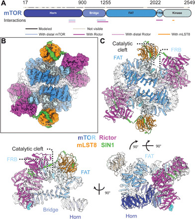
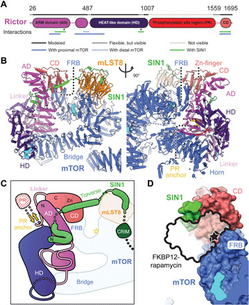
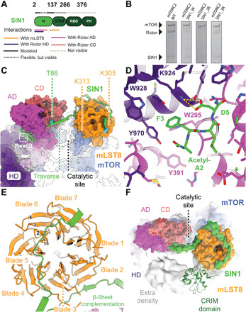
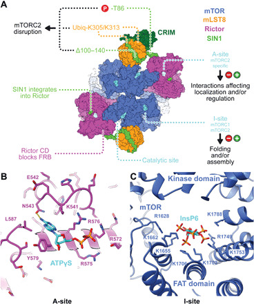
Verification
Claims the paper makes
The claims and statistics of this paper haven’t been checked yet.
Run the check — free, no account needed. It reads the open-access text and checks the sources it cites.
Claims other papers make about this one
Each is checked against this paper’s own text: does it say what the citing paper says it does?
The cited source says what the claim attributes to it.
In the citing paper
“The reasons for this difference in rapamycin sensitivity is structural; the mTOR interacting protein RICTOR, which is present in mTORC2 but not mTORC1, masks the rapamycin-interacting domain of mTOR29–31.”
From Targeting the biology of aging with mTOR inhibitors (2023) · cites this paper as [29]
Self-check
Percentages, sums and reported differences re-computed from the paper’s own numbers.
The self-check needs the paper’s full text.
Citations
Cites
Loewith, R., Jacinto, E., Wullschleger, S., Lorberg, A., Crespo, J. L., Bonenfant, D., Oppliger, W., Jenoe, P., & Hall, M. N. (2002) · Molecular Cell · doi:10.1016/s1097-2765(02)00636-6 · Cited by 1,886
Self-citationmTOR kinase structure, mechanism and regulation
Yang, H., Rudge, D. G., Koos, J. D., Vaidialingam, B., Yang, H. J., & Pavletich, N. P. (2013) · Nature · doi:10.1038/nature12122 · Cited by 1,075
Mechanisms of mTORC1 activation by RHEB and inhibition by PRAS40
Yang, H., Jiang, X., Li, B., Yang, H. J., Miller, M., Yang, A., Dhar, A., & Pavletich, N. P. (2017) · Nature · doi:10.1038/nature25023 · Cited by 548
Dazert, E., & Hall, M. N. (2011) · Current Opinion in Cell Biology · doi:10.1016/j.ceb.2011.09.003 · Cited by 460
Architecture of human mTOR complex 1
Aylett, C. H. S., Sauer, E., Imseng, S., Boehringer, D., Hall, M. N., Ban, N., & Maier, T. (2016) · Science · doi:10.1126/science.aaa3870 · Cited by 344
Self-citationStructural Basis for Regulation of Phosphoinositide Kinases and Their Involvement in Human Disease
Burke, J. E. (2018) · Molecular Cell · doi:10.1016/j.molcel.2018.08.005 · Cited by 276
MTOR inhibitors in cancer therapy
Xie, J., Wang, X., & Proud, C. G. (2016) · F1000Research · doi:10.12688/f1000research.9207.1 · Cited by 266 · cited 1×
“…proliferation ( 4 ) by phosphoryl ating the AGC family kinases: AKT, PKC, and SGK ( 1 , 4 , 11 – 13 ). mTORC2 also promotes tumorigenesis via up-regulation of lipid biosynthesis ( 14 ). mTOR inhibitors played a major role in the elucidation of mTOR signaling and are used in cancer treatment ( 15 ).”
Research works printed but not linked: 55
- 10.1038/s41568-018-0074-8 · DOI · Paper page
- 10.1016/j.sbi.2018.03.010 · DOI
- 10.1126/science.270.5233.50 · DOI
- 10.1016/S0092-8674(02)00833-4 · DOI
- 10.1016/S1097-2765(03)00114-X · DOI
- 10.1016/j.cub.2004.06.054 · DOI · Paper page
- 10.1101/gad.1461206 · DOI
- 10.1042/BJ20070540 · DOI
- 10.1042/BJ20081668 · DOI · Paper page
- 10.1038/emboj.2008.119 · DOI
- 10.1126/science.1106148 · DOI · Paper page
- 10.1016/j.ccell.2017.11.011 · DOI
- 10.1038/nrd3531 · DOI
- 10.1371/journal.pone.0176599 · DOI
- 10.1038/ncomms11016 · DOI
- 10.1038/s41422-018-0029-3 · DOI
- 10.1038/s41467-017-01862-0 · DOI
- 10.7554/eLife.33101 · DOI
- 10.1107/S1744309112007178 · DOI
- 10.7554/eLife.19594 · DOI
- 10.1007/s10858-014-9882-7 · DOI
- 10.1016/j.molcel.2015.04.031 · DOI · Paper page
- 10.1128/MCB.00735-09 · DOI
- 10.1152/ajpheart.00293.2009 · DOI
- 10.1039/C6MT00286B · DOI
- 10.1038/ncb2860 · DOI
- 10.1158/0008-5472.CAN-18-3658 · DOI
- 10.1074/jbc.M505553200 · DOI
- 10.1038/nature22344 · DOI
- 10.1038/s41594-019-0342-7 · DOI
- 10.1016/S0092-8674(00)00061-1 · DOI
- 10.1038/s41422-019-0143-x · DOI
- 10.1016/j.molcel.2019.07.014 · DOI
- 10.1016/j.molcel.2016.02.033 · DOI
- 10.1038/nmeth983 · DOI · Paper page
- 10.1074/jbc.272.50.31515 · DOI · Paper page
- 10.1073/pnas.1716109115 · DOI
- 10.1128/mBio.02323-18 · DOI
- 10.1038/nmeth.3047 · DOI · Paper page
- 10.1021/acs.jproteome.6b00066 · DOI
- 10.1021/ac0341261 · DOI
- 10.1016/j.jsb.2005.07.007 · DOI
- 10.1038/nmeth.4193 · DOI
- 10.1016/j.jsb.2015.11.003 · DOI
- 10.1006/jsbi.1999.4174 · DOI
- 10.1107/S205225251801463X · DOI
- 10.1038/nmeth.4169 · DOI · Paper page
- 10.1002/jcc.20084 · DOI
- 10.1107/S0907444909052925 · DOI · Paper page
- 10.1107/S0907444910007493 · DOI · Paper page
- 10.1002/pro.3235 · DOI · Paper page
- 10.1073/pnas.181342398 · DOI
- 10.1038/msb.2011.75 · DOI
- 10.1093/bioinformatics/15.4.305 · DOI
- 10.1093/bioinformatics/17.8.700 · DOI
Cited by
Sorted by citations
Panwar, V., Singh, A., Bhatt, M., Tonk, R. K., Azizov, S., Raza, A. S., Sengupta, S., Kumar, D., & Garg, M. (2023) · Signal Transduction and Targeted Therapy · doi:10.1038/s41392-023-01608-z · Cited by 967
Molecular mechanisms of dietary restriction promoting health and longevity
Green, C. L., Lamming, D. W., & Fontana, L. (2022) · Nature Reviews Molecular Cell Biology · doi:10.1038/s41580-021-00411-4 · Cited by 714
Targeting the biology of aging with mTOR inhibitors
Mannick, J. B., & Lamming, D. W. (2023) · Nature Aging · doi:10.1038/s43587-023-00416-y · Cited by 372
Burnett, G. L., Yang, Y. C., Aggen, J. B., Pitzen, J., Gliedt, M. K., Semko, C. M., Marquez, A., Evans, J. W., Wang, G., Won, W. S., et al. (2023) · Journal of Medicinal Chemistry · doi:10.1021/acs.jmedchem.2c01658 · Cited by 72
From Obscurity to Prominence: IPMK’s Expanding Role in Cellular Signaling, Physiology, and Disease
Mishra, S. H., Chatterjee, S., Viera-Preval, L., & Guha, P. (2025) · Biomolecules · doi:10.3390/biom15091266 · Cited by 1
“Recent cryo-electron microscopy studies have elucidated the structures of mTORC1 and mTORC2, identifying a conserved InsP6 binding site in mTOR [67,68].”
Metadata
- Authors
- Scaiola, A., Mangia, F., Imseng, S., Boehringer, D., Berneiser, K., Shimobayashi, M., Stuttfeld, E., Hall, M. N., Ban, N., & Maier, T.
- Affiliations
- Institute for Molecular Biology and Biophysics, ETH Zurich, Zurich, Switzerland.
- Biozentrum, University of Basel, Klingelbergstrasse 50/70, 4056 Basel, Switzerland.
- Cryo-EM Knowledge Hub, ETH Zurich, 8093 Zurich, Switzerland.
- Venue
- Science Advances
- Published
- 2020-11-06
- Volume
- 6
- Issue
- 45
- ISSN
- 2375-2548
- PMC
- PMC7673708
- Cited by
- 105
- References
- 7
- Language
- en
- Funders
- Swiss National Science Foundation (177084)
- Fellowship for Excellence from the Biozentrum Basel International PhD program
- National Center of Excellence in RNA and Disease (138262)
- Swiss National Science Foundation SNSF (179323)
- Fellowship of the Biozentrum International PhD Program
About these checks
Registry, retraction, Cites and statistics results are computed, not judged, with one exception: a printed reference that no index lists is read once by a model before it counts as a citation error. Claims are extracted by a model and kept only when their quote appears verbatim in the text; each cited source is then checked, with a model’s help, for whether it says what the claim relies on. Those verdicts can be wrong — read the claim’s quote and the cited source before relying on one.
Statistics and arithmetic are recomputed, not judged.
Confidence bands are shown only for fields calibrated on our annotated test set. None is calibrated yet, so no band is shown.
What counts as a claim ›How we measure confidence ›How we check this ›
Are you an author, or do you see a problem with this page? Request a review
How to cite
Cite CoChat for the checks on this page, and the paper for its own findings.
- This page
- CoChat. (2026). Citation check: The 3.2-Å resolution structure of human mTORC2. https://app.cochat.ai/papers/10.1126/sciadv.abc1251
- The paper
- Scaiola, A., Mangia, F., Imseng, S., Boehringer, D., Berneiser, K., Shimobayashi, M., Stuttfeld, E., Hall, M. N., Ban, N., & Maier, T. (2020). The 3.2-Å resolution structure of human mTORC2. Science Advances. https://doi.org/10.1126/sciadv.abc1251
