Cited by 101

The DOI matches its Crossref record; 4 of the 4 references it cites were found in a registry or an index.

High-Risk Human Papillomavirus E7 Proteins Target PTPN14 for Degradation

White, Elizabeth A., Münger, Karl, Howley, Peter M.·2016·mBio

Abstract

The major transformation activity of the high-risk human papillomaviruses (HPV) is associated with the E7 oncoprotein. The interaction of HPV E7 with retinoblastoma family proteins is important for several E7 activities; however, this interaction does not fully account for the high-risk E7-specific cellular immortalization and transformation activities. We have determined that the cellular non-receptor protein tyrosine phosphatase PTPN14 interacts with HPV E7 from many genus alpha and beta HPV types. We find that high-risk genus alpha HPV E7, but not low-risk genus alpha or beta HPV E7, is necessary and sufficient to reduce the steady-state level of PTPN14 in cells. High-risk E7 proteins target PTPN14 for proteasome-mediated degradation, which requires the ubiquitin ligase UBR4, and PTPN14 is degraded by the proteasome in HPV-positive cervical cancer cell lines. Residues in the C terminus of E7 interact with the C-terminal phosphatase domain of PTPN14, and interference with the E7-PTPN14 interaction restores PTPN14 levels in cells. Finally, PTPN14 degradation correlates with the retinoblastoma-independent transforming activity of high-risk HPV E7. IMPORTANCE High-risk human papillomaviruses (HPV) are the cause of cervical cancer, some other anogenital cancers, and a growing fraction of oropharyngeal carcinomas. The high-risk HPV E6 and E7 oncoproteins enable these viruses to cause cancer, and the mechanistic basis of their carcinogenic activity has been the subject of intense study. The high-risk E7 oncoprotein is especially important in the immortalization and transformation of human cells, which makes it a central component of HPV-associated cancer development. E7 oncoproteins interact with retinoblastoma family proteins, but for several decades, it has been recognized that high-risk HPV E7 oncoproteins have additional cancer-associated activities. We have determined that high-risk E7 proteins target the proteolysis of the cellular protein tyrosine phosphatase PTPN14 and find that this activity is correlated with the retinoblastoma-independent transforming activity of E7.

Overview

The claims and statistics of this paper haven’t been checked yet.

Run the check — free, no account needed. It reads the open-access text and checks the sources it cites.

A few free checks a day · papers already verified open instantly · results are public

Integrity signals

Registry

Crossref · 10.1128/mbio.01530-16

Cites

4 of 4 references verified0 citation errors · 0 not checkable

Statistics

Figures

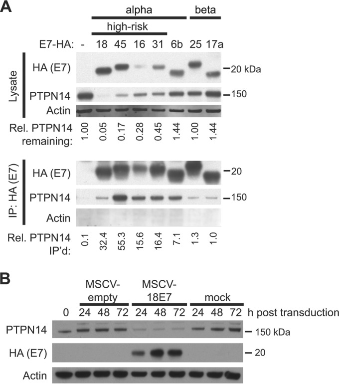
FIG 1 HPV E7 proteins bind to PTPN14, and high-risk HPV E7 proteins reduce PTPN14 levels. (A) Lysates from N/Tert-1 cells expressing E7-Flag-HA were used in immunoprecipitations with HA antibody. Immunoprecipitates were separated by SDS-PAGE and Western blotted using antibodies to HA, PTPN14, and actin. (Top panels) Protein levels in input lysates. Band intensity was quantified with ImageJ, and the numbers indicate the intensity of the PTPN14 band in each lysate lane relative to the actin intensity, normalized to the amount of PTPN14 in the empty vector cells. (Bottom panels) PTPN14 recovered after immunoprecipitation with anti-HA antibody. HA and PTPN14 were detected on film, and actin was detected using the LI-COR imaging system. Band intensity was quantified with ImageJ, and numbers indicate the amount of PTPN14 recovered in the immunoprecipitation relative to the amount of PTPN14 in the lysates, normalized to the amount recovered in the HPV17a E7 IP. (B) N/Tert-1 cells were transduced with a retrovirus encoding HPV18 E7 or with an empty vector control retrovirus or mock transduced and harvested at indicated time points. Steady-state levels of PTPN14, HPV18-Flag-HA, and actin were measured by Western blotting.
FIG 1 HPV E7 proteins bind to PTPN14, and high-risk HPV E7 proteins reduce PTPN14 levels. (A) Lysates from N/Tert-1 cells expressing E7-Flag-HA were used in immunoprecipitations with HA antibody. Immunoprecipitates were separated by SDS-PAGE and Western blotted using antibodies to HA, PTPN14, and actin. (Top panels) Protein levels in input lysates. Band intensity was quantified with ImageJ, and the numbers indicate the intensity of the PTPN14 band in each lysate lane relative to the actin intensity, normalized to the amount of PTPN14 in the empty vector cells. (Bottom panels) PTPN14 recovered after immunoprecipitation with anti-HA antibody. HA and PTPN14 were detected on film, and actin was detected using the LI-COR imaging system. Band intensity was quantified with ImageJ, and numbers indicate the amount of PTPN14 recovered in the immunoprecipitation relative to the amount of PTPN14 in the lysates, normalized to the amount recovered in the HPV17a E7 IP. (B) N/Tert-1 cells were transduced with a retrovirus encoding HPV18 E7 or with an empty vector control retrovirus or mock transduced and harvested at indicated time points. Steady-state levels of PTPN14, HPV18-Flag-HA, and actin were measured by Western blotting.CC BY · PubMed Central
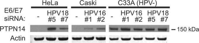
FIG 2 PTPN14 protein levels in cervical cancer cells depend on HPV oncoprotein expression. Cells of the HPV18-positive HeLa, HPV16-positive Caski, and HPV-negative C33A cervical cancer cell lines were transfected with siRNAs targeting the HPV16 E6/E7 and/or HPV18 E6/E7 transcripts at a final concentration of 40 nM. Cells were harvested 72 h posttransfection, and lysates were used for Western blotting with antibodies to PTPN14 and actin.
FIG 2 PTPN14 protein levels in cervical cancer cells depend on HPV oncoprotein expression. Cells of the HPV18-positive HeLa, HPV16-positive Caski, and HPV-negative C33A cervical cancer cell lines were transfected with siRNAs targeting the HPV16 E6/E7 and/or HPV18 E6/E7 transcripts at a final concentration of 40 nM. Cells were harvested 72 h posttransfection, and lysates were used for Western blotting with antibodies to PTPN14 and actin.CC BY · PubMed Central
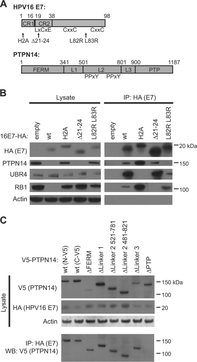
FIG 3 Identification of protein binding sites on HPV16 E7 and on PTPN14. (A) Schematic of HPV16 and PTPN14 domain organization and mutants used in the study (39). (B) N/Tert-1 cells expressing wild-type (wt) HPV16 E7-Flag-HA, one of several mutants of HPV16 (E7), or the empty vector were subjected to immunoprecipitation with HA antibody. Immunoprecipitates were separated by SDS-PAGE and Western blotted using antibodies to HA, PTPN14, UBR4, RB1, and actin. (Left panels) Protein levels in input lysates. (Right panels) Proteins recovered after immunoprecipitation with anti-HA antibody. (C) N/Tert-1 cells expressing wild-type HPV16 E7-Flag-HA were transduced with retroviruses expressing V5-tagged PTPN14 or one of several deletion mutant forms of PTPN14 and were harvested 48 h posttransduction. Cell lysates were subjected to immunoprecipitation with HA antibody. Immunoprecipitates were separated by SDS-PAGE and Western blotted using antibodies to HA, V5, and actin. (Top panels) Protein levels in input lysates. (Bottom panel) V5-tagged PTPN14 recovered after immunoprecipitation with anti-HA antibody.
FIG 3 Identification of protein binding sites on HPV16 E7 and on PTPN14. (A) Schematic of HPV16 and PTPN14 domain organization and mutants used in the study (39). (B) N/Tert-1 cells expressing wild-type (wt) HPV16 E7-Flag-HA, one of several mutants of HPV16 (E7), or the empty vector were subjected to immunoprecipitation with HA antibody. Immunoprecipitates were separated by SDS-PAGE and Western blotted using antibodies to HA, PTPN14, UBR4, RB1, and actin. (Left panels) Protein levels in input lysates. (Right panels) Proteins recovered after immunoprecipitation with anti-HA antibody. (C) N/Tert-1 cells expressing wild-type HPV16 E7-Flag-HA were transduced with retroviruses expressing V5-tagged PTPN14 or one of several deletion mutant forms of PTPN14 and were harvested 48 h posttransduction. Cell lysates were subjected to immunoprecipitation with HA antibody. Immunoprecipitates were separated by SDS-PAGE and Western blotted using antibodies to HA, V5, and actin. (Top panels) Protein levels in input lysates. (Bottom panel) V5-tagged PTPN14 recovered after immunoprecipitation with anti-HA antibody.CC BY · PubMed Central
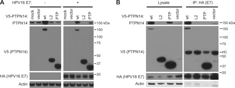
FIG 4 Overexpression of the PTPN14 PTP domain can block the interaction of E7 with full-length PTPN14 and restore PTPN14 levels. (A) N/Tert-1 cells expressing wild-type (wt) HPV16 E7-Flag-HA or the empty vector were mock transduced, transduced with a retrovirus packaged with the empty vector, or transduced with retroviruses expressing wild-type V5-tagged PTPN14, the PTPN14 L2 domain, or the PTPN14 PTP domain and were harvested 48 h posttransduction. Steady-state levels of full-length PTPN14, V5-tagged PTPN14 fragments, HA-tagged E7, and actin were measured by Western blotting. (B) N/Tert-1 cells expressing wild-type HPV18 E7-Flag-HA were transduced with retroviruses expressing wild-type V5-tagged PTPN14, the PTPN14 L2 domain, the PTPN14 PTP domain, or the empty vector and were harvested 48 h posttransduction. Cell lysates were subjected to immunoprecipitation with HA antibody. Immunoprecipitates were separated by SDS-PAGE and Western blotted using antibodies to PTPN14, HA, V5, and actin. (Left panels) Protein levels in input lysates. (Right panels) Proteins recovered after immunoprecipitation with anti-HA antibody. HC, antibody heavy chain.
FIG 4 Overexpression of the PTPN14 PTP domain can block the interaction of E7 with full-length PTPN14 and restore PTPN14 levels. (A) N/Tert-1 cells expressing wild-type (wt) HPV16 E7-Flag-HA or the empty vector were mock transduced, transduced with a retrovirus packaged with the empty vector, or transduced with retroviruses expressing wild-type V5-tagged PTPN14, the PTPN14 L2 domain, or the PTPN14 PTP domain and were harvested 48 h posttransduction. Steady-state levels of full-length PTPN14, V5-tagged PTPN14 fragments, HA-tagged E7, and actin were measured by Western blotting. (B) N/Tert-1 cells expressing wild-type HPV18 E7-Flag-HA were transduced with retroviruses expressing wild-type V5-tagged PTPN14, the PTPN14 L2 domain, the PTPN14 PTP domain, or the empty vector and were harvested 48 h posttransduction. Cell lysates were subjected to immunoprecipitation with HA antibody. Immunoprecipitates were separated by SDS-PAGE and Western blotted using antibodies to PTPN14, HA, V5, and actin. (Left panels) Protein levels in input lysates. (Right panels) Proteins recovered after immunoprecipitation with anti-HA antibody. HC, antibody heavy chain.CC BY · PubMed Central
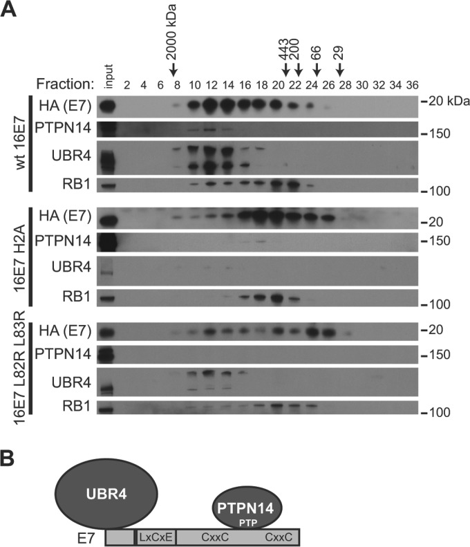
FIG 5 E7 interacts with UBR4 and PTPN14 in a complex distinct from the E7-RB1 complex. (A) Lysates from N/Tert-1 cells expressing wild-type (wt) HPV16 (E7), HPV16 E7 H2A, or HPV16 E7 L82R L83R were fractionated on a Superose 6 column using a GE Akta Pure high-performance liquid chromatography (HPLC) column. Five hundred-microliter fractions were collected, and alternate fractions were used for anti-HA immunoprecipitation. Immunoprecipitates were separated by SDS-PAGE and Western blotted using antibodies to PTPN14, HA, UBR4, and RB1. Unfractionated lysate (input) is included on each gel as a control. (B) Schematic of E7/PTPN14/UBR4 complex and approximate interaction sites.
FIG 5 E7 interacts with UBR4 and PTPN14 in a complex distinct from the E7-RB1 complex. (A) Lysates from N/Tert-1 cells expressing wild-type (wt) HPV16 (E7), HPV16 E7 H2A, or HPV16 E7 L82R L83R were fractionated on a Superose 6 column using a GE Akta Pure high-performance liquid chromatography (HPLC) column. Five hundred-microliter fractions were collected, and alternate fractions were used for anti-HA immunoprecipitation. Immunoprecipitates were separated by SDS-PAGE and Western blotted using antibodies to PTPN14, HA, UBR4, and RB1. Unfractionated lysate (input) is included on each gel as a control. (B) Schematic of E7/PTPN14/UBR4 complex and approximate interaction sites.CC BY · PubMed Central
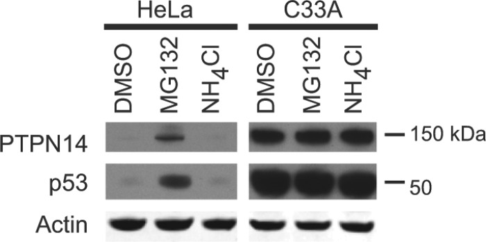
FIG 6 PTPN14 is degraded by the proteasome in HPV-positive cervical cancer cells. HPV18-positive HeLa cells and HPV-negative C33A cervical cancer cells were treated for 8 h with 30 µM MG132, 10 mM NH4Cl, or DMSO as a control. Lysates were separated by SDS-PAGE and Western blotted using antibodies to PTPN14, p53, and actin.
FIG 6 PTPN14 is degraded by the proteasome in HPV-positive cervical cancer cells. HPV18-positive HeLa cells and HPV-negative C33A cervical cancer cells were treated for 8 h with 30 µM MG132, 10 mM NH4Cl, or DMSO as a control. Lysates were separated by SDS-PAGE and Western blotted using antibodies to PTPN14, p53, and actin.CC BY · PubMed Central
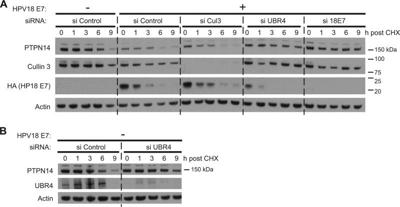
FIG 7 HPV18 E7-mediated PTPN14 degradation requires the E3 ubiquitin ligase UBR4. (A) N/Tert cells were transfected with siRNAs targeting cullin 3, UBR4, or HPV18 E7 or with a nontargeting control (si Control). Twenty-four hours posttransfection, cells were transduced with a retrovirus expressing HPV18 E7 or with the empty vector control retrovirus. Forty-eight hours post-retrovirus transduction, cells were treated with 40 µg/ml cycloheximide (CHX) and harvested at indicated time points post-cycloheximide treatment. Equal amounts of protein lysates were separated by SDS-PAGE and Western blotted using antibodies to PTPN14, cullin 3, HA, and actin. (B) N/Tert cells were transfected with siRNA targeting UBR4 or with a nontargeting control (si Control). Twenty-four hours posttransfection, cells were transduced with an empty vector control retrovirus. Forty-eight hours post-retrovirus transduction, cells were treated with 40 µg/ml cycloheximide and harvested at indicated time points post-cycloheximide treatment. Lysates were separated by SDS-PAGE and Western blotted using antibodies to PTPN14, UBR4, and actin. The same lysates were run in the first five lanes of the gels in panels A and B. The experiment was repeated four times, and a representative experiment is shown.
FIG 7 HPV18 E7-mediated PTPN14 degradation requires the E3 ubiquitin ligase UBR4. (A) N/Tert cells were transfected with siRNAs targeting cullin 3, UBR4, or HPV18 E7 or with a nontargeting control (si Control). Twenty-four hours posttransfection, cells were transduced with a retrovirus expressing HPV18 E7 or with the empty vector control retrovirus. Forty-eight hours post-retrovirus transduction, cells were treated with 40 µg/ml cycloheximide (CHX) and harvested at indicated time points post-cycloheximide treatment. Equal amounts of protein lysates were separated by SDS-PAGE and Western blotted using antibodies to PTPN14, cullin 3, HA, and actin. (B) N/Tert cells were transfected with siRNA targeting UBR4 or with a nontargeting control (si Control). Twenty-four hours posttransfection, cells were transduced with an empty vector control retrovirus. Forty-eight hours post-retrovirus transduction, cells were treated with 40 µg/ml cycloheximide and harvested at indicated time points post-cycloheximide treatment. Lysates were separated by SDS-PAGE and Western blotted using antibodies to PTPN14, UBR4, and actin. The same lysates were run in the first five lanes of the gels in panels A and B. The experiment was repeated four times, and a representative experiment is shown.CC BY · PubMed Central
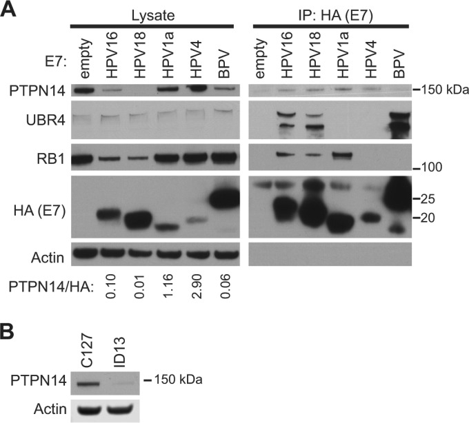
FIG 8 Papillomavirus E7 proteins vary in their ability to bind PTPN14 and target PTPN14 for degradation. (A) N/Tert-1 cells expressing one of several HPV E7 proteins or BPV1 E7 protein tagged with Flag and HA were subjected to immunoprecipitation with HA antibody. Immunoprecipitates were separated by SDS-PAGE and Western blotted using antibodies to HA, PTPN14, UBR4, RB1, and actin. (Left panels) Protein levels in input lysates. (Right panels) Proteins recovered after immunoprecipitation with anti-HA antibody. PTPN14 and HA protein amounts were quantified using ImageJ. The ratio of PTPN14 protein amount relative to E7-HA protein amount in the E7-expressing cell lines is indicated. (B) PTPN14 and actin protein levels in C127 and ID13 cell lysates were measured by Western blotting.
FIG 8 Papillomavirus E7 proteins vary in their ability to bind PTPN14 and target PTPN14 for degradation. (A) N/Tert-1 cells expressing one of several HPV E7 proteins or BPV1 E7 protein tagged with Flag and HA were subjected to immunoprecipitation with HA antibody. Immunoprecipitates were separated by SDS-PAGE and Western blotted using antibodies to HA, PTPN14, UBR4, RB1, and actin. (Left panels) Protein levels in input lysates. (Right panels) Proteins recovered after immunoprecipitation with anti-HA antibody. PTPN14 and HA protein amounts were quantified using ImageJ. The ratio of PTPN14 protein amount relative to E7-HA protein amount in the E7-expressing cell lines is indicated. (B) PTPN14 and actin protein levels in C127 and ID13 cell lysates were measured by Western blotting.CC BY · PubMed Central

Verification

Claims the paper makes

The claims and statistics of this paper haven’t been checked yet.

Run the check — free, no account needed. It reads the open-access text and checks the sources it cites.

A few free checks a day · papers already verified open instantly · results are public

Claims other papers make about this one

Each is checked against this paper’s own text: does it say what the citing paper says it does?

Self-check

Percentages, sums and reported differences re-computed from the paper’s own numbers.

The self-check needs the paper’s full text.

Citations

Cites

Research works printed but not linked: 64

  1. Howley PM, Schiller JT, Lowy DR. 2013. Papillomaviruses, p 1662–1703. In Knipe DM, Howley PM (ed), Fields virology, 6th ed. Lippincott Williams & Wilkins, Philadelphia, PA.
  2. 10.1056/NEJMoa021641 · DOI · Paper page
  3. 10.1016/j.virol.2005.04.002 · DOI · Paper page
  4. 10.1016/j.virol.2008.12.006 · DOI
  5. 10.1016/j.virol.2013.04.013 · DOI
  6. 10.1128/jvi.66.12.6893-6902.1992 · DOI · Paper page
  7. 10.1002/j.1460-2075.1989.tb08594.x · DOI · Paper page
  8. Berezutskaya E, Yu B, Morozov A, Raychaudhuri P, Bagchi S. 1997. Differential regulation of the pocket domains of the retinoblastoma family proteins by the HPV16 E7 oncoprotein. Cell Growth Differ 8:1277–1286.
  9. Boyer SN, Wazer DE, Band V. 1996. E7 protein of human papillomavirus-16 induces degradation of retinoblastoma protein through the ubiquitin-proteasome pathway. Cancer Res 56:4620–4624.
  10. 10.1128/JVI.75.16.7583-7591.2001 · DOI · Paper page
  11. 10.1128/jvi.71.4.2905-2912.1997 · DOI · Paper page
  12. 10.1016/0092-8674(90)90409-8 · DOI · Paper page
  13. 10.1126/science.2157286 · DOI · Paper page
  14. 10.3390/pathogens5010008 · DOI · Paper page
  15. Duensing S, Münger K. 2002. The human papillomavirus type 16 E6 and E7 oncoproteins independently induce numerical and structural chromosome instability. Cancer Res 62:7075–7082.
  16. 10.1002/j.1460-2075.1989.tb08570.x · DOI · Paper page
  17. 10.1128/jvi.65.1.473-478.1991 · DOI · Paper page
  18. 10.1128/jvi.63.10.4417-4421.1989 · DOI · Paper page
  19. 10.1099/0022-1317-71-1-165 · DOI
  20. 10.1128/jvi.65.1.292-298.1991 · DOI · Paper page
  21. 10.1073/pnas.89.10.4442 · DOI
  22. 10.1128/JVI.75.15.6737-6747.2001 · DOI · Paper page
  23. 10.1128/jvi.66.3.1329-1335.1992 · DOI
  24. 10.1128/jvi.66.4.2418-2427.1992 · DOI · Paper page
  25. Banks L, Edmonds C, Vousden KH. 1990. Ability of the HPV16 E7 protein to bind RB and induce DNA synthesis is not sufficient for efficient transforming activity in NIH3T3 cells. Oncogene 5:1383–1389.
  26. 10.1073/pnas.0505337102 · DOI · Paper page
  27. 10.1128/jvi.67.6.3142-3150.1993 · DOI · Paper page
  28. 10.1158/0008-5472.CAN-12-2083 · DOI
  29. 10.1016/j.virol.2012.09.046 · DOI · Paper page
  30. 10.1128/JVI.02172-12 · DOI · Paper page
  31. 10.1038/nrm2039 · DOI
  32. 10.1126/science.1096096 · DOI
  33. 10.1158/2159-8290.CD-12-0095 · DOI · Paper page
  34. 10.1038/ng.3525 · DOI
  35. 10.1038/ng.3349 · DOI
  36. 10.1128/MCB.20.4.1436-1447.2000 · DOI · Paper page
  37. 10.1128/jvi.70.10.6862-6869.1996 · DOI
  38. 10.1073/pnas.0505322102 · DOI · Paper page
  39. 10.1016/0092-8674(93)90384-3 · DOI · Paper page
  40. 10.1128/JVI.00422-07 · DOI
  41. Ciccolini F, Di Pasquale G, Carlotti F, Crawford L, Tommasino M. 1994. Functional studies of E7 proteins from different HPV types. Oncogene 9:2633–2638.
  42. 10.1007/BF01702398 · DOI
  43. 10.1128/JVI.75.1.513-521.2001 · DOI
  44. 10.1128/jvi.63.1.259-266.1989 · DOI
  45. 10.1128/jvi.52.2.377-388.1984 · DOI
  46. 10.1128/JVI.00881-07 · DOI · Paper page
  47. 10.1128/JVI.03282-14 · DOI
  48. 10.1002/j.1460-2075.1988.tb03185.x · DOI · Paper page
  49. 10.1128/JVI.03486-14 · DOI
  50. 10.1128/JVI.03263-13 · DOI
  51. 10.18632/oncotarget.3465 · DOI
  52. 10.1371/journal.ppat.1004712 · DOI · Paper page
  53. Kong Q, Wang W, Li P. 2015. Regulator role of HPV E7 protein on miR-21 expression in cervical carcinoma cells and its functional implication. Int J Clin Exp Pathol 8:15808–15813.
  54. 10.1371/journal.pone.0002557 · DOI
  55. 10.1016/j.bbagrm.2011.05.005 · DOI
  56. 10.1038/onc.2012.231 · DOI
  57. 10.1038/onc.2012.147 · DOI · Paper page
  58. 10.1101/gad.173435.111 · DOI
  59. 10.1016/j.tcb.2015.05.002 · DOI
  60. 10.1074/jbc.M114.627968 · DOI
  61. 10.1128/JVI.01197-14 · DOI
  62. 10.1073/pnas.0914818107 · DOI
  63. 10.1073/pnas.0507458102 · DOI
  64. 10.1128/MCB.00201-12 · DOI

Metadata

Authors
White, E. A., Münger, K., & Howley, P. M.
Affiliations
  • Department of Microbiology and Immunobiology, Harvard Medical School, Boston, Massachusetts, USA
  • Department of Developmental, Molecular and Chemical Biology, Tufts University School of Medicine, Boston, Massachusetts, USA
Venue
mBio
Published
2016-11-02
Volume
7
Issue
5
ISSN
  • 2161-2129
  • 2150-7511
Cited by
101
References
4
Language
en
Funders
  • HHS | NIH | National Cancer Institute (R35CA197262)
  • Alexander and Margaret Stewart Trust

About these checks

Registry, retraction, Cites and statistics results are computed, not judged, with one exception: a printed reference that no index lists is read once by a model before it counts as a citation error. Claims are extracted by a model and kept only when their quote appears verbatim in the text; each cited source is then checked, with a model’s help, for whether it says what the claim relies on. Those verdicts can be wrong — read the claim’s quote and the cited source before relying on one.

Statistics and arithmetic are recomputed, not judged.

Confidence bands are shown only for fields calibrated on our annotated test set. None is calibrated yet, so no band is shown.

What counts as a claim ›How we measure confidence ›How we check this ›

Are you an author, or do you see a problem with this page? Request a review