

The DOI matches its Crossref record; 71 of the 71 references it cites were found in a registry or an index.
ATP-Dependent Persister Formation in Escherichia coli
Open accessAbstract
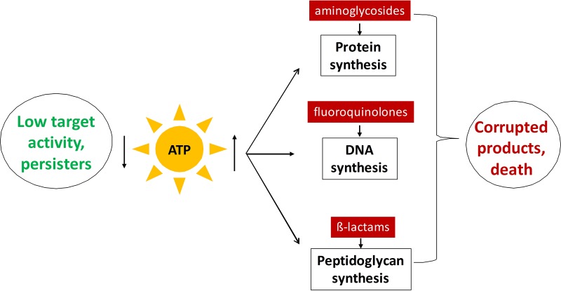
Persisters are dormant variants that form a subpopulation of cells tolerant to antibiotics. Persisters are largely responsible for the recalcitrance of chronic infections to therapy. In Escherichia coli , one widely accepted model of persister formation holds that stochastic accumulation of ppGpp causes activation of the Lon protease that degrades antitoxins; active toxins then inhibit translation, resulting in dormant, drug-tolerant persisters. We found that various stresses induce toxin-antitoxin (TA) expression but that induction of TAs does not necessarily increase persisters. The 16S rRNA promoter rrnB P1 was proposed to be a persister reporter and an indicator of toxin activation regulated by ppGpp. Using fluorescence-activated cell sorting (FACS), we confirmed the enrichment for persisters in the fraction of rrnB P1 -gfp dim cells; however, this is independent of toxin-antitoxins. rrnB P1 is coregulated by ppGpp and ATP. We show that rrnB P1 can report persisters in a relA / spoT deletion background, suggesting that rrnB P1 is a persister marker responding to ATP. Consistent with this finding, decreasing the level of ATP by arsenate treatment causes drug tolerance. Lowering ATP slows translation and prevents the formation of DNA double-strand breaks upon fluoroquinolone treatment. We conclude that variation in ATP levels leads to persister formation by decreasing the activity of antibiotic targets. IMPORTANCE Persisters are a subpopulation of antibiotic-tolerant cells responsible for the recalcitrance of chronic infections. Our current understanding of persister formation is primarily based on studies of E. coli . The activation of toxin-antitoxin systems by ppGpp has become a widely accepted model for persister formation. In this study, we found that stress-induced activation of mRNA interferase-type toxins does not necessarily cause persister formation. We also found that the persister marker rrnB P1 reports persister cells because it detects a drop in cellular ATP levels. Consistent with this, lowering the ATP level decreases antibiotic target activity and, thus, leads to persister formation. We conclude that stochastic variation in ATP is the main mechanism of persister formation. A decrease in ATP provides a satisfactory explanation for the drug tolerance of persisters, since bactericidal antibiotics act by corrupting energy-dependent targets.
Overview
The claims and statistics of this paper haven’t been checked yet.
Run the check — free, no account needed. It reads the open-access text and checks the sources it cites.
Integrity signals
Registry
Cites
Statistics
Figures
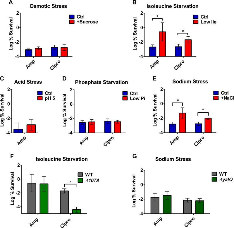
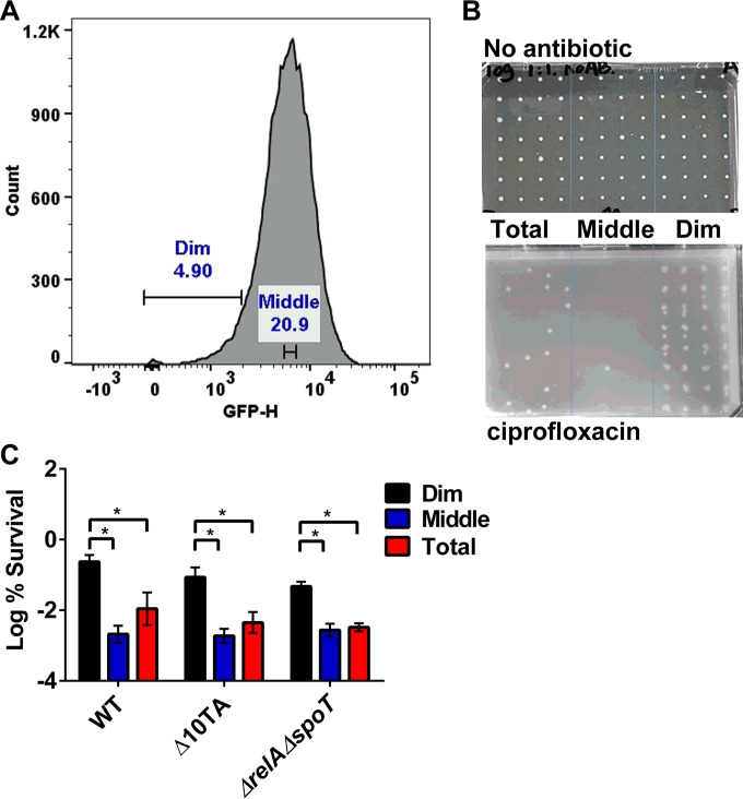
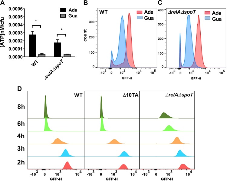
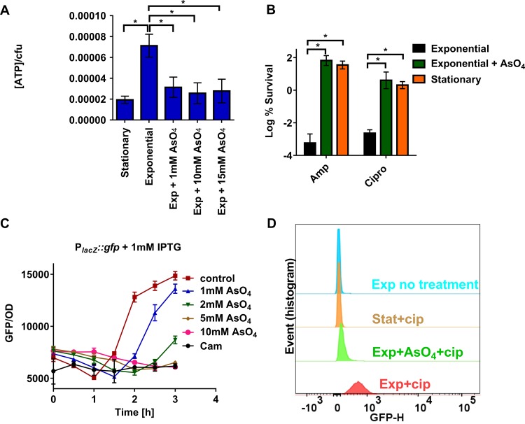
Verification
Claims the paper makes
The claims and statistics of this paper haven’t been checked yet.
Run the check — free, no account needed. It reads the open-access text and checks the sources it cites.
Claims other papers make about this one
Each is checked against this paper’s own text: does it say what the citing paper says it does?
The source backs part of the claim, not all of it — or backs it for a different population, condition or comparison.
In the citing paper
“It has been well studied that dormant state reduces the exposure of antibiotic targets and helps bacterial cells tolerate a variety of antibiotics, a process linked to the toxin-antitoxin system [18,54].”
From Antifungal persistence: Clinical relevance and mechanisms (2025) · cites this paper as [54]
Experimental — accuracy not yet measured
Self-check
Percentages, sums and reported differences re-computed from the paper’s own numbers.
The self-check needs the paper’s full text.
Citations
Cites
2 retracted
RETRACTED: Bacterial persistence by RNA endonucleases
Maisonneuve, E., Shakespeare, L. J., Jørgensen, M. G., & Gerdes, K. (2011) · Proceedings of the National Academy of Sciences · doi:10.1073/pnas.1100186108 · Cited by 473
RetractedMaisonneuve, E., Castro-Camargo, M., & Gerdes, K. (2013) · Cell · doi:10.1016/j.cell.2013.07.048 · Cited by 439
RetractedBacterial Persistence as a Phenotypic Switch
Balaban, N. Q., Merrin, J., Chait, R., Kowalik, L., & Leibler, S. (2004) · Science · doi:10.1126/science.1099390 · Cited by 2,952
Lewis, K. (2010) · Annual Review of Microbiology · doi:10.1146/annurev.micro.112408.134306 · Cited by 1,858
Self-citationIntracellular Bacterial Biofilm-Like Pods in Urinary Tract Infections
Anderson, G. G., Palermo, J. J., Schilling, J. D., Roth, R. A., Heuser, J., & Hultgren, S. J. (2003) · Science · doi:10.1126/science.1084550 · Cited by 1,175
Persister cells and tolerance to antimicrobials
Keren, I., Kaldalu, N., Spoering, A. L., Wang, Y., & Lewis, K. (2003) · FEMS Microbiology Letters · doi:10.1016/s0378-1097(03)00856-5 · Cited by 1,094
Self-citationProkaryotic toxin–antitoxin stress response loci
Gerdes, K., Christensen, S. K., & Løbner‐Olesen, A. (2005) · Nature Reviews Microbiology · doi:10.1038/nrmicro1147 · Cited by 1,033
Active Starvation Responses Mediate Antibiotic Tolerance in Biofilms and Nutrient-Limited Bacteria
Nguyen, D. M., Joshi-Datar, A., Lepine, F. L., Bauerle, E., Olakanmi, O., Beer, K. D., McKay, G. A., Siehnel, R., Schafhauser, J., Wang, Y., et al. (2011) · Science · doi:10.1126/science.1211037 · Cited by 1,008
The RpoS-Mediated General Stress Response in Escherichia coli
Battesti, A., Majdalani, N., & Gottesman, S. (2010) · Annual Review of Microbiology · doi:10.1146/annurev-micro-090110-102946 · Cited by 922
Specialized Persister Cells and the Mechanism of Multidrug Tolerance in Escherichia coli
Keren, I., Shah, D., Spoering, A. L., Kaldalu, N., & Lewis, K. (2004) · Journal of Bacteriology · doi:10.1128/jb.186.24.8172-8180.2004 · Cited by 848
Self-citation
Research works printed but not linked: 1
- 10.1016/j.cell.2013.07.048 · DOI
Cited by
Sorted by citations
Molecular mechanisms of antibiotic resistance revisited
Darby, E. M., Trampari, E., Siasat, P., Gaya, M. S., Alav, I., Webber, M. A., & Blair, J. M. A. (2023) · Nature Reviews Microbiology · doi:10.1038/s41579-022-00820-y · Cited by 1,398
Persistent bacterial infections and persister cells
Fisher, R. A., Gollan, B., & Helaine, S. (2017) · Nature Reviews Microbiology · doi:10.1038/nrmicro.2017.42 · Cited by 1,243
The value of antimicrobial peptides in the age of resistance
Magana, M., Muthuirulan, P., Santos, A. L., Leanse, L. G., Fernández, M., Ioannidis, A., Giulianotti, M. A., Apidianakis, Y., Bradfute, S. B., Ferguson, A. L., et al. (2020) · The Lancet Infectious Diseases · doi:10.1016/s1473-3099(20)30327-3 · Cited by 1,193
Antibiotic resistance and persistence—implications for human health and treatment perspectives
Huemer, M., Mairpady Shambat, S., Brugger, S. D., & Zinkernagel, A. S. (2020) · The EMBO Reports · doi:10.15252/embr.202051034 · Cited by 850
The Science of Antibiotic Discovery
Lewis, K. (2020) · Cell · doi:10.1016/j.cell.2020.02.056 · Cited by 848
Self-citationStaphylococcal Biofilm Development: Structure, Regulation, and Treatment Strategies
Schilcher, K., & Horswill, A. R. (2020) · Microbiology and Molecular Biology Reviews · doi:10.1128/mmbr.00026-19 · Cited by 762
Understanding the Mechanism of Bacterial Biofilms Resistance to Antimicrobial Agents
Singh, S., Singh, S. K., Chowdhury, I., & Singh, R. (2017) · The Open Microbiology Journal · doi:10.2174/1874285801711010053 · Cited by 696
Mechanisms of antimicrobial resistance in biofilms
Liu, H. Y., Prentice, E. L., & Webber, M. A. (2024) · npj Antimicrobials and Resistance · doi:10.1038/s44259-024-00046-3 · Cited by 688
Toxins, Targets, and Triggers: An Overview of Toxin-Antitoxin Biology
Harms, A., Brodersen, D. E., Mitarai, N., & Gerdes, K. (2018) · Molecular Cell · doi:10.1016/j.molcel.2018.01.003 · Cited by 622
Bacterial Metabolism and Antibiotic Efficacy
Stokes, J. M., Lopatkin, A. J., Lobritz, M. A., & Collins, J. J. (2019) · Cell Metabolism · doi:10.1016/j.cmet.2019.06.009 · Cited by 633
Pu, Y., Li, Y., Jin, X., Tian, T., Ma, Q., Zhao, Z., Lin, S.-Y., Chen, Z., Li, B., Yao, G., et al. (2018) · Molecular Cell · doi:10.1016/j.molcel.2018.10.022 · Cited by 387
Salmonella persisters undermine host immune defenses during antibiotic treatment
Stapels, D. A. C., Hill, P. W. S., Westermann, A. J., Fisher, R. A., Thurston, T. L. M., Saliba, A., Blommestein, I., Vogel, J., & Hélaine, S. (2018) · Science · doi:10.1126/science.aat7148 · Cited by 341
Bacterial persisters: molecular mechanisms and therapeutic development
Niu, H., Gu, J., & Zhang, Y. (2024) · Signal Transduction and Targeted Therapy · doi:10.1038/s41392-024-01866-5 · Cited by 348
A White-Box Machine Learning Approach for Revealing Antibiotic Mechanisms of Action
Yang, J. H., Wright, S. N., Hamblin, M., McCloskey, D., Alcantar, M. A., Schrübbers, L. C., Lopatkin, A. J., Satish, S., Nili, A., Palsson, B. Ø., et al. (2019) · Cell · doi:10.1016/j.cell.2019.04.016 · Cited by 321
Relationship between the Viable but Nonculturable State and Antibiotic Persister Cells
Ayrapetyan, M., Williams, T. C., & Oliver, J. D. (2018) · Journal of Bacteriology · doi:10.1128/jb.00249-18 · Cited by 286
Bacterial Persisters and Infection: Past, Present, and Progressing
Gollan, B., Grabe, G. J., Michaux, C., & Hélaine, S. (2019) · Annual Review of Microbiology · doi:10.1146/annurev-micro-020518-115650 · Cited by 287
Bacterial metabolic state more accurately predicts antibiotic lethality than growth rate
Lopatkin, A. J., Stokes, J. M., Zheng, E. J., Yang, J. H., Takahashi, M. K., You, L., & Collins, J. J. (2019) · Nature Microbiology · doi:10.1038/s41564-019-0536-0 · Cited by 274
Inhibitors of bacterial H 2 S biogenesis targeting antibiotic resistance and tolerance
Shatalin, K. Y., Nuthanakanti, A., Kaushik, A., Shishov, D., Peselis, A., Shamovsky, I., Pani, B., Lechpammer, M., Vasilyev, N., Shatalina, E., et al. (2021) · Science · doi:10.1126/science.abd8377 · Cited by 260
Prophages and Growth Dynamics Confound Experimental Results with Antibiotic-Tolerant Persister Cells
Harms, A., Fino, C., Sørensen, M. A., Semsey, S., & Gerdes, K. (2017) · mBio · doi:10.1128/mbio.01964-17 · Cited by 229
Dewachter, L., Fauvart, M., & Michiels, J. J. (2019) · Molecular Cell · doi:10.1016/j.molcel.2019.09.028 · Cited by 236
Goormaghtigh, F., Fraikin, N., Putrinš, M., Hallaert, T., Hauryliuk, V., García-Pino, A., Sjödin, A., Kasvandik, S., Udekwu, K. I., Tenson, T., et al. (2018) · mBio · doi:10.1128/mbio.00640-18 · Cited by 210
Slow growth determines nonheritable antibiotic resistance in Salmonella enterica
Pontes, M. H., & Groisman, E. A. (2019) · Science Signaling · doi:10.1126/scisignal.aax3938 · Cited by 216
Pseudomonas aeruginosa exoproducts determine antibiotic efficacy against Staphylococcus aureus
Radlinski, L. C., Rowe, S. E., Kartchner, L. B., Maile, R., Cairns, B. A., Vitko, N. P., Gode, C. J., Lachiewicz, A. M., Wolfgang, M. C., & Conlon, B. P. (2017) · PLoS Biology · doi:10.1371/journal.pbio.2003981 · Cited by 194
Self-citationGeneral Mechanisms Leading to Persister Formation and Awakening
Wilmaerts, D., Windels, E. M., Verstraeten, N., & Michiels, J. J. (2019) · Trends in Genetics · doi:10.1016/j.tig.2019.03.007 · Cited by 186
Trastoy, R., Manso, T., Fernández-García, L., Blasco, L., Ambroa, A., del Molino, M. L. P., Bou, G., García‐Contreras, R., Wood, T. K., & Tomás, M. (2018) · Clinical Microbiology Reviews · doi:10.1128/cmr.00023-18 · Cited by 184
Groisman, E. A., Duprey, A., & Choi, J. (2021) · Microbiology and Molecular Biology Reviews · doi:10.1128/mmbr.00176-20 · Cited by 166
Understanding and Sensitizing Density-Dependent Persistence to Quinolone Antibiotics
Gutierrez, A., Jain, S. R., Bhargava, P., Hamblin, M., Lobritz, M. A., & Collins, J. J. (2017) · Molecular Cell · doi:10.1016/j.molcel.2017.11.012 · Cited by 162
Bacterial persisters are a stochastically formed subpopulation of low-energy cells
Manuse, S., Shan, Y., Cañas-Duarte, S. J., Bakshi, S., Sun, W.-S. W., Mori, H., Paulsson, J., & Lewis, K. (2021) · PLoS Biology · doi:10.1371/journal.pbio.3001194 · Cited by 154
Self-citation(p)ppGpp and Its Role in Bacterial Persistence: New Challenges
Pacios, O., Blasco, L., Bleriot, I., Fernández-García, L., Ambroa, A., López, M. C., Bou, G., Cantón, R., García‐Contreras, R., Wood, T. K., et al. (2020) · Antimicrobial Agents and Chemotherapy · doi:10.1128/aac.01283-20 · Cited by 151
Grassi, L. C., Luca, M. D., Maisetta, G., Rinaldi, A. C., Esin, S., Trampuž, A., & Batoni, G. (2017) · Frontiers in Microbiology · doi:10.3389/fmicb.2017.01917 · Cited by 138
Stochastic Variation in Expression of the Tricarboxylic Acid Cycle Produces Persister Cells
Zalis, E. A., Nuxoll, A. S., Manuse, S., Clair, G., Radlinski, L. C., Conlon, B. P., Adkins, J., & Lewis, K. (2019) · mBio · doi:10.1128/mbio.01930-19 · Cited by 136
Self-citationStress Can Induce Transcription of Toxin-Antitoxin Systems without Activating Toxin
LeRoux, M., Culviner, P. H., Liu, Y. J., Littlehale, M. L., & Laub, M. T. (2020) · Molecular Cell · doi:10.1016/j.molcel.2020.05.028 · Cited by 131
Microbial Metabolism Modulates Antibiotic Susceptibility within the Murine Gut Microbiome
Cabral, D. J., Penumutchu, S., Reinhart, E. M., Zhang, C., Korry, B. J., Wurster, J. I., Nilson, R., Guang, A., Sano, W. H., Rowan-Nash, A. D., et al. (2019) · Cell Metabolism · doi:10.1016/j.cmet.2019.08.020 · Cited by 132
Clarifying the Link between Toxin–Antitoxin Modules and Bacterial Persistence
Ronneau, S., & Hélaine, S. (2019) · Journal of Molecular Biology · doi:10.1016/j.jmb.2019.03.019 · Cited by 125
Lee, J. J., Lee, S.-K., Song, N., Nathan, T. O., Swarts, B. M., Eum, S.-Y., Ehrt, S., Cho, S.-N., & Eoh, H. (2019) · Nature Communications · doi:10.1038/s41467-019-10975-7 · Cited by 122
Endogenous rRNA Sequence Variation Can Regulate Stress Response Gene Expression and Phenotype
Kurylo, C. M., Parks, M. M., Juette, M. F., Zinshteyn, B., Altman, R. B., Thibado, J. K., Vincent, C. T., & Blanchard, S. C. (2018) · Cell Reports · doi:10.1016/j.celrep.2018.08.093 · Cited by 117
Radlinski, L. C., Rowe, S. E., Brzozowski, R., Wilkinson, A. D., Huang, R., Eswara, P. J., & Conlon, B. P. (2019) · Cell chemical biology · doi:10.1016/j.chembiol.2019.07.009 · Cited by 114
Self-citationAzeem, K., Fatima, S., Ali, A., Ubaid, A., Husain, F. M., & Abid, M. (2025) · Life · doi:10.3390/life15010049 · Cited by 112
Wang, Y., Bojer, M. S., George, S. E., Wang, Z., Jensen, P. R., Wolz, C., & Ingmer, H. (2018) · Scientific Reports · doi:10.1038/s41598-018-29123-0 · Cited by 111
Targeting bacterial energetics to produce new antimicrobials
Hards, K., & Cook, G. M. (2017) · Drug Resistance Updates · doi:10.1016/j.drup.2017.11.001 · Cited by 102
The Persistence-Inducing Toxin HokB Forms Dynamic Pores That Cause ATP Leakage
Wilmaerts, D., Bayoumi, M., Dewachter, L., Knapen, W., Mika, J. T., Hofkens, J., Dedecker, P., Maglia, G., Verstraeten, N., & Michiels, J. J. (2018) · mBio · doi:10.1128/mbio.00744-18 · Cited by 98
Dewachter, L., Bollen, C., Wilmaerts, D., Louwagie, E., Herpels, P., Matthay, P., Khodaparast, L., Khodaparast, L., Rousseau, F., Schymkowitz, J. W. H., et al. (2021) · mBio · doi:10.1128/mbio.00703-21 · Cited by 102
Antibiotic efficacy — context matters
Yang, J. H., Bening, S. C., & Collins, J. J. (2017) · Current Opinion in Microbiology · doi:10.1016/j.mib.2017.09.002 · Cited by 93
In Vitro Studies of Persister Cells
Kaldalu, N., Hauryliuk, V., Turnbull, K. J., Mensa, A. L., Putrinš, M., & Tenson, T. (2020) · Microbiology and Molecular Biology Reviews · doi:10.1128/mmbr.00070-20 · Cited by 92
Birth and Resuscitation of (p)ppGpp Induced Antibiotic Tolerant Persister Cells
Svenningsen, M. S., Veress, A., Harms, A., Mitarai, N., & Semsey, S. (2019) · Scientific Reports · doi:10.1038/s41598-019-42403-7 · Cited by 86
Deng, Y., Beahm, D. R., Ionov, S., & Sarpeshkar, R. (2021) · BMC Biology · doi:10.1186/s12915-021-01023-2 · Cited by 90
A Genetic Determinant of Persister Cell Formation in Bacterial Pathogens
Cameron, D. R., Shan, Y., Zalis, E. A., Isabella, V. M., & Lewis, K. (2018) · Journal of Bacteriology · doi:10.1128/jb.00303-18 · Cited by 85
Self-citationLink Between Antibiotic Persistence and Antibiotic Resistance in Bacterial Pathogens
Eisenreich, W., Rudel, T., Heesemann, J., & Goebel, W. (2022) · Frontiers in Cellular and Infection Microbiology · doi:10.3389/fcimb.2022.900848 · Cited by 89
Antibiotic persistence and tolerance: not just one and the same
Ronneau, S., Hill, P. W. S., & Hélaine, S. (2021) · Current Opinion in Microbiology · doi:10.1016/j.mib.2021.09.017 · Cited by 83
The vulnerable versatility of Salmonella antibiotic persisters during infection
Hill, P. W. S., Moldoveanu, A. L., Sargen, M. R., Ronneau, S., Glegola-Madejska, I., Beetham, C. M., Fisher, R. A., & Hélaine, S. (2021) · Cell Host & Microbe · doi:10.1016/j.chom.2021.10.002 · Cited by 84
Fodor, A., Abate, B. A., Deák, P., Fodor, L., Gyenge, E., Klein, M. G., Koncz–Kálmán, Z., Muvevi, J., Otvos, L. J., Székely, G., et al. (2020) · Pathogens · doi:10.3390/pathogens9070522 · Cited by 78
HokB Monomerization and Membrane Repolarization Control Persister Awakening
Wilmaerts, D., Dewachter, L., Loose, P.-J. D., Bollen, C., Verstraeten, N., & Michiels, J. J. (2019) · Molecular Cell · doi:10.1016/j.molcel.2019.06.015 · Cited by 77
Antibiotic efficacy in the complex infection environment
Radlinski, L. C., & Conlon, B. P. (2017) · Current Opinion in Microbiology · doi:10.1016/j.mib.2017.09.007 · Cited by 77
Self-citationXu, T., Wang, X.-Y., Cui, P., Zhang, Y.-M., Zhang, W.-H., & Zhang, Y. (2017) · Frontiers in Microbiology · doi:10.3389/fmicb.2017.02189 · Cited by 72
Maffei, E., Woischnig, A.-K., Burkolter, M., Heyer, Y., Humolli, D., Thürkauf, N., Bock, T., Schmidt, A., Manfredi, P., Egli, A., et al. (2024) · Nature Communications · doi:10.1038/s41467-023-44157-3 · Cited by 72
Metabolic Rewiring of Bacterial Pathogens in Response to Antibiotic Pressure—A Molecular Perspective
Acierno, C., Barletta, F., Nevola, R., Rinaldi, L., Sasso, F. C., Adinolfi, L. E., & Caturano, A. (2025) · International Journal of Molecular Sciences · doi:10.3390/ijms26125574 · Cited by 69
Forming and waking dormant cells: The ppGpp ribosome dimerization persister model
Wood, T. K., & Song, S. (2020) · Biofilm · doi:10.1016/j.bioflm.2019.100018 · Cited by 68
Bacteria primed by antimicrobial peptides develop tolerance and persist
Rodríguez-Rojas, A., Baeder, D. Y., Johnston, P. R., Regoes, R. R., & Rolff, J. (2021) · PLoS Pathogens · doi:10.1371/journal.ppat.1009443 · Cited by 72
Persister cells: formation, resuscitation and combative therapies
Wainwright, J., Hobbs, G., & Nakouti, I. (2021) · Archives of Microbiology · doi:10.1007/s00203-021-02585-z · Cited by 67
Variable Persister Gene Interactions with (p)ppGpp for Persister Formation in Escherichia coli
Liu, S., Wu, N., Zhang, S., Yuan, Y., Zhang, W., & Zhang, Y. (2017) · Frontiers in Microbiology · doi:10.3389/fmicb.2017.01795 · Cited by 64
Treatment strategies targeting persister cell formation in bacterial pathogens
Khan, F., Pham, D. T. N., Tabassum, N., Oloketuyi, S. F., & Kim, Y. (2020) · Critical Reviews in Microbiology · doi:10.1080/1040841x.2020.1822278 · Cited by 63
A Physiological Basis for Nonheritable Antibiotic Resistance
Pontes, M. H., & Groisman, E. A. (2020) · mBio · doi:10.1128/mbio.00817-20 · Cited by 64
Küehl, R., Morata, L., Meylan, S., Mensa, J., & Soriano, À. (2019) · Journal of Antimicrobial Chemotherapy · doi:10.1093/jac/dkz559 · Cited by 69
Multiform antimicrobial resistance from a metabolic mutation
Schrader, S. M., Botella, H., Jansen, R. S., Ehrt, S., Rhee, K. Y., Nathan, C., & Vaubourgeix, J. (2021) · Science Advances · doi:10.1126/sciadv.abh2037 · Cited by 61
Deng, W., Fu, T., Zhang, Z., Jiang, X. Y., Xie, J., Sun, H., Hu, P., Ren, H., Zhou, P., Liu, Q., et al. (2020) · Emerging Microbes & Infections · doi:10.1080/22221751.2020.1740611 · Cited by 60
Novel Glycopolymer Eradicates Antibiotic- and CCCP-Induced Persister Cells in Pseudomonas aeruginosa
Narayanaswamy, V. P., Keagy, L. L., Duris, K., Wiesmann, W. P., Loughran, A. J., Townsend, S. M., & Baker, S. (2018) · Frontiers in Microbiology · doi:10.3389/fmicb.2018.01724 · Cited by 60
Yu, X., Gao, X., Zhu, K., Yin, H., Mao, X., Wojdyla, J. A., Qin, B., Huang, H., Wang, M., Sun, Y., et al. (2020) · Communications Biology · doi:10.1038/s42003-020-0941-1 · Cited by 58
Ecology and evolution of antibiotic persistence
Verstraete, L., den Bergh, B. V., Verstraeten, N., & Michiels, J. J. (2021) · Trends in Microbiology · doi:10.1016/j.tim.2021.10.001 · Cited by 61
Targeting Type II Toxin–Antitoxin Systems as Antibacterial Strategies
Równicki, M., Lasek, R., Trylska, J., & Bartosik, D. (2020) · Toxins · doi:10.3390/toxins12090568 · Cited by 60
Are Bacterial Persisters Dormant Cells Only?
Zou, J., Peng, B., Qu, J., & Zheng, J. (2022) · Frontiers in Microbiology · doi:10.3389/fmicb.2021.708580 · Cited by 62
Decline in nitrosative stress drives antibiotic persister regrowth during infection
Ronneau, S., Michaux, C., & Hélaine, S. (2023) · Cell Host & Microbe · doi:10.1016/j.chom.2023.05.002 · Cited by 58
Stochastic modelling reveals mechanisms of metabolic heterogeneity
Tonn, M. K., Thomas, P., Barahona, M., & Oyarzún, D. A. (2019) · Communications Biology · doi:10.1038/s42003-019-0347-0 · Cited by 58
Antimicrobial resistance three ways: healthcare crisis, major concepts and the relevance of biofilms
Jorge, P., Magalhães, A. P., Grainha, T., Alves, D. F., Sousa, A. M., Lopes, S. P., & Pereira, M. O. (2019) · FEMS Microbiology Ecology · doi:10.1093/femsec/fiz115 · Cited by 59
Quorum sensing modulates the formation of virulent Legionella persisters within infected cells
Personnic, N., Striednig, B., Lezan, E., Manske, C., Welin, A., Schmidt, A., & Hilbi, H. (2019) · Nature Communications · doi:10.1038/s41467-019-13021-8 · Cited by 60
Scheper, H., Wubbolts, J. M., Verhagen, J. A. M., de Visser, A. W., van der Wal, R. J. P., Visser, L. G., de Boer, M. G. J., & Nibbering, P. H. (2021) · Frontiers in Microbiology · doi:10.3389/fmicb.2021.625952 · Cited by 58
den Bergh, B. V., Schramke, H., Michiels, J. E., Kimkes, T. E. P., Radzikowski, J. L., Schimpf, J., Vedelaar, S. R., Burschel, S., Dewachter, L., Lončar, N., et al. (2022) · Nature Communications · doi:10.1038/s41467-022-28141-x · Cited by 55
Structural conservation of the PIN domain active site across all domains of life
Senissar, M., Manav, M. C., & Brodersen, D. E. (2017) · Protein Science · doi:10.1002/pro.3193 · Cited by 54
Nabb, D. L., Song, S., Kluthe, K. E., Daubert, T., Luedtke, B. E., & Nuxoll, A. S. (2019) · Frontiers in Microbiology · doi:10.3389/fmicb.2019.02803 · Cited by 53
Bioenergetic Inhibitors: Antibiotic Efficacy and Mechanisms of Action in Mycobacterium tuberculosis
Hasenoehrl, E., Wiggins, T. J., & Berney, M. (2021) · Frontiers in Cellular and Infection Microbiology · doi:10.3389/fcimb.2020.611683 · Cited by 54
Mortaji, L. E., Tejada-Arranz, A., Rifflet, A., Boneca, I. G., Pehau‐Arnaudet, G., Radicella, J. P., Marsin, S., & Reuse, H. D. (2020) · Proceedings of the National Academy of Sciences · doi:10.1073/pnas.2016195117 · Cited by 55
Recalcitrant Staphylococcus aureus Infections: Obstacles and Solutions
Rowe, S. E., Beam, J. E., & Conlon, B. P. (2021) · Infection and Immunity · doi:10.1128/iai.00694-20 · Cited by 50
Self-citationRNA-based regulation in type I toxin–antitoxin systems and its implication for bacterial persistence
Berghoff, B. A., & Wagner, E. G. H. (2017) · Current Genetics · doi:10.1007/s00294-017-0710-y · Cited by 50
Antibiotic tolerance is associated with a broad and complex transcriptional response in E. coli
Deter, H. S., Hossain, T., & Butzin, N. C. (2021) · Scientific Reports · doi:10.1038/s41598-021-85509-7 · Cited by 50
Kitzenberg, D., Lee, J. S., Mills, K. B., Kim, J.-S., Liu, L., Vázquez‐Torres, A., Colgan, S. P., & Kao, D. J. (2022) · mBio · doi:10.1128/mbio.00480-22 · Cited by 52
Toxin-antitoxin systems in bacterial pathogenesis
Sonika, S., Singh, S., Mishra, S., & Verma, S. (2023) · Heliyon · doi:10.1016/j.heliyon.2023.e14220 · Cited by 49
Increased energy demand from anabolic-catabolic processes drives β-lactam antibiotic lethality
Lobritz, M. A., Andrews, I. W., Braff, D., Porter, C. B. M., Gutierrez, A., Furuta, Y., Cortes, L. B. G., Ferrante, T. C., Bening, S. C., Wong, F. J., et al. (2022) · Cell chemical biology · doi:10.1016/j.chembiol.2021.12.010 · Cited by 55
Environmental, mechanistic and evolutionary landscape of antibiotic persistence
Bollen, C., Louwagie, E., Verstraeten, N., Michiels, J. J., & Ruelens, P. (2023) · EMBO Reports · doi:10.15252/embr.202357309 · Cited by 52
Novel toxins from type II toxin-antitoxin systems with acetyltransferase activity
Jurėnas, D., Garcia‐Pino, A., & Melderen, L. V. (2017) · Plasmid · doi:10.1016/j.plasmid.2017.08.005 · Cited by 48
Digital Insights Into Nucleotide Metabolism and Antibiotic Treatment Failure
Lopatkin, A. J., & Yang, J. H. (2021) · Frontiers in Digital Health · doi:10.3389/fdgth.2021.583468 · Cited by 47
Recent Advances and Current Trends in Nucleotide Second Messenger Signaling in Bacteria
Hengge, R., Häußler, S., Pruteanu, M., Stülke, J., Tschowri, N., & Turgay, K. (2019) · Journal of Molecular Biology · doi:10.1016/j.jmb.2019.01.014 · Cited by 49
Discovery and Therapeutic Targeting of Differentiated Biofilm Subpopulations
Bisht, K., & Wakeman, C. A. (2019) · Frontiers in Microbiology · doi:10.3389/fmicb.2019.01908 · Cited by 52
Flow-cytometry analysis reveals persister resuscitation characteristics
Mohiuddin, S. G., Kavousi, P., & Orman, M. A. (2020) · BMC Microbiology · doi:10.1186/s12866-020-01888-3 · Cited by 47
Persistence of Intracellular Bacterial Pathogens—With a Focus on the Metabolic Perspective
Eisenreich, W., Rudel, T., Heesemann, J., & Goebel, W. (2021) · Frontiers in Cellular and Infection Microbiology · doi:10.3389/fcimb.2020.615450 · Cited by 49
Single-Cell Technologies to Study Phenotypic Heterogeneity and Bacterial Persisters
Hare, P. J., LaGree, T. J., Byrd, B. A., DeMarco, A. M., & Mok, W. W. K. (2021) · Microorganisms · doi:10.3390/microorganisms9112277 · Cited by 46
A genome-wide atlas of antibiotic susceptibility targets and pathways to tolerance
Leshchiner, D. R., Rosconi, F., Sundaresh, B., Rudmann, E., Ramírez, L. M. N., Nishimoto, A. T., Wood, S. J., Jana, B., Buján, N., Li, K., et al. (2022) · Nature Communications · doi:10.1038/s41467-022-30967-4 · Cited by 50
Demarre, G., Prudent, V., Schenk, H., Rousseau, E., Bringer, M.-A., Barnich, N., Nhieu, G. T. V., Rimsky, S., Monte, S. D., & Espéli, O. (2019) · PLoS Pathogens · doi:10.1371/journal.ppat.1008123 · Cited by 58
Sun, L., Rasmussen, P. K., Bai, Y., Chen, X., Cai, T., Wang, J., Guo, X., Xie, Z., Ding, X., Niu, L., et al. (2020) · Antimicrobial Agents and Chemotherapy · doi:10.1128/aac.02200-19 · Cited by 45
Staphylococcus aureus PhoU Homologs Regulate Persister Formation and Virulence
Shang, Y., Wang, X., Zhong, C., Lyu, Z., Lin, Z., Zheng, J., Wu, Y., Deng, Q., Yu, Z., Zhang, Y., et al. (2020) · Frontiers in Microbiology · doi:10.3389/fmicb.2020.00865 · Cited by 45
Impact of bacterial persisters on their host
Moldoveanu, A. L., Rycroft, J. A., & Helaine, S. (2021) · Current Opinion in Microbiology · doi:10.1016/j.mib.2020.07.006 · Cited by 44
Choudhary, E., Sharma, R., Kumar, Y., & Agarwal, N. (2019) · Frontiers in Cellular and Infection Microbiology · doi:10.3389/fcimb.2019.00070 · Cited by 44
Metadata
- Authors
- Shan, Y., Brown Gandt, A., Rowe, S. E., Deisinger, J. P., Conlon, B. P., & Lewis, K.
- Affiliations
- Northeastern University (US)
- Department of Biology, Antimicrobial Discovery Center, Northeastern University, Boston, Massachusetts, USA
- Corresponding author
- Lewis, Kim
- Venue
- mBio
- Publisher
- American Society for Microbiology
- Published
- 2017-03-08
- Volume
- 8
- Issue
- 1
- ISSN
- 2150-7511 (ISSN-L)
- 2161-2129
- PMC
- PMC5296605
- OpenAlex
- W2586386056
- Cited by
- 508
- References
- 71
- Citations by year
- 2026: 55 · 2025: 61 · 2024: 47 · 2023: 42 · 2022: 54 · 2021: 61 · 2020: 56 · 2019: 69 · 2018: 37 · 2017: 26
- FWCI
- 34.04
- Type
- journal article
- Language
- en
- Open access
- gold
- doi.org
- Licence
- Locations
- mBio (publishedVersion, cc-by)
- Europe PMC (PubMed Central) (publishedVersion, cc-by)
- PubMed Central (publishedVersion, cc-by)
- PubMed (publishedVersion)
- Carolina Digital Repository (University of North Carolina at Chapel Hill) (submittedVersion)
- DOAJ (DOAJ: Directory of Open Access Journals) (submittedVersion, cc-by-sa)
- DOAJ (DOAJ: Directory of Open Access Journals) (submittedVersion)
- UNC Libraries
- Funders
- DH | National Institute for Health Research (R01AI110578)
- Charles A. King Trust (Fellowship)
- Topics
- Life Sciences › Biochemistry, Genetics and Molecular Biology › Genetics › Bacterial Genetics and Biotechnology
- Health Sciences › Medicine › Infectious Diseases › Antimicrobial Resistance in Staphylococcus
- Life Sciences › Biochemistry, Genetics and Molecular Biology › Molecular Biology › Bacterial biofilms and quorum sensing
- Keywords
- persister cells, antibiotic tolerance, Escherichia coli, ATP levels, toxin-antitoxin systems, ppGpp, Lon protease, arsenate, protein synthesis inhibition, DNA double-strand breaks, fluoroquinolones, chronic infection
- MeSH terms
- Adenosine Triphosphate, Drug Tolerance, Escherichia coli, Gene Expression Regulation, Bacterial, Microbial Viability
About these checks
Registry, retraction, Cites and statistics results are computed, not judged, with one exception: a printed reference that no index lists is read once by a model before it counts as a citation error. Claims are extracted by a model and kept only when their quote appears verbatim in the text; each cited source is then checked, with a model’s help, for whether it says what the claim relies on. Those verdicts can be wrong — read the claim’s quote and the cited source before relying on one.
Statistics and arithmetic are recomputed, not judged.
Confidence bands are shown only for fields calibrated on our annotated test set. None is calibrated yet, so no band is shown.
What counts as a claim ›How we measure confidence ›How we check this ›
Are you an author, or do you see a problem with this page? Request a review
