

The DOI matches its Crossref record; 16 of the 16 references it cites were found in a registry or an index.
Macrophage Heterogeneity in Respiratory Diseases
Abstract
Macrophages are among the most abundant cells in the respiratory tract, and they can have strikingly different phenotypes within this environment. Our knowledge of the different phenotypes and their functions in the lung is sketchy at best, but they appear to be linked to the protection of gas exchange against microbial threats and excessive tissue responses. Phenotypical changes of macrophages within the lung are found in many respiratory diseases including asthma, chronic obstructive pulmonary disease (COPD), and pulmonary fibrosis. This paper will give an overview of what macrophage phenotypes have been described, what their known functions are, what is known about their presence in the different obstructive and restrictive respiratory diseases (asthma, COPD, pulmonary fibrosis), and how they are thought to contribute to the etiology and resolution of these diseases.
Overview
The claims and statistics of this paper haven’t been checked yet.
Run the check — free, no account needed. It reads the open-access text and checks the sources it cites.
Integrity signals
Registry
Cites
Statistics
Figures
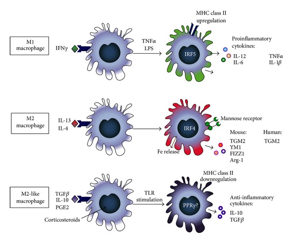
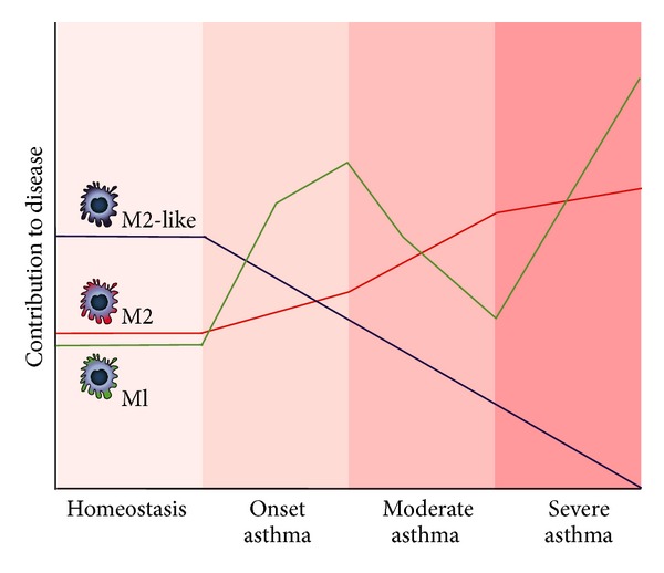
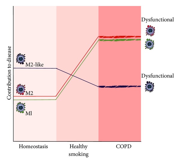
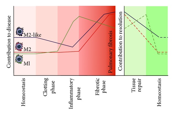
Verification
Claims the paper makes
The claims and statistics of this paper haven’t been checked yet.
Run the check — free, no account needed. It reads the open-access text and checks the sources it cites.
Claims other papers make about this one
Each is checked against this paper’s own text: does it say what the citing paper says it does?
In the citing paper
“However, observations obtained from macrophages involved in chronic inflammation, chronic infection, or cancer strongly suggest that the myeloid compartment has a much broader transcriptional repertoire depending on the different environmental signals received”
From Transcriptome-Based Network Analysis Reveals a Spectrum Model of Human Macrophage Activation (2014) · cites this paper as Boorsma et al., 2013
Self-check
Percentages, sums and reported differences re-computed from the paper’s own numbers.
The self-check needs the paper’s full text.
Citations
Cites
Projections of Global Mortality and Burden of Disease from 2002 to 2030
Mathers, C. D., & Loncar, D. (2006) · PLoS Medicine · doi:10.1371/journal.pmed.0030442 · Cited by 11,386
Macrophage plasticity and polarization: in vivo veritas
Sica, A., & Mantovani, A. (2012) · Journal of Clinical Investigation · doi:10.1172/jci59643 · Cited by 6,335
Alternative activation of macrophages
Gordon, S. (2003) · Nature Reviews Immunology · doi:10.1038/nri978 · Cited by 6,189
Monocyte and macrophage heterogeneity
Gordon, S., & Taylor, P. R. (2005) · Nature Reviews Immunology · doi:10.1038/nri1733 · Cited by 5,190
Rabe, K. F., Hurd, S., Anzueto, A., Barnes, P. J., Buist, S. A., Calverley, P., Fukuchi, Y., Jenkins, C., Rodriguez-Roisin, R., van Weel, C., et al. (2007) · American Journal of Respiratory and Critical Care Medicine · doi:10.1164/rccm.200703-456so · Cited by 4,925
Transcriptional regulation of macrophage polarization: enabling diversity with identity
Lawrence, T., & Natoli, G. (2011) · Nature Reviews Immunology · doi:10.1038/nri3088 · Cited by 2,284
Selective depletion of macrophages reveals distinct, opposing roles during liver injury and repair
Duffield, J. S., Forbes, S. J., Constandinou, C. M., Clay, S., Partolina, M., Vuthoori, S., Wu, S., Lang, R., & Iredale, J. P. (2005) · Journal of Clinical Investigation · doi:10.1172/jci200522675 · Cited by 1,649
Bleomycin and IL-1β–mediated pulmonary fibrosis is IL-17A dependent
Wilson, M. S., Madala, S. K., Ramalingam, T. R., Gochuico, B. R., Rosas, I. O., Cheever, A. W., & Wynn, T. A. (2010) · Journal of Experimental Medicine · doi:10.1084/jem.20092121 · Cited by 730
Holt, P. G., Oliver, J., Bilyk, N., McMenamin, C., McMenamin, P. G., Kraal, G., & Thepen, T. (1993) · The Journal of experimental medicine · doi:10.1084/jem.177.2.397 · Cited by 531
Kim, E. Y., Battaile, J. T., Patel, A. C., You, Y., Agapov, E., Grayson, M. H., Benoit, L. A., Byers, D. E., Alevy, Y., Tucker, J., et al. (2008) · Nature Medicine · doi:10.1038/nm1770 · Cited by 523
Research works printed but not linked: 213
- 10.1016/j.molmed.2011.07.009 · DOI
- 10.1002/eji.200737638 · DOI
- 10.2741/2692 · DOI · Paper page
- 10.1038/nri2448 · DOI · Paper page
- 10.1038/nri2784 · DOI
- 10.1007/s00441-010-1032-2 · DOI
- 10.1172/JCI39717 · DOI · Paper page
- 1994, Journal of Leukocyte Biology, 55, 141, 10.1002/jlb.55.2.141 · DOI
- 10.1038/ni.1990 · DOI
- 10.1038/ni.1937 · DOI · Paper page
- 1985, Infection and Immunity, 49, 61, 10.1128/IAI.49.1.61-66.1985 · DOI
- 1992, Clinical and Experimental Immunology, 88, 492
- 10.1006/cimm.2000.1687 · DOI · Paper page
- 10.1074/jbc.M111.290676 · DOI
- 10.1084/jem.176.1.287 · DOI · Paper page
- 10.1038/ni.1920 · DOI · Paper page
- 10.1146/annurev.immunol.021908.132532 · DOI · Paper page
- 10.1182/blood-2012-06-436212 · DOI
- 10.1016/j.coi.2007.07.002 · DOI
- 10.1016/S0165-2478(02)00225-0 · DOI
- 10.1016/S1471-4906(02)02302-5 · DOI · Paper page
- 10.1074/jbc.M110.208033 · DOI
- 10.1016/j.jim.2009.02.007 · DOI
- 10.1002/hep.22950 · DOI · Paper page
- 1999, American Journal of Pathology, 155, 1749, 10.1016/S0002-9440(10)65490-9 · DOI
- 1997, American Journal of Pathology, 150, 59
- 10.1111/j.1365-2222.2008.02971.x · DOI
- 10.1164/rccm.200409-1181PP · DOI
- 10.1016/S0140-6736(08)61452-X · DOI
- 10.1111/j.1398-9995.2010.02528.x · DOI
- 10.1165/rcmb.f279 · DOI
- 10.1155/2013/632049 · DOI
- 2007, The Journal of Immunology, 178, 5375, 10.4049/jimmunol.178.8.5375 · DOI
- 10.1378/chest.07-1881 · DOI
- 10.1056/NEJMoa050580 · DOI
- 10.1183/09031936.98.12030546 · DOI
- 10.1016/S0140-6736(05)17704-6 · DOI
- 10.1073/pnas.0706378104 · DOI
- 10.4049/jimmunol.0803468 · DOI
- 10.1136/thorax.57.9.774 · DOI
- 1995, The Journal of Immunology, 154, 5411, 10.4049/jimmunol.154.10.5411 · DOI
- 1996, American Journal of Respiratory and Critical Care Medicine, 154, 1641, 10.1164/ajrccm.154.6.8970348 · DOI
- 10.1164/rccm.200505-758OC · DOI
- 1989, Journal of Applied Physiology, 66, 1059, 10.1152/jappl.1989.66.3.1059 · DOI
- 1989, American Review of Respiratory Disease, 140, 981, 10.1164/ajrccm/140.4.981 · DOI
- 10.1016/j.gene.2012.05.021 · DOI
- 10.1136/thx.2005.043182 · DOI
- 10.1016/j.jaci.2008.07.007 · DOI
- 10.1164/rccm.200507-1175OC · DOI
- 2001, The Journal of Immunology, 166, 1471, 10.4049/jimmunol.166.3.1471 · DOI
- 2006, The Journal of Immunology, 177, 6460, 10.4049/jimmunol.177.9.6460 · DOI
- 10.1056/NEJMoa073600 · DOI
- 10.1126/science.1095336 · DOI
- 10.1016/j.jaci.2010.10.045 · DOI
- 10.4049/jimmunol.0900695 · DOI
- 10.1165/rcmb.2009-0016OC · DOI
- 10.1186/1471-2172-13-6 · DOI
- 10.1016/j.jaci.2010.06.010 · DOI
- 10.1111/j.1476-5381.2010.01033.x · DOI
- 2004, American Journal of Pathology, 165, 2045, 10.1016/S0002-9440(10)63255-5 · DOI
- 10.1164/rccm.200710-1588OC · DOI
- 10.1016/j.jaci.2012.03.011 · DOI
- 10.1016/j.jaci.2007.10.011 · DOI
- 10.1016/j.jaci.2010.01.048 · DOI
- 10.2174/156652408785160907 · DOI
- 10.1186/1465-9921-5-21 · DOI
- 10.1056/NEJMoa1106955 · DOI
- 10.1146/annurev.pathol.4.110807.092145 · DOI
- 10.1016/S0165-6147(00)01600-X · DOI
- 10.1097/00000478-200205000-00011 · DOI
- 10.1164/rccm.200608-1113OC · DOI
- 10.1016/S0140-6736(11)60968-9 · DOI
- 10.1016/S0140-6736(11)60988-4 · DOI
- 10.1183/09031936.95.08081398 · DOI
- 1997, American Journal of Respiratory and Critical Care Medicine, 156, 1633, 10.1164/ajrccm.156.5.9701081 · DOI
- 10.1136/thx.2004.024463 · DOI · Paper page
- 1995, American Journal of Respiratory and Critical Care Medicine, 152, 1666, 10.1164/ajrccm.152.5.7582312 · DOI
- 1999, American Journal of Physiology, 277, L97
- 10.1126/science.277.5334.2002 · DOI
- 10.1038/bjp.2008.180 · DOI
- 10.4049/jimmunol.0900473 · DOI · Paper page
- 10.1165/rcmb.2009-0459OC · DOI
- 1968, Chemical Reviews, 68, 153, 10.1021/cr60252a002 · DOI
- 2000, American Journal of Respiratory and Critical Care Medicine, 162, 701, 10.1164/ajrccm.162.2.9908132 · DOI
- 1998, Modern Pathology, 11, 648
- 2003, European Respiratory Journal, 21, 971, 10.1183/09031936.03.00098203 · DOI
- 10.1378/chest.08-1419 · DOI
- 2000, American Journal of Respiratory and Critical Care Medicine, 162, 369, 10.1164/ajrccm.162.2.9909025 · DOI
- 2000, American Journal of Respiratory and Critical Care Medicine, 162, 1175, 10.1164/ajrccm.162.3.2001063 · DOI
- 10.1164/rccm.2110101 · DOI
- 2008, The Journal of Immunology, 180, 1169, 10.4049/jimmunol.180.2.1169 · DOI
- 10.1165/rcmb.2007-0130OC · DOI
- 10.1016/j.bbadis.2006.04.004 · DOI
- 10.1124/mol.105.012591 · DOI
- 10.1152/ajplung.00241.2005 · DOI
- 10.1186/1465-9921-7-66 · DOI
- 10.1007/s10875-009-9286-8 · DOI
- 10.1136/thx.2006.059808 · DOI
- 10.1136/thorax.55.2.114 · DOI
- 10.1016/j.rmed.2003.07.008 · DOI
- 1997, Chest, 112, 505, 10.1378/chest.112.2.505 · DOI
- 10.1034/j.1399-3003.2000.15b09.x · DOI
- 10.1183/09031936.01.99114901 · DOI
- 1994, American Journal of Respiratory and Critical Care Medicine, 150, 1453, 10.1164/ajrccm.150.5.7952575 · DOI
- 1996, American Journal of Respiratory and Critical Care Medicine, 153, 530, 10.1164/ajrccm.153.2.8564092 · DOI
- 10.1016/S0954-6111(96)90202-4 · DOI
- 2005, Brazilian Journal of Medical and Biological Research, 38, 1359, 10.1590/S0100-879X2005000900010 · DOI
- 10.1165/rcmb.2003-0062OC · DOI
- 10.4049/jimmunol.0803154 · DOI
- 10.1165/rcmb.2004-0309OC · DOI
- 10.4049/jimmunol.0900614 · DOI
- 10.1084/jem.192.11.1587 · DOI
- 10.1164/rccm.200404-511OC · DOI
- 2001, American Journal of Physiology, 280, L39
- 10.1016/j.ajpath.2011.12.020 · DOI
- 10.1016/j.pupt.2009.10.007 · DOI
- 1999, American Journal of Respiratory and Critical Care Medicine, 160, 711, 10.1164/ajrccm.160.2.9812020 · DOI
- 1997, American Journal of Respiratory and Critical Care Medicine, 155, 852, 10.1164/ajrccm.155.3.9117016 · DOI
- 2002, American Journal of Respiratory Cell and Molecular Biology, 26, 602, 10.1165/ajrcmb.26.5.4685 · DOI
- 10.1152/ajplung.00481.2007 · DOI
- 10.1056/NEJMoa0909883 · DOI
- 10.1183/09031936.00036709 · DOI
- 2001, American Journal of Respiratory and Critical Care Medicine, 163, 1578, 10.1164/ajrccm.163.7.2002015 · DOI
- 10.1086/508428 · DOI
- 10.1164/rccm.200711-1666OC · DOI
- 2000, Journal of Clinical Investigation, 106, 1081, 10.1172/JCI10458 · DOI
- 2002, Journal of Leukocyte Biology, 71, 597, 10.1189/jlb.71.4.597 · DOI
- 10.1182/blood-2005-07-2843 · DOI
- 10.2353/ajpath.2010.090455 · DOI
- 10.1165/2009-0122RC · DOI
- 10.1165/rcmb.2010-0081OC · DOI
- 10.1371/journal.pone.0024177 · DOI
- 2008, The Journal of Immunology, 181, 5167, 10.4049/jimmunol.181.7.5167 · DOI
- 10.1002/eji.200535496 · DOI
- 10.1164/rccm.200505-686OC · DOI
- 10.1164/rccm.200212-1396OC · DOI
- 10.1378/chest.125.2.466 · DOI
- 10.1080/01926230490431466 · DOI
- 10.1159/000088439 · DOI
- 10.1080/01902140500324828 · DOI
- 10.1165/rcmb.2006-0345OC · DOI
- 10.1186/1465-9921-8-81 · DOI
- 10.1182/blood-2007-12-129080 · DOI
- 10.1186/1465-9921-9-47 · DOI
- 1999, European Respiratory Journal, 14, 309
- 10.1111/j.1365-2613.2006.00515.x · DOI
- 10.1038/nrd2958 · DOI
- 10.1164/rccm.200602-163OC · DOI · Paper page
- 10.1016/j.rmed.2011.08.015 · DOI
- 10.1111/j.1365-2222.2007.02829.x · DOI
- 10.1152/ajplung.00099.2011 · DOI
- 1996, Journal of Clinical Investigation, 97, 232, 10.1172/JCI118396 · DOI
- 10.1016/0049-3848(95)00025-9 · DOI
- 2011, Critical Reviews in Immunology, 31, 379, 10.1615/CritRevImmunol.v31.i5.20 · DOI
- 10.1378/chest.128.5_suppl_1.526S · DOI
- 10.1513/pats.200601-003TK · DOI
- 10.1038/nm.2807 · DOI · Paper page
- 2003, Cochrane Database of Systematic Reviews
- 2001, Annals of Internal Medicine, 134, 136, 10.7326/0003-4819-134-2-200101160-00015 · DOI
- 10.1074/jbc.M610764200 · DOI
- 2002, Chest, 122
- 10.1378/chest.125.2.754 · DOI
- 10.1183/09031936.00027411 · DOI
- 2000, American Journal of Physiology, 279, L562
- 10.1111/j.1365-2249.2010.04182.x · DOI
- 10.1182/blood-2008-04-150789 · DOI
- 10.3389/fimmu.2011.00065 · DOI
- 10.1111/j.1365-2249.2011.04468.x · DOI
- 10.1371/journal.pone.0040789 · DOI
- 1993, The Journal of Immunology, 150, 4188, 10.4049/jimmunol.150.9.4188 · DOI
- 1998, Journal of Investigative Medicine, 46, 223
- 1990, American Review of Respiratory Disease, 141, 124, 10.1164/ajrccm/141.1.124 · DOI
- 1991, European Respiratory Journal, 4, 400, 10.1183/09031936.93.04040400 · DOI
- 1993, American Review of Respiratory Disease, 147, 1507
- 1993, Chest, 104, 47, 10.1378/chest.104.1.47 · DOI
- 10.1164/rccm.200812-1837OC · DOI
- 10.1183/09031936.02.00022302 · DOI
- 1999, American Journal of Respiratory Cell and Molecular Biology, 20, 903, 10.1165/ajrcmb.20.5.3260 · DOI
- 10.1053/rmed.2003.1493 · DOI
- 10.1385/IR:30:3:339 · DOI
- 1999, The Journal of Immunology, 162, 920, 10.4049/jimmunol.162.2.920 · DOI
- 10.1046/j.1365-2222.2002.01420.x · DOI
- 10.1038/ni1544 · DOI
- 10.2353/ajpath.2006.050975 · DOI
- 2007, The Journal of Immunology, 178, 511, 10.4049/jimmunol.178.1.511 · DOI
- 2004, The Journal of Immunology, 172, 4068, 10.4049/jimmunol.172.7.4068 · DOI
- 10.1016/j.cyto.2004.08.007 · DOI
- 1998, American Journal of Respiratory Cell and Molecular Biology, 18, 60, 10.1165/ajrcmb.18.1.2627 · DOI
- 10.1016/j.clim.2010.06.017 · DOI
- 1998, American Journal of Respiratory and Critical Care Medicine, 158, 1626, 10.1164/ajrccm.158.5.9804025 · DOI
- 1991, American journal of respiratory cell and molecular biology, 5, 531, 10.1165/ajrcmb/5.6.531 · DOI
- 10.1038/nm.2628 · DOI
- 2008, The Journal of Immunology, 180, 2650, 10.4049/jimmunol.180.4.2650 · DOI
- 10.2332/allergolint.O-06-449 · DOI
- 10.1164/rccm.200509-1518OC · DOI · Paper page
- 10.1016/j.rmed.2010.02.026 · DOI
- 10.1165/rcmb.2006-0121OC · DOI
- 10.1038/labinvest.2010.73 · DOI
- 10.1165/rcmb.2006-0471OC · DOI
- 10.4049/jimmunol.0903008 · DOI
200 of 213 shown
Cited by
Sorted by citations
Transcriptome-Based Network Analysis Reveals a Spectrum Model of Human Macrophage Activation
Xue, J., Schmidt, S. V., Sander, J., Draffehn, A., Krebs, W., Quester, I., De Nardo, D., Gohel, T. D., Emde, M., Schmidleithner, L., et al. (2014) · Immunity · doi:10.1016/j.immuni.2014.01.006 · Cited by 2,210
Leus, N. G. J., van der Wouden, P. E., van den Bosch, T., Hooghiemstra, W. T. R., Ourailidou, M. E., Kistemaker, L. E. M., Bischoff, R., Gosens, R., Haisma, H. J., & Dekker, F. J. (2016) · Biochemical Pharmacology · doi:10.1016/j.bcp.2016.03.010 · Cited by 131
Collagen morphology influences macrophage shape and marker expression in?vitro
Vasse, G. F., Kühn, P. T., Zhou, Q., Bhusari, S. A., Reker-Smit, C., Melgert, B. N., & van Rijn, P. (2018) · Journal of Immunology and Regenerative Medicine · doi:10.1016/j.regen.2018.01.002 · Cited by 25
Self-citation
Metadata
- Authors
- Boorsma, C. E., Draijer, C., & Melgert, B. N.
- Affiliations
- Department of Pharmacokinetics, Toxicology and Targeting, Groningen Research Institute for Pharmacy, University of Groningen, Antonius Deusinglaan 1, 9713 AV Groningen, The Netherlands
- Venue
- Mediators of Inflammation
- Publisher
- Hindawi Limited
- Published
- 2013
- Volume
- 2013
- Pages
- 1-19
- ISSN
- 0962-9351
- 1466-1861
- PMC
- PMC3600198
- Cited by
- 126
- References
- 16
- Language
- en
About these checks
Registry, retraction, Cites and statistics results are computed, not judged, with one exception: a printed reference that no index lists is read once by a model before it counts as a citation error. Claims are extracted by a model and kept only when their quote appears verbatim in the text; each cited source is then checked, with a model’s help, for whether it says what the claim relies on. Those verdicts can be wrong — read the claim’s quote and the cited source before relying on one.
Statistics and arithmetic are recomputed, not judged.
Confidence bands are shown only for fields calibrated on our annotated test set. None is calibrated yet, so no band is shown.
What counts as a claim ›How we measure confidence ›How we check this ›
Are you an author, or do you see a problem with this page? Request a review
