

The DOI matches its Crossref record; 48 of the 48 references it cites were found in a registry or an index; 14 of the 50 sources its claims cite were checked for support.
miR-155 induction in microglial cells suppresses Japanese encephalitis virus replication and negatively modulates innate immune responses
Open accessAbstract
BACKGROUND: Microglial cells, which are resident macrophages of the central nervous system, play important roles in immune responses and pathogenesis. Japanese encephalitis virus (JEV) is a neurotropic virus that infects microglial cells in brain. Several microRNAs including miR-155 and miR-146a play an important role in defining the microglia inflammatory profile. In this study, we have investigated the effect of miR-155 and miR-146a modulation on JEV infection as well as innate immune responses in human microglial cells. METHODS: In vitro studies were performed in JEV-infected human microglial CHME3 cells. miR-155 or miR-146a were overexpressed and total RNA and protein were extracted following JEV-infection. Expression of genes involved in innate immune responses was studied by PCR array, quantitative real-time PCR (qPCR), western blot and Fluorescence activated cell sorter (FACS). JEV replication was monitored by studying the viral RNA by qPCR, protein by western blot, and titres by plaque assay. RESULTS: Overexpression of miR-155 in CHME3 cells resulted in significantly reduced JEV replication whereas miR-146a overexpression had an insignificant effect. Additionally, interferon regulatory factor 8 (IRF8) and complement factor H (CFH) were induced during JEV infection; however, this induction was attenuated in miR-155 overexpressing cells following JEV infection. Further, JEV-induced NF-κB regulated downstream gene expression was attenuated. Interestingly, an increased level of CD45, a negative regulator of microglia activation and a reduced phosphorylated-Signal Transducers and Activators of Transcription (p-STAT1) expression was observed in miR-155 overexpressing cells upon JEV infection. CONCLUSION: Induction of miR-155 in human microglial cells may negatively modulate JEV-induced innate immune gene expression and may have a beneficial role in limiting JEV replication in human microglial cells.
Overview
AI snapshot
Written from the paper’s full text.
TL;DR
In human microglial CHME3 cells, JEV infection induces miR-155, and miR-155 overexpression suppresses JEV replication while attenuating JEV-induced innate immune gene expression. miR-146a overexpression had little effect on JEV replication or the measured responses.
Key findings
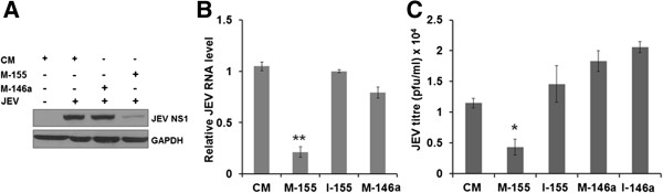
- miR-155 overexpression significantly reduced JEV NS1 protein, JEV RNA, and virus titre at 24 h post-infection, with approximately 1 log2 suppression of JEV titres; miR-146a overexpression had no significant effect.
- JEV infection induced IRF8 and complement factor H (CFH); miR-155 overexpression attenuated these inductions and also reduced JEV-induced IFN-β and interferon-stimulated gene (ISG) expression, effects reversed by a miR-155 inhibitor.
- In an NF-κB PCR array covering 84 genes, control mimic + JEV cells had 26 genes upregulated >2-fold, while miR-155 overexpressing + JEV cells had 32 upregulated; 19 were common, and 16 of these 19 showed attenuated expression in miR-155 cells.
- miR-155 overexpression increased CD45 surface expression, reduced p-STAT1, and reduced IL-1β, TNF-α, and IL-10 mRNA, while IL-4 was not affected.
Methods
- In vitro studies used human microglial CHME3 cells transfected with miRNA mimics or inhibitors (25 pmol), then infected with JEV strain P20778 at MOI = 5; PS cells were used for virus propagation and plaque assay.
- RNA and protein were extracted at different times post-infection and analysed by qPCR, TaqMan miRNA assays, RT2 Profiler NF-κB PCR array, western blot, and FACS.
- Experiments were performed in triplicate; statistical analysis used one-way ANOVA with Bonferroni’s multiple comparison tests and unpaired Student’s t-test, with P < 0.05 considered significant.
Limitations
- The paper states that further in vivo study is needed and that additional in vitro and in vivo studies are required to understand miR-155’s role in switching microglial activation toward a neuroprotective state.
- The paper reports that IRF8 3′-UTR luciferase target validation showed no change in miR-155 overexpressing cells (data not shown), and in silico analysis found no potential miR-155 target sites in JEV RNA.
- Evident from the text: all results come from a single in vitro human microglial cell line (CHME3) using overexpression (approximately 100-fold), and the authors note a discrepancy with a mouse BV-2 study where miR-155 rose approximately 4-fold and appeared pro-inflammatory, so cell type, expression level, and virus strain may limit generalizability.
Model-written summary. Check it against the paper before relying on it.
Integrity signals
Registry
Cites
Statistics
Figures
Verification
Claims the paper makes
59 Own result · 22 Own interpretation · 16 Not checked yet · 6 Partly supported · 6 Own method · 5 No full text of the sources · 3 Supported · 2 Sources not found · accuracy not yet measured
Every source the paper cites for this claim was checked and supports it.
In the paper
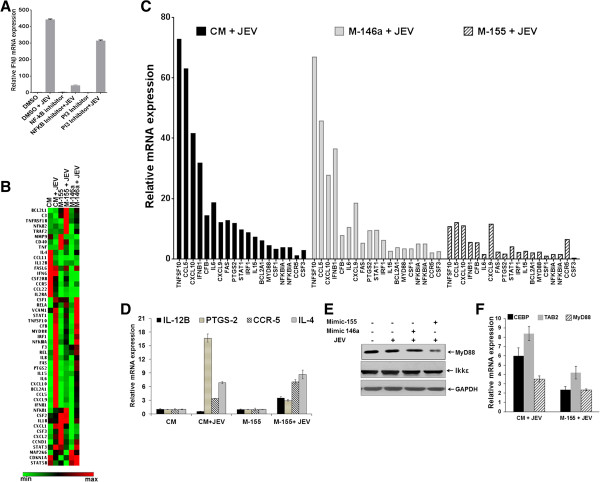
“CCR-5, which has an important role in JEV infection, [27] was up-regulated in these cells.”
§ miR-155 induction in microglial cells suppresses Japanese encephalitis virus replication and negatively modulates innate immune responses > Results > miR-155 attenuates JEV-induced NF-κB pathway gene expression in microglial cells
What the sources say
- Supportedcites [27] Larena M, Regner M, Lobigs M: The chemokine receptor CCR5, a therapeutic target for HIV/AIDS antagonists. Is critical for recovery in a mouse model of Japanese encephalitis. PLoS One. 2012, 7: e44834-10.1371/journal.pone.0044834. source
The citation is attached to the statement that CCR5 has an important role in JEV infection, not to the citing authors’ observation of up-regulation in their cells. The Results section (‘CCR5 is required for protection from Japanese encephalitis’) concludes that CCR5 is critically required for recovery from JEV infection in mice. (LLM, conf=0.92)
plaindirect- Asserted: passes — The authors put it forward as true.
- Checkable: passes — It can be checked against evidence.
- Load-bearing: unclear — It is unclear whether the paper’s conclusions depend on it.
- Based: passes — The paper gives a reason why it holds.
- Supported
A result the paper reports from its own data — it cites no other source for it, so there is none to check. The quote shows it is in the paper.
In the paper
“whereas PTGS2 (COX2), an inflammatory mediator, was significantly suppressed in miR-155 overexpressing cells.”
§ miR-155 induction in microglial cells suppresses Japanese encephalitis virus replication and negatively modulates innate immune responses > Results > miR-155 attenuates JEV-induced NF-κB pathway gene expression in microglial cells
plaindirect- Asserted: passes — The authors put it forward as true.
- Checkable: passes — It can be checked against evidence.
- Load-bearing: passes — The paper’s conclusions depend on it.
- Based: passes — The paper gives a reason why it holds.
A result the paper reports from its own data — it cites no other source for it, so there is none to check. The quote shows it is in the paper.
In the paper
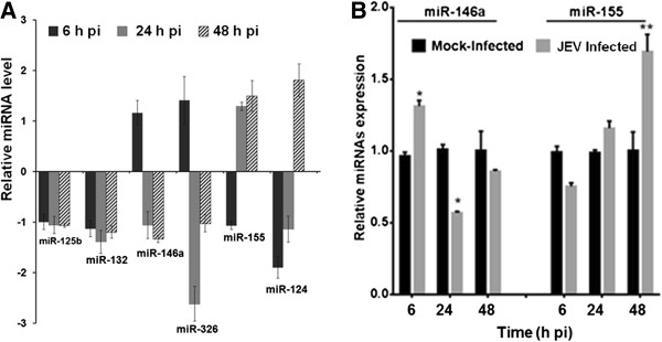
“We observed that miR-125b and miR-132 remained down-regulated throughout the course of infection.”
§ miR-155 induction in microglial cells suppresses Japanese encephalitis virus replication and negatively modulates innate immune responses > Results > JEV infection modulates expression of NeurimmiRs in microglial cells
plain- Asserted: passes — The authors put it forward as true.
- Checkable: passes — It can be checked against evidence.
- Load-bearing: unclear — It is unclear whether the paper’s conclusions depend on it.
- Based: passes — The paper gives a reason why it holds.
A result the paper reports from its own data — it cites no other source for it, so there is none to check. The quote shows it is in the paper.
In the paper
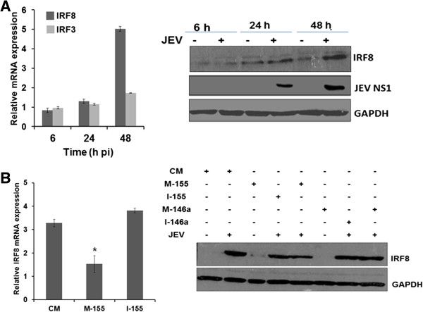
“However, there was relatively little increase in IRF3 expression during JEV infection (Figure 4A, left panel).”
§ miR-155 induction in microglial cells suppresses Japanese encephalitis virus replication and negatively modulates innate immune responses > Results > miR-155 attenuates JEV-induced IRF8 expression in microglial cells
plain- Asserted: passes — The authors put it forward as true.
- Checkable: passes — It can be checked against evidence.
- Load-bearing: unclear — It is unclear whether the paper’s conclusions depend on it.
- Based: passes — The paper gives a reason why it holds.
A result the paper reports from its own data — it cites no other source for it, so there is none to check. The quote shows it is in the paper.
In the paper
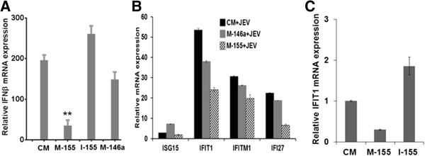
“while only a marginal effect was observed in miR-146a overexpressing cells (Figure 3A).”
§ miR-155 induction in microglial cells suppresses Japanese encephalitis virus replication and negatively modulates innate immune responses > Results > miR-155 modulates JEV-induced IFN-β expression in microglial cells
plain- Asserted: passes — The authors put it forward as true.
- Checkable: passes — It can be checked against evidence.
- Load-bearing: unclear — It is unclear whether the paper’s conclusions depend on it.
- Based: passes — The paper gives a reason why it holds.
A result the paper reports from its own data — it cites no other source for it, so there is none to check. The quote shows it is in the paper.
In the paper
“For miR-155 however, the expression levels showed a small decrease at 6 hours pi that was followed by a moderate (approximately 1.8-fold) increase at 48 hours pi (Figure 1B).”
§ miR-155 induction in microglial cells suppresses Japanese encephalitis virus replication and negatively modulates innate immune responses > Results > JEV infection modulates expression of NeurimmiRs in microglial cells
plaindirect- Asserted: passes — The authors put it forward as true.
- Checkable: passes — It can be checked against evidence.
- Load-bearing: passes — The paper’s conclusions depend on it.
- Based: passes — The paper gives a reason why it holds.
A result the paper reports from its own data — it cites no other source for it, so there is none to check. The quote shows it is in the paper.
In the paper
“Figure 3B shows that JEV-induced ISG expression was significantly repressed in miR-155 overexpressing cells”
§ miR-155 induction in microglial cells suppresses Japanese encephalitis virus replication and negatively modulates innate immune responses > Results > miR-155 modulates JEV-induced IFN-β expression in microglial cells
plaindirect- Asserted: passes — The authors put it forward as true.
- Checkable: passes — It can be checked against evidence.
- Load-bearing: passes — The paper’s conclusions depend on it.
- Based: passes — The paper gives a reason why it holds.
A result the paper reports from its own data — it cites no other source for it, so there is none to check. The quote shows it is in the paper.
In the paper
“Thus cells transfected with the miR-155 inhibitor prior to JEV infection showed an enhanced IFN-β mRNA expression when compared to control mimic-transfected cells infected with JEV (Figure 3A).”
§ miR-155 induction in microglial cells suppresses Japanese encephalitis virus replication and negatively modulates innate immune responses > Results > miR-155 modulates JEV-induced IFN-β expression in microglial cells
plaindirect- Asserted: passes — The authors put it forward as true.
- Checkable: passes — It can be checked against evidence.
- Load-bearing: passes — The paper’s conclusions depend on it.
- Based: passes — The paper gives a reason why it holds.
A result the paper reports from its own data — it cites no other source for it, so there is none to check. The quote shows it is in the paper.
In the paper
“At 24 hours pi, levels of the JEV NS1 protein and JEV RNA were significantly reduced in miR-155 overexpressing microglial cells”
§ miR-155 induction in microglial cells suppresses Japanese encephalitis virus replication and negatively modulates innate immune responses > Results > miR-155 suppresses JEV replication in microglial cells
plaindirect- Asserted: passes — The authors put it forward as true.
- Checkable: passes — It can be checked against evidence.
- Load-bearing: passes — The paper’s conclusions depend on it.
- Based: passes — The paper gives a reason why it holds.
A result the paper reports from its own data — it cites no other source for it, so there is none to check. The quote shows it is in the paper.
In the paper
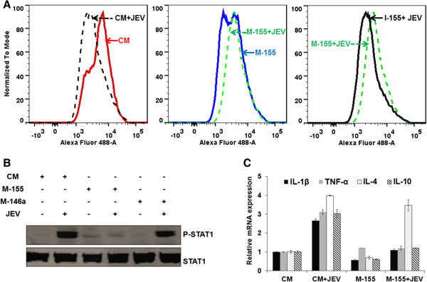
“We observed that JEV infection in miR-155 overexpressing human microglial cells up-regulated CD45 expression on cell surface (Figure 6A).”
§ miR-155 induction in microglial cells suppresses Japanese encephalitis virus replication and negatively modulates innate immune responses > Results > miR-155 enhances JEV-induced CD45 expression on microglial cells
plaindirect- Asserted: passes — The authors put it forward as true.
- Checkable: passes — It can be checked against evidence.
- Load-bearing: passes — The paper’s conclusions depend on it.
- Based: passes — The paper gives a reason why it holds.
- Probably not a substantial claim. 33
- It may state two separate claims as one. 33
- The quote only partly states this claim. 3
The cited sources support only part of this claim — read each source’s verdict below.
In the paper
“Thus, miR-155 and miR-146a were shown to increase in the microglial cells in response to stimulation with Lipopolysaccharides (LPS) and Polyinosinic-polycytidylic acid (poly(I:C), respectively, and they seem to play a fundamental role in the microglial inflammatory profile [13,14].”
§ miR-155 induction in microglial cells suppresses Japanese encephalitis virus replication and negatively modulates innate immune responses > Background
What the sources say
- Not checked — no full text of the sourcecites [13] Cardoso AL, Guedes JR, Pereira de Almeida L, Pedroso de Lima MC: miR-155 modulates microglia-mediated immune response by down-regulating SOCS-1 and promoting cytokine and nitric oxide production. Immunology. 2012, 135: 73-88. 10.1111/j.1365-2567.2011.03514.x. source
- Partly supportedcites [14] Saba R, Gushue S, Huzarewich RLCH, Manguiat K, Medina S, Robertson C, Booth SA: MicroRNA 146a (miR-146a) is overexpressed during prion disease and modulates the innate immune response and the microglial activation state. PLoS One. 2012, 7: e30832-10.1371/journal.pone.0030832. sourceExperimental — accuracy not yet measured
The Results section reports that miR-146a increases in microglial-derived cells after LPS stimulation, and that altering miR-146a expression changes immune-response genes. It does not support the attributed pairing of miR-146a with poly(I:C) stimulation. The paired citation may address the separate miR-155 claim, so that claim is not assessed against this paper. (LLM, conf=0.87)
hedgeddirect- Asserted: passes — The authors put it forward as true.
- Checkable: passes — It can be checked against evidence.
- Load-bearing: fails — The paper’s conclusions may not depend on it.
- Based: passes — The paper gives a reason why it holds.
Probably not a substantial claim. The paper’s conclusions may not depend on it. It may state two separate claims as one.
The cited sources support only part of this claim — read each source’s verdict below.
In the paper
“whereas overexpression of miR-155 significantly suppressed human immunodeficiency virus (HIV) infection in activated macrophages [21].”
§ miR-155 induction in microglial cells suppresses Japanese encephalitis virus replication and negatively modulates innate immune responses > Background
What the sources say
- Partly supportedcites [21] Swaminathan G, Rossi F, Sierra LJ, Gupta A, Navas-Martín S, Martín-García J: A role for microRNA-155 modulation in the anti-HIV-1 effects of Toll-like receptor 3 stimulation in macrophages. PLoS Pathog. 2012, 8: e1002937-10.1371/journal.ppat.1002937. sourceExperimental — accuracy not yet measured
The Results section ('Ectopic expression of miR-155 reduces HIV-1 infection in MDMs and cell lines'; page unavailable) reports markedly lower HIV-1 infection after miR-155 overexpression in monocyte-derived macrophages, and the Discussion calls the reduction significant. The overexpression experiment does not identify those macrophages as activated; the paper examines TLR-stimulated macrophages separately. (LLM, conf=0.85)
plaindirect- Asserted: passes — The authors put it forward as true.
- Checkable: passes — It can be checked against evidence.
- Load-bearing: fails — The paper’s conclusions may not depend on it.
- Based: passes — The paper gives a reason why it holds.
Probably not a substantial claim. The paper’s conclusions may not depend on it.
- Partly supported
The cited sources support only part of this claim — read each source’s verdict below.
In the paper
“The M1 phenotype is a pro-inflammatory state and induces neuropathology, whereas M2 is anti-inflammatory state that could have a neuroprotective role [36].”
§ miR-155 induction in microglial cells suppresses Japanese encephalitis virus replication and negatively modulates innate immune responses > Discussion
What the sources say
- Partly supportedcites [36] Salemi J, Obregon DF, Cobb A, Reed S, Sadic E, Jin J, Fernandez F, Tan J, Giunta B: Flipping the switches: CD40 and CD45 modulation of microglial activation states in HIV associated dementia (HAD). Mol Neurodegener. 2011, 6: 3-10.1186/1750-1326-6-3. sourceExperimental — accuracy not yet measured
In “The Role of Microglia in HIV Associated Dementia (HAD),” the paper describes M1 as pro-inflammatory and M2 as dampening inflammation and clearing cellular debris, consistent with a possible neuroprotective role. It says increased M1 polarization *may play a role* in disease pathophysiology, rather than establishing that M1 induces neuropathology. (LLM, conf=0.85)
hedgeddirect- Asserted: passes — The authors put it forward as true.
- Checkable: passes — It can be checked against evidence.
- Load-bearing: passes — The paper’s conclusions depend on it.
- Based: passes — The paper gives a reason why it holds.
Probably not a substantial claim. It may state two separate claims as one.
- Partly supported
Every source the paper cites for this claim was checked and supports it.
In the paper
“Swaminathan et al.[21] have shown that miR-155 exerts an anti-HIV-1 effect by targeting several HIV-1 dependency factors involved in post-entry, pre-integration events, leading to severely diminished HIV-1 infection.”
§ miR-155 induction in microglial cells suppresses Japanese encephalitis virus replication and negatively modulates innate immune responses > Discussion
What the sources say
- Supportedcites [21] Swaminathan G, Rossi F, Sierra LJ, Gupta A, Navas-Martín S, Martín-García J: A role for microRNA-155 modulation in the anti-HIV-1 effects of Toll-like receptor 3 stimulation in macrophages. PLoS Pathog. 2012, 8: e1002937-10.1371/journal.ppat.1002937. source
The Results section reports that miR-155 over-expression reduced HIV-1 infection in primary macrophages, with 60–96% inhibition in 10 of 12 donors. The Discussion identifies several HIV-1 dependency factors whose levels fell with increased miR-155 and links them to trafficking or nuclear import of pre-integration complexes. It presents the multiple-target mechanism as a proposed explanation rather than definitive proof. (LLM, conf=0.93)
boosteddirect- Asserted: passes — The authors put it forward as true.
- Checkable: passes — It can be checked against evidence.
- Load-bearing: fails — The paper’s conclusions may not depend on it.
- Based: passes — The paper gives a reason why it holds.
Probably not a substantial claim. The paper’s conclusions may not depend on it.
- Supported
The cited sources support only part of this claim — read each source’s verdict below.
In the paper
“Specific induction of active SHP2 phosphatase dephosphorylates IRF8, which in turn becomes an active repressor and down-regulates TLR-mediated gene expression [35].”
§ miR-155 induction in microglial cells suppresses Japanese encephalitis virus replication and negatively modulates innate immune responses > Discussion
What the sources say
- Partly supportedcites [35] Fragale A, Stellacci E, Ilari R, Remoli AL, Lanciotti A, Perrotti E, Shytaj I, Orsatti R, Lawrence HR, Lawrence NJ, Wu J, Rehli M, Ozato K, Battistini A: Critical role of IRF-8 in negative regulation of TLR3 expression by Src homology 2 domain-containing protein tyrosine phosphatase-2 activity in human myeloid dendritic cells. J Immunol. 2011, 186: 1951-1962. 10.4049/jimmunol.1000918. sourceExperimental — accuracy not yet measured
The Discussion describes active SHP2 acting on IRF-8, after which dephosphorylated IRF-8 becomes a repressor that downregulates TLR3 gene expression. The claim broadens this specific TLR3 finding to TLR-mediated gene expression generally; the cited passage does not establish that broader scope. No page number is provided in our copy. (LLM, conf=0.86)
plaindirect- Asserted: passes — The authors put it forward as true.
- Checkable: passes — It can be checked against evidence.
- Load-bearing: fails — The paper’s conclusions may not depend on it.
- Based: passes — The paper gives a reason why it holds.
Probably not a substantial claim. The paper’s conclusions may not depend on it. It may state two separate claims as one.
- Partly supported
The cited sources support only part of this claim — read each source’s verdict below.
In the paper
“The miR-155 is a multifunctional microRNA and it can modulate inflammatory responses in both a positive and negative way [40-48].”
§ miR-155 induction in microglial cells suppresses Japanese encephalitis virus replication and negatively modulates innate immune responses > Discussion
What the sources say
- Partly supportedcites [40] Thounaojam MC, Kundu K, Kaushik DK, Swaroop S, Mahadevan A, Shankar SK, Basu A: MicroRNA-155 regulates Japanese encephalitis virus induced inflammatory response by targeting src homology 2-containing inositol phosphatase-1 (SHIP1). J Virol. 2014, 88: 4798-4810. 10.1128/JVI.02979-13. sourceExperimental — accuracy not yet measured
The IMPORTANCE section describes miR-155 as a widely studied regulator of inflammation, and the DISCUSSION reports that it promotes inflammation during JEV infection. The paper does not establish the claim that miR-155 can also reduce inflammatory responses; that direction appears only indirectly in a cited reference title. (LLM, conf=0.84)
- Partly supportedcites [41] Ceppi M, Pereira PM, Dunand-Sauthier I, Barras E, Reith W, Santos MA, Pierre P: MicroRNA-155 modulates the interleukin-1 signaling pathway in activated human monocyte-derived dendritic cells. Proc Natl Acad Sci USA. 2009, 106: 2735-2740. 10.1073/pnas.0811073106. sourceExperimental — accuracy not yet measured
The paper’s opening section (p. 1) and Results (p. 3) support a negative regulatory role: miR-155 down-modulates inflammatory cytokine production in activated human dendritic cells. The cited passages do not establish that miR-155 also positively modulates inflammatory responses, so they support only one direction of the claim. (LLM, conf=0.86)
- Not checked — no full text of the sourcecites [42] Xiao B, Liu Z, Li BS, Tang B, Li W, Guo G, Shi Y, Wang F, Wu Y, Tong WD, Guo H, Mao XH, Zou QM: Induction of microRNA-155 during Helicobacter pylori infection and its negative regulatory role in the inflammatory response. J Infect Dis. 2009, 200: 916-925. 10.1086/605443. source
- Partly supportedcites [43] Xu C, Ren G, Cao G, Chen Q, Shou P, Zheng C, Du L, Han X, Jiang M, Yang Q, Lin L, Wang G, Yu P, Zhang X, Cao W, Brewer G, Wang Y, Shi Y: miR-155 regulates immune modulatory properties of mesenchymal stem cells by targeting TAK1-binding protein 2. J Biol Chem. 2013, 288: 11074-11079. 10.1074/jbc.M112.414862. sourceExperimental — accuracy not yet measured
The Abstract describes miR-155 as having roles in cancer, immune responses and hematopoiesis. The Discussion reports that it reduces the immunosuppressive capacity of cytokine-activated MSCs, but our copy does not establish that miR-155 modulates inflammatory responses in both positive and negative directions. (LLM, conf=0.84)
- Supportedcites [44] Sullivan RP, Fogel LA, Leong JW, Schneider SE, Wong R, Romee R, Thai TH, Sexl V, Matkovich SJ, Dorn GW, French AR, Fehniger TA: MicroRNA-155 tunes both the threshold and extent of NK cell activation via targeting of multiple signaling pathways. J Immunol. 2013, 12: 5904-5913. source
The Introduction reports that both loss and overexpression of miR-155 augment NK-cell IFN-γ responses, through different mechanisms. The Discussion describes this as a dual role in regulating NK-cell activation and cytokine production, supporting the claim that miR-155 can modulate an inflammatory response in opposing ways. The paper's direct evidence is specific to NK cells. (LLM, conf=0.84)
- Not checked — no full text of the sourcecites [45] Zhou H, Huang X, Cui H, Luo X, Tang Y, Chen S, Wu L, Shen N: miR-155 and its star-form partner miR-155* cooperatively regulate type I interferon production by human plasmacytoid dendritic cells. Blood. 2010, 116: 5885-5894. 10.1182/blood-2010-04-280156. source
- Partly supportedcites [46] Lu F, Weidmer A, Liu CG, Volinia S, Croce CM, Lieberman PM: Epstein-Barr virus-induced miR-155 attenuates NF-kappaB signaling and stabilizes latent virus persistence. J Virol. 2008, 82: 10436-10443. 10.1128/JVI.00752-08. sourceExperimental — accuracy not yet measured
The Results show that miR-155 reduces NF-κB activity in B cells and modulates an IFN-responsive gene, supporting a negative effect on inflammatory signaling. The Discussion suggests it may limit the innate immune response to viral infection. Our copy does not show miR-155 positively modulating an inflammatory response, so the claim's bidirectional part is unsupported by this source. (LLM, conf=0.87)
- Not checked — no full text of the sourcecites [47] Tang B, Xiao B, Liu Z, Li N, Zhu ED, Li BS, Xie QH, Zhuang Y, Zou QM, Mao XH: Identification of MyD88 as a novel target of miR-155, involved in negative regulation of Helicobacter pylori-induced inflammation. FEBS Lett. 2010, 584: 1481-1486. 10.1016/j.febslet.2010.02.063. source
- Supportedcites [48] Ma X, Becker Buscaglia LE, Barker JR, Li Y: MicroRNAs in NF-kappaB signaling. J Mol Cell Biol. 2011, 3: 159-166. 10.1093/jmcb/mjr007. source
The Introduction describes miR-155 as contributing to increased TNF-α production in macrophages, supporting a positive effect on an inflammatory response. It also describes miR-155 as part of a negative feedback loop that limits NF-κB activation. Together, these passages support the claim that miR-155 can modulate inflammatory responses in both directions. (LLM, conf=0.88)
plaindirect- Asserted: passes — The authors put it forward as true.
- Checkable: passes — It can be checked against evidence.
- Load-bearing: fails — The paper’s conclusions may not depend on it.
- Based: passes — The paper gives a reason why it holds.
Probably not a substantial claim. The paper’s conclusions may not depend on it. It may state two separate claims as one.
- Partly supported
The cited sources support only part of this claim — read each source’s verdict below.
In the paper
“It is reported that CD45 can negatively regulate CD40L-CD40-induced microglial M1 activation; an effect leading to the promotion of the M2 phenotype.”
§ miR-155 induction in microglial cells suppresses Japanese encephalitis virus replication and negatively modulates innate immune responses > Discussion
What the sources say
- Partly supportedcites [36] Salemi J, Obregon DF, Cobb A, Reed S, Sadic E, Jin J, Fernandez F, Tan J, Giunta B: Flipping the switches: CD40 and CD45 modulation of microglial activation states in HIV associated dementia (HAD). Mol Neurodegener. 2011, 6: 3-10.1186/1750-1326-6-3. sourceExperimental — accuracy not yet measured
The section on CD40 and CD45 modulation says CD45 antagonizes CD40L/CD40-mediated microglial activation and reduces TNF-α production. It does not establish that this promotes an M2 phenotype; its summary instead calls the CD40-promoted phenotype “M2 type.” No page number is available in our copy. (LLM, conf=0.83)
plaindirect- Asserted: passes — The authors put it forward as true.
- Checkable: passes — It can be checked against evidence.
- Load-bearing: passes — The paper’s conclusions depend on it.
- Based: passes — The paper gives a reason why it holds.
Probably not a substantial claim. It may state two separate claims as one.
- Partly supported
Every source the paper cites for this claim was checked and supports it.
In the paper
“inducible miR-155 in response to the vesicular stomatitis virus (an RNA virus) infection in macrophage was shown to induce type I IFN signaling and inhibit viral replication [16].”
§ miR-155 induction in microglial cells suppresses Japanese encephalitis virus replication and negatively modulates innate immune responses > Discussion
What the sources say
- Supportedcites [16] Wang P, Hou J, Lin L, Wang C, Liu X, Li D, Ma F, Wang Z, Cao X: Inducible microRNA-155 feedback promotes type I IFN signaling in antiviral innate immunity by targeting suppressor of cytokine signaling 1. J Immunol. 2010, 185: 6226-6233. 10.4049/jimmunol.1000491. source
The paper reports that VSV infection induces miR-155 in macrophages (The Journal of Immunology, pp. 6226–6233). In “miR-155 feedback attenuates the viral replication in macrophages,” inhibition of miR-155 increased VSV replication, while overexpression suppressed it; the section on viral propagation reports that miR-155 promotes type I IFN signaling. (LLM, conf=0.94)
plaindirect- Asserted: passes — The authors put it forward as true.
- Checkable: passes — It can be checked against evidence.
- Load-bearing: fails — The paper’s conclusions may not depend on it.
- Based: passes — The paper gives a reason why it holds.
Probably not a substantial claim. The paper’s conclusions may not depend on it. It may state two separate claims as one.
- Supported
A result the paper reports from its own data — it cites no other source for it, so there is none to check. The quote shows it is in the paper.
In the paper
“PCR array data showed that JEV-induced MyD88 mRNA expression was suppressed in miR-155 overexpressing cells.”
§ miR-155 induction in microglial cells suppresses Japanese encephalitis virus replication and negatively modulates innate immune responses > Results > miR-155 attenuates JEV-induced NF-κB pathway gene expression in microglial cells
plaindirect- Asserted: passes — The authors put it forward as true.
- Checkable: passes — It can be checked against evidence.
- Load-bearing: passes — The paper’s conclusions depend on it.
- Based: passes — The paper gives a reason why it holds.
The quote only partly states this claim.
A result the paper reports from its own data — it cites no other source for it, so there is none to check. The quote shows it is in the paper.
In the paper
“Our results suggested that IRF8 was induced during JEV infection and significantly up-regulated mRNA levels were observed at 48 hours pi.”
§ miR-155 induction in microglial cells suppresses Japanese encephalitis virus replication and negatively modulates innate immune responses > Results > miR-155 attenuates JEV-induced IRF8 expression in microglial cells
hedgeddirect- Asserted: passes — The authors put it forward as true.
- Checkable: passes — It can be checked against evidence.
- Load-bearing: passes — The paper’s conclusions depend on it.
- Based: passes — The paper gives a reason why it holds.
It may state two separate claims as one.
A result the paper reports from its own data — it cites no other source for it, so there is none to check. The quote shows it is in the paper.
In the paper
“While miR-146a and miR-326 were up-regulated at an early time point (6 hours pi), these were down-regulated as the infection progressed.”
§ miR-155 induction in microglial cells suppresses Japanese encephalitis virus replication and negatively modulates innate immune responses > Results > JEV infection modulates expression of NeurimmiRs in microglial cells
plaindirect- Asserted: passes — The authors put it forward as true.
- Checkable: passes — It can be checked against evidence.
- Load-bearing: unclear — It is unclear whether the paper’s conclusions depend on it.
- Based: passes — The paper gives a reason why it holds.
It may state two separate claims as one.
A result the paper reports from its own data — it cites no other source for it, so there is none to check. The quote shows it is in the paper.
In the paper
“Interestingly, JEV-induced IRF8 expression was suppressed in miR-155 overexpressing cells”
§ miR-155 induction in microglial cells suppresses Japanese encephalitis virus replication and negatively modulates innate immune responses > Results > miR-155 attenuates JEV-induced IRF8 expression in microglial cells
plaindirect- Asserted: passes — The authors put it forward as true.
- Checkable: passes — It can be checked against evidence.
- Load-bearing: passes — The paper’s conclusions depend on it.
- Based: passes — The paper gives a reason why it holds.
The quote only partly states this claim.
A result the paper reports from its own data — it cites no other source for it, so there is none to check. The quote shows it is in the paper.
In the paper
“On the other hand, miR-124 and miR-155 were suppressed at early time points, but were up-regulated at later time points.”
§ miR-155 induction in microglial cells suppresses Japanese encephalitis virus replication and negatively modulates innate immune responses > Results > JEV infection modulates expression of NeurimmiRs in microglial cells
plaindirect- Asserted: passes — The authors put it forward as true.
- Checkable: passes — It can be checked against evidence.
- Load-bearing: passes — The paper’s conclusions depend on it.
- Based: passes — The paper gives a reason why it holds.
It may state two separate claims as one.
A result the paper reports from its own data — it cites no other source for it, so there is none to check. The quote shows it is in the paper.
In the paper
“The reduced MyD88 protein level was confirmed by western blot.”
§ miR-155 induction in microglial cells suppresses Japanese encephalitis virus replication and negatively modulates innate immune responses > Results > miR-155 attenuates JEV-induced NF-κB pathway gene expression in microglial cells
plaindirect- Asserted: passes — The authors put it forward as true.
- Checkable: passes — It can be checked against evidence.
- Load-bearing: passes — The paper’s conclusions depend on it.
- Based: passes — The paper gives a reason why it holds.
The quote only partly states this claim.
A result the paper reports from its own data — it cites no other source for it, so there is none to check. The quote shows it is in the paper.
In the paper
“No change was seen in miR-212 levels during JEV infection.”
§ miR-155 induction in microglial cells suppresses Japanese encephalitis virus replication and negatively modulates innate immune responses > Results > JEV infection modulates expression of NeurimmiRs in microglial cells
plain- Asserted: passes — The authors put it forward as true.
- Checkable: passes — It can be checked against evidence.
- Load-bearing: fails — The paper’s conclusions may not depend on it.
- Based: passes — The paper gives a reason why it holds.
Probably not a substantial claim. The paper’s conclusions may not depend on it.
A result the paper reports from its own data — it cites no other source for it, so there is none to check. The quote shows it is in the paper.
In the paper
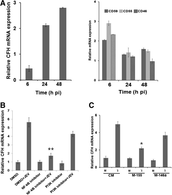
“Further, overexpression of miR-155, but not miR-146a, attenuated the CFH expression in microglial cells during JEV infection (Figure 7C).”
§ miR-155 induction in microglial cells suppresses Japanese encephalitis virus replication and negatively modulates innate immune responses > Results > miR-155 suppresses JEV-induced complement factor H expression in microglial cells
plaindirect- Asserted: passes — The authors put it forward as true.
- Checkable: passes — It can be checked against evidence.
- Load-bearing: passes — The paper’s conclusions depend on it.
- Based: passes — The paper gives a reason why it holds.
It may state two separate claims as one.
A result the paper reports from its own data — it cites no other source for it, so there is none to check. The quote shows it is in the paper.
In the paper
“Interestingly, bioinformatics analysis predicted IRF8 as one of the potential targets for miR-155.”
§ miR-155 induction in microglial cells suppresses Japanese encephalitis virus replication and negatively modulates innate immune responses > Discussion
hedgedcorrelational- Asserted: passes — The authors put it forward as true.
- Checkable: passes — It can be checked against evidence.
- Load-bearing: fails — The paper’s conclusions may not depend on it.
- Based: passes — The paper gives a reason why it holds.
Probably not a substantial claim. The paper’s conclusions may not depend on it.
A result the paper reports from its own data — it cites no other source for it, so there is none to check. The quote shows it is in the paper.
In the paper
“Expression of other complement regulators (CD46, CD55, or CD59) showed a marginal increase during the early phase of JEV infection (6 hours pi) but at the later time points it was same as in uninfected control cells (Figure 7A, right panel).”
§ miR-155 induction in microglial cells suppresses Japanese encephalitis virus replication and negatively modulates innate immune responses > Results > miR-155 suppresses JEV-induced complement factor H expression in microglial cells
plain- Asserted: passes — The authors put it forward as true.
- Checkable: passes — It can be checked against evidence.
- Load-bearing: unclear — It is unclear whether the paper’s conclusions depend on it.
- Based: passes — The paper gives a reason why it holds.
It may state two separate claims as one.
A result the paper reports from its own data — it cites no other source for it, so there is none to check. The quote shows it is in the paper.
In the paper
“In this study, we observed an increased CD45 expression in JEV-infected miR-155 overexpressing cells and relatively reduced p-STAT1 expression.”
§ miR-155 induction in microglial cells suppresses Japanese encephalitis virus replication and negatively modulates innate immune responses > Discussion
plaincorrelational- Asserted: passes — The authors put it forward as true.
- Checkable: passes — It can be checked against evidence.
- Load-bearing: passes — The paper’s conclusions depend on it.
- Based: passes — The paper gives a reason why it holds.
It may state two separate claims as one.
A result the paper reports from its own data — it cites no other source for it, so there is none to check. The quote shows it is in the paper.
In the paper
“We have shown that JEV infection can induce CFH expression in human and mouse microglial cells and this induction is attenuated in miR-155 overexpressing cells.”
§ miR-155 induction in microglial cells suppresses Japanese encephalitis virus replication and negatively modulates innate immune responses > Discussion
boosteddirect- Asserted: passes — The authors put it forward as true.
- Checkable: passes — It can be checked against evidence.
- Load-bearing: passes — The paper’s conclusions depend on it.
- Based: passes — The paper gives a reason why it holds.
It may state two separate claims as one.
A claim is checked only against the cited paper’s full text, never its abstract, and none is available for its sources.
In the paper
“Among them, miR-155 and miR-146a are multifunctional and widely reported to modulate different stages of innate immune response during inflammation and infection [10-12].”
§ miR-155 induction in microglial cells suppresses Japanese encephalitis virus replication and negatively modulates innate immune responses > Background
What the sources say
- Not checked — no full text of the sourcecites [10] Lindsay MA: microRNAs and the immune response. Trends Immunol. 2011, 29: 343-351. source
- Not checked — no full text of the sourcecites [11] Elton TS, Selemon H, Elton SM, Parinandi NL: Regulation of the MIR155 host gene in physiological and pathological processes. Gene. 2013, 532: 1-12. 10.1016/j.gene.2012.12.009. source
- Not checked — no full text of the sourcecites [12] O’Connell RM, Rao DS, Chaudhuri AA, Baltimore D: Physiological and pathological roles for microRNAs in the immune system. Nat Rev Immunol. 2010, 10: 111-122. 10.1038/nri2708. source
boosteddirect- Asserted: passes — The authors put it forward as true.
- Checkable: passes — It can be checked against evidence.
- Load-bearing: fails — The paper’s conclusions may not depend on it.
- Based: passes — The paper gives a reason why it holds.
Probably not a substantial claim. The paper’s conclusions may not depend on it.
None of the sources cited for this claim have been checked yet.
In the paper
“CFH expression can be regulated by miR-155 or miR-146 as both of them have matched seed sequences on the 3’-untranslated region (UTR) of the CFH gene [17,18].”
§ miR-155 induction in microglial cells suppresses Japanese encephalitis virus replication and negatively modulates innate immune responses > Results > miR-155 suppresses JEV-induced complement factor H expression in microglial cells
What the sources say
- Not matchedcites [17] Lukiw WJ, Surjyadipta B, Dua P, Alexandrov PN: Common micro RNAs (miRNAs) target complement factor H (CFH) regulation in Alzheimer’s disease (AD) and in age-related macular degeneration (AMD). Int J Biochem Mol Biol. 2012, 3: 105-116.
Couldn’t be matched to a paper in Crossref, so it can’t be checked.
- Not checked yetcites [18] Lukiw WJ, Alexandrov PN: Regulation of complement factor H (CFH) by multiple miRNAs in Alzheimer’s disease (AD) brain. Mol Neurobiol. 2012, 46: 11-19. 10.1007/s12035-012-8234-4. source
Checked before — its earlier verdict is held back until a new check runs.
plaindirect- Asserted: passes — The authors put it forward as true.
- Checkable: passes — It can be checked against evidence.
- Load-bearing: unclear — It is unclear whether the paper’s conclusions depend on it.
- Based: passes — The paper gives a reason why it holds.
It may state two separate claims as one.
- Not matched
What the paper did — it cites no source for this, so there is none to check. The quote shows it is in the paper.
In the paper
“Expression levels of miRNAs were normalized to that of U6 snRNA.”
§ miR-155 induction in microglial cells suppresses Japanese encephalitis virus replication and negatively modulates innate immune responses > Materials and methods > Quantitation of miRNA expression
plain- Asserted: passes — The authors put it forward as true.
- Checkable: passes — It can be checked against evidence.
- Load-bearing: fails — The paper’s conclusions may not depend on it.
- Based: passes — The paper gives a reason why it holds.
Probably not a substantial claim. The paper’s conclusions may not depend on it.
What the paper did — it cites no source for this, so there is none to check. The quote shows it is in the paper.
In the paper
“All experiments were performed in triplicate.”
§ miR-155 induction in microglial cells suppresses Japanese encephalitis virus replication and negatively modulates innate immune responses > Materials and methods > Statistical analysis
plain- Asserted: passes — The authors put it forward as true.
- Checkable: passes — It can be checked against evidence.
- Load-bearing: fails — The paper’s conclusions may not depend on it.
- Based: passes — The paper gives a reason why it holds.
Probably not a substantial claim. The paper’s conclusions may not depend on it.
None of the sources cited for this claim have been checked yet.
In the paper
“Therefore, downregulation of cytokine production can serve to dampen the inflammatory response and can contribute to better virus clearance and increased protection against JEV [3].”
§ miR-155 induction in microglial cells suppresses Japanese encephalitis virus replication and negatively modulates innate immune responses > Background
What the sources say
- Not checked yetcites [3] Das S, Dutta K, Kumawat KL, Ghoshal A, Adhya D, Basu A: Abrogated inflammatory response promotes neurogenesis in a murine model of Japanese encephalitis. PLoS One. 2011, 6: e17225-10.1371/journal.pone.0017225. source
Checked before — its earlier verdict is held back until a new check runs.
plainconditional- Asserted: passes — The authors put it forward as true.
- Checkable: passes — It can be checked against evidence.
- Load-bearing: passes — The paper’s conclusions depend on it.
- Based: passes — The paper gives a reason why it holds.
It may state two separate claims as one.
- Rests on an assumption: JEV infection of microglia leads to neuronal cell death through uncontrolled production of pro-inflammatory cytokines.
- Not checked yet
What the paper did — it cites no source for this, so there is none to check. The quote shows it is in the paper.
In the paper
“The protein levels were normalized with the GAPDH levels.”
§ miR-155 induction in microglial cells suppresses Japanese encephalitis virus replication and negatively modulates innate immune responses > Materials and methods > Western blot analysis
plain- Asserted: passes — The authors put it forward as true.
- Checkable: passes — It can be checked against evidence.
- Load-bearing: fails — The paper’s conclusions may not depend on it.
- Based: passes — The paper gives a reason why it holds.
Probably not a substantial claim. The paper’s conclusions may not depend on it.
A conclusion the paper draws from its own results — it cites no other source for it, so there is none to check. The quote shows it is in the paper.
In the paper
“Together, these data show that overexpression of miR-155 during JEV infection enhances CD45 expression on the microglial cell surface and negatively modulates cytokine expression.”
§ miR-155 induction in microglial cells suppresses Japanese encephalitis virus replication and negatively modulates innate immune responses > Results > miR-155 enhances JEV-induced CD45 expression on microglial cells
plaindirect- Asserted: passes — The authors put it forward as true.
- Checkable: passes — It can be checked against evidence.
- Load-bearing: passes — The paper’s conclusions depend on it.
- Based: passes — The paper gives a reason why it holds.
It may state two separate claims as one.
A conclusion the paper draws from its own results — it cites no other source for it, so there is none to check. The quote shows it is in the paper.
In the paper
“Induction of miR-155 in human microglial cells may negatively modulate JEV-induced innate immune gene expression and may have a beneficial role in limiting JEV replication in human microglial cells.”
§ miR-155 induction in microglial cells suppresses Japanese encephalitis virus replication and negatively modulates innate immune responses > Abstract > Conclusion
hedgedconditional- Asserted: passes — The authors put it forward as true.
- Checkable: passes — It can be checked against evidence.
- Load-bearing: passes — The paper’s conclusions depend on it.
- Based: passes — The paper gives a reason why it holds.
It may state two separate claims as one.
- Rests on an assumption: NeurimmiRs play an important role in JEV replication and immunopathology.
A conclusion the paper draws from its own results — it cites no other source for it, so there is none to check. The quote shows it is in the paper.
In the paper
“These results indicate that miR-155 can differentially target multiple genes in the NF-κB signaling pathway, resulting in significant modulation of the expression of innate immune genes in JEV-infected human microglial cells.”
§ miR-155 induction in microglial cells suppresses Japanese encephalitis virus replication and negatively modulates innate immune responses > Results > miR-155 attenuates JEV-induced NF-κB pathway gene expression in microglial cells
hedgeddirect- Asserted: passes — The authors put it forward as true.
- Checkable: passes — It can be checked against evidence.
- Load-bearing: passes — The paper’s conclusions depend on it.
- Based: passes — The paper gives a reason why it holds.
It may state two separate claims as one.
- Rests on an assumption: NF-κB-mediated pro-inflammatory gene expression plays an important role in innate immune response against viral infection.
What the paper did — it cites no source for this, so there is none to check. The quote shows it is in the paper.
In the paper
“Each quantitative PCR reaction was performed in triplicate and the mean threshold cycle (Ct) value for each sample was used for data analysis.”
- Also stated in: § Quantitation of miRNA expression · plain
“Each qPCR reaction was performed in triplicate and the mean Ct value for each sample was used for data analysis.”
§ miR-155 induction in microglial cells suppresses Japanese encephalitis virus replication and negatively modulates innate immune responses > Materials and methods > Quantitation of mRNA expression
plain- Asserted: passes — The authors put it forward as true.
- Checkable: passes — It can be checked against evidence.
- Load-bearing: fails — The paper’s conclusions may not depend on it.
- Based: passes — The paper gives a reason why it holds.
Probably not a substantial claim. The paper’s conclusions may not depend on it. It may state two separate claims as one.
- Also stated in: § Quantitation of miRNA expression · plain
What the paper did — it cites no source for this, so there is none to check. The quote shows it is in the paper.
In the paper
“The RNA transcript levels were normalized to that of GAPDH.”
§ miR-155 induction in microglial cells suppresses Japanese encephalitis virus replication and negatively modulates innate immune responses > Materials and methods > Quantitation of mRNA expression
plain- Asserted: passes — The authors put it forward as true.
- Checkable: passes — It can be checked against evidence.
- Load-bearing: passes — The paper’s conclusions depend on it.
- Based: passes — The paper gives a reason why it holds.
Probably not a substantial claim.
What the paper did — it cites no source for this, so there is none to check. The quote shows it is in the paper.
In the paper
“statistical significance of difference (P value) for two means was assessed using an unpaired Student’s t-test using the GraphPad Prism 5 software (GraphPad, San Diego, CA, USA), and P < 0.05 was considered significant.”
§ miR-155 induction in microglial cells suppresses Japanese encephalitis virus replication and negatively modulates innate immune responses > Materials and methods > Statistical analysis
plain- Asserted: passes — The authors put it forward as true.
- Checkable: passes — It can be checked against evidence.
- Load-bearing: passes — The paper’s conclusions depend on it.
- Based: passes — The paper gives a reason why it holds.
Probably not a substantial claim. It may state two separate claims as one.
None of the sources cited for this claim have been checked yet.
In the paper
“Wu et al.[20] reported an increased dengue virus 2 (DENV2) replication in miR-146a overexpressing cells”
§ miR-155 induction in microglial cells suppresses Japanese encephalitis virus replication and negatively modulates innate immune responses > Background
What the sources say
- Not checked yetcites Wu et al. [20] Wu S, He L, Li Y, Wang T, Feng L, Jiang L, Zhang P, Huang X: miR-146a facilitates replication of dengue virus by dampening interferon induction by targeting TRAF6. J Infect. 2013, 67: 329-341. 10.1016/j.jinf.2013.05.003. source
Checked before — its earlier verdict is held back until a new check runs.
plaindirect- Asserted: passes — The authors put it forward as true.
- Checkable: passes — It can be checked against evidence.
- Load-bearing: fails — The paper’s conclusions may not depend on it.
- Based: passes — The paper gives a reason why it holds.
Probably not a substantial claim. The paper’s conclusions may not depend on it.
- Not checked yet
A conclusion the paper draws from its own results — it cites no other source for it, so there is none to check. The quote shows it is in the paper.
In the paper
“These data demonstrate that miR-155 can suppress JEV-induced IRF8 expression and this might result in the reduced IFN-ß levels seen above.”
§ miR-155 induction in microglial cells suppresses Japanese encephalitis virus replication and negatively modulates innate immune responses > Results > miR-155 attenuates JEV-induced IRF8 expression in microglial cells
hedgedconditional- Asserted: passes — The authors put it forward as true.
- Checkable: passes — It can be checked against evidence.
- Load-bearing: passes — The paper’s conclusions depend on it.
- Based: passes — The paper gives a reason why it holds.
It may state two separate claims as one.
- Rests on an assumption: IRF8 plays an essential role in microglial activation and is involved in inducing innate immune response.
A conclusion the paper draws from its own results — it cites no other source for it, so there is none to check. The quote shows it is in the paper.
In the paper
“It can enhance CD45 expression, reduce pro-inflammatory cytokines and CFH expression by targeting several key genes, and suppress JEV replication in microglial cells.”
§ miR-155 induction in microglial cells suppresses Japanese encephalitis virus replication and negatively modulates innate immune responses > Discussion
plaindirect- Asserted: passes — The authors put it forward as true.
- Checkable: passes — It can be checked against evidence.
- Load-bearing: passes — The paper’s conclusions depend on it.
- Based: passes — The paper gives a reason why it holds.
It may state two separate claims as one.
A conclusion the paper draws from its own results — it cites no other source for it, so there is none to check. The quote shows it is in the paper.
In the paper
“A major difference between these two studies is that while results reported by Thounaojam et al.[40] are derived from mouse BV-2 cells and the mouse brain where various different kinds of cells may get infected with JEV, our results are derived entirely from in vitro cultured human microglial cells CHME3.”
§ miR-155 induction in microglial cells suppresses Japanese encephalitis virus replication and negatively modulates innate immune responses > Discussion
plain- Asserted: passes — The authors put it forward as true.
- Checkable: passes — It can be checked against evidence.
- Load-bearing: passes — The paper’s conclusions depend on it.
- Based: passes — The paper gives a reason why it holds.
It may state two separate claims as one.
None of the sources cited for this claim have been checked yet.
In the paper
“IRF8 as it plays a major role in IFN signaling, response to infection, and maturation of myeloid lineages cells [33].”
§ miR-155 induction in microglial cells suppresses Japanese encephalitis virus replication and negatively modulates innate immune responses > Discussion
What the sources say
- Not checked yetcites [33] Berghout J, Langlais D, Radovanovic I, Tam M, MacMicking JD, Stevenson MM, Gros P: Irf8-regulated genomic responses drive pathological inflammation during cerebral malaria. PLoS Pathog. 2013, 9: e1003491-10.1371/journal.ppat.1003491. source
Checked before — its earlier verdict is held back until a new check runs.
plaindirect- Asserted: passes — The authors put it forward as true.
- Checkable: passes — It can be checked against evidence.
- Load-bearing: passes — The paper’s conclusions depend on it.
- Based: passes — The paper gives a reason why it holds.
Probably not a substantial claim. It may state two separate claims as one.
- Not checked yet
A conclusion the paper draws from its own results — it cites no other source for it, so there is none to check. The quote shows it is in the paper.
In the paper
“Overall our study suggests that miR-155 modulation can act on multiple levels to control JEV infection of microglial cells and induce innate immune responses that may be beneficial to the host.”
§ miR-155 induction in microglial cells suppresses Japanese encephalitis virus replication and negatively modulates innate immune responses > Discussion
hedgedconditional- Asserted: passes — The authors put it forward as true.
- Checkable: passes — It can be checked against evidence.
- Load-bearing: passes — The paper’s conclusions depend on it.
- Based: passes — The paper gives a reason why it holds.
It may state two separate claims as one.
None of the sources cited for this claim have been checked yet.
In the paper
“Ceppi et al.[41] showed that low level miR-155 expression enables the activation of p38 MAPK pathway, favoring IL-1β expression, which induces inflammation in an autocrine manner.”
§ miR-155 induction in microglial cells suppresses Japanese encephalitis virus replication and negatively modulates innate immune responses > Discussion
What the sources say
- Not checked yetcites [41] Ceppi M, Pereira PM, Dunand-Sauthier I, Barras E, Reith W, Santos MA, Pierre P: MicroRNA-155 modulates the interleukin-1 signaling pathway in activated human monocyte-derived dendritic cells. Proc Natl Acad Sci USA. 2009, 106: 2735-2740. 10.1073/pnas.0811073106. source
Checked before — its earlier verdict is held back until a new check runs.
plaindirect- Asserted: passes — The authors put it forward as true.
- Checkable: passes — It can be checked against evidence.
- Load-bearing: fails — The paper’s conclusions may not depend on it.
- Based: passes — The paper gives a reason why it holds.
Probably not a substantial claim. The paper’s conclusions may not depend on it.
- Not checked yet
None of the sources cited for this claim have been checked yet.
In the paper
“Moreover, this CD45-mediated activation state appears to decrease harmful cytokine production [36].”
§ miR-155 induction in microglial cells suppresses Japanese encephalitis virus replication and negatively modulates innate immune responses > Discussion
What the sources say
- Not checked yetcites [36] Salemi J, Obregon DF, Cobb A, Reed S, Sadic E, Jin J, Fernandez F, Tan J, Giunta B: Flipping the switches: CD40 and CD45 modulation of microglial activation states in HIV associated dementia (HAD). Mol Neurodegener. 2011, 6: 3-10.1186/1750-1326-6-3. source
Checked before — its earlier verdict is held back until a new check runs.
hedgedconditional- Asserted: passes — The authors put it forward as true.
- Checkable: passes — It can be checked against evidence.
- Load-bearing: passes — The paper’s conclusions depend on it.
- Based: passes — The paper gives a reason why it holds.
Probably not a substantial claim.
- Not checked yet
None of the sources cited for this claim have been checked yet.
In the paper
“In hematopoietic cell lineage CD45 (a Src-homology 2 domain (SH2)-containing protein tyrosine phosphatase) plays an important role in regulating cytokine receptor mediated signal [36].”
§ miR-155 induction in microglial cells suppresses Japanese encephalitis virus replication and negatively modulates innate immune responses > Discussion
What the sources say
- Not checked yetcites [36] Salemi J, Obregon DF, Cobb A, Reed S, Sadic E, Jin J, Fernandez F, Tan J, Giunta B: Flipping the switches: CD40 and CD45 modulation of microglial activation states in HIV associated dementia (HAD). Mol Neurodegener. 2011, 6: 3-10.1186/1750-1326-6-3. source
Checked before — its earlier verdict is held back until a new check runs.
plaindirect- Asserted: passes — The authors put it forward as true.
- Checkable: passes — It can be checked against evidence.
- Load-bearing: fails — The paper’s conclusions may not depend on it.
- Based: passes — The paper gives a reason why it holds.
Probably not a substantial claim. The paper’s conclusions may not depend on it.
- Not checked yet
None of the sources cited for this claim have been checked yet.
In the paper
“Another reason for these differences may be related to the smaller increase (approximately 4-fold) of miR-155 during JEV infection in BV2 cells [40], compared with the super maximal concentration in CHME3 cells (approximately 100-fold) that occurred during miR-155 overexpression in the present study.”
§ miR-155 induction in microglial cells suppresses Japanese encephalitis virus replication and negatively modulates innate immune responses > Discussion
What the sources say
- Not checked yetcites [40] Thounaojam MC, Kundu K, Kaushik DK, Swaroop S, Mahadevan A, Shankar SK, Basu A: MicroRNA-155 regulates Japanese encephalitis virus induced inflammatory response by targeting src homology 2-containing inositol phosphatase-1 (SHIP1). J Virol. 2014, 88: 4798-4810. 10.1128/JVI.02979-13. source
Checked before — its earlier verdict is held back until a new check runs.
hedgedconditional- Asserted: passes — The authors put it forward as true.
- Checkable: passes — It can be checked against evidence.
- Load-bearing: fails — The paper’s conclusions may not depend on it.
- Based: passes — The paper gives a reason why it holds.
Probably not a substantial claim. The paper’s conclusions may not depend on it.
- Not checked yet
In the paper
“CD45 has also been shown to down-regulate NF-κB, an important mediator of pro-inflammatory cytokines [38].”
§ miR-155 induction in microglial cells suppresses Japanese encephalitis virus replication and negatively modulates innate immune responses > Discussion
What the sources say
- Not checked — no full text of the sourcecites [38] Baur A, Garber S, Peterlin BM: Effects of CD45 on NF-kappa B. Implications for replication of HIV-1. J Immunol. 1994, 152: 976-983. source
plaindirect- Asserted: passes — The authors put it forward as true.
- Checkable: passes — It can be checked against evidence.
- Load-bearing: passes — The paper’s conclusions depend on it.
- Based: passes — The paper gives a reason why it holds.
Probably not a substantial claim.
None of the sources cited for this claim have been checked yet.
In the paper
“IRF8 is also involved in TLR-mediated NF-κB activation [26].”
§ miR-155 induction in microglial cells suppresses Japanese encephalitis virus replication and negatively modulates innate immune responses > Discussion
What the sources say
- Not checked yetcites [26] Tsujimura H, Tamura T, Kong HJ, Nishiyama A, Ishii KJ, Klinman DM, Ozato K: Toll-like receptor 9 signaling activates NF-kappaB through IFN regulatory factor-8/IFN consensus sequence binding protein in dendritic cells. J Immunol. 2004, 172: 6820-6827. 10.4049/jimmunol.172.11.6820. source
Checked before — its earlier verdict is held back until a new check runs.
plaindirect- Asserted: passes — The authors put it forward as true.
- Checkable: passes — It can be checked against evidence.
- Load-bearing: fails — The paper’s conclusions may not depend on it.
- Based: passes — The paper gives a reason why it holds.
Probably not a substantial claim. The paper’s conclusions may not depend on it.
- Not checked yet
None of the sources cited for this claim have been checked yet.
In the paper
“However, the same pathway was inhibited when the miR-155 expression level went up significantly, ultimately reflecting an altered immune profile.”
§ miR-155 induction in microglial cells suppresses Japanese encephalitis virus replication and negatively modulates innate immune responses > Discussion
What the sources say
- Not checked yetcites [41] Ceppi M, Pereira PM, Dunand-Sauthier I, Barras E, Reith W, Santos MA, Pierre P: MicroRNA-155 modulates the interleukin-1 signaling pathway in activated human monocyte-derived dendritic cells. Proc Natl Acad Sci USA. 2009, 106: 2735-2740. 10.1073/pnas.0811073106. source
Checked before — its earlier verdict is held back until a new check runs.
plaindirect- Asserted: passes — The authors put it forward as true.
- Checkable: passes — It can be checked against evidence.
- Load-bearing: unclear — It is unclear whether the paper’s conclusions depend on it.
- Based: passes — The paper gives a reason why it holds.
It may state two separate claims as one.
- Not checked yet
A conclusion the paper draws from its own results — it cites no other source for it, so there is none to check. The quote shows it is in the paper.
In the paper
“Therefore, miR-155 could modulate inflammation depending on various factors including its expression level, the cell type, and the environmental stimuli.”
§ miR-155 induction in microglial cells suppresses Japanese encephalitis virus replication and negatively modulates innate immune responses > Discussion
hedgedconditional- Asserted: passes — The authors put it forward as true.
- Checkable: passes — It can be checked against evidence.
- Load-bearing: fails — The paper’s conclusions may not depend on it.
- Based: passes — The paper gives a reason why it holds.
Probably not a substantial claim. The paper’s conclusions may not depend on it.
In the paper
“CFH can be induced by NF-κB activation and can be regulated through miRNAs [17].”
§ miR-155 induction in microglial cells suppresses Japanese encephalitis virus replication and negatively modulates innate immune responses > Discussion
What the sources say
- Not matchedcites [17] Lukiw WJ, Surjyadipta B, Dua P, Alexandrov PN: Common micro RNAs (miRNAs) target complement factor H (CFH) regulation in Alzheimer’s disease (AD) and in age-related macular degeneration (AMD). Int J Biochem Mol Biol. 2012, 3: 105-116.
Couldn’t be matched to a paper in Crossref, so it can’t be checked.
plaindirect- Asserted: passes — The authors put it forward as true.
- Checkable: passes — It can be checked against evidence.
- Load-bearing: passes — The paper’s conclusions depend on it.
- Based: passes — The paper gives a reason why it holds.
Probably not a substantial claim. It may state two separate claims as one.
- Not matched
In the paper
“Additionally, the GP78 strain of JEV used by Thounaojam et al.[40] is a slow growing virus, both in cultured cells and the mouse brain, with lesions in the virus-cell fusion process [22].”
§ miR-155 induction in microglial cells suppresses Japanese encephalitis virus replication and negatively modulates innate immune responses > Discussion
What the sources say
- Not checked — no full text of the sourcecites [22] Vrati S, Agarwal V, Malik P, Wani SA, Saini M: Molecular characterization of an Indian isolate of Japanese encephalitis virus that shows an extended lag phase during growth. J Gen Virol. 1999, 80: 1665-1671. source
plain- Asserted: passes — The authors put it forward as true.
- Checkable: passes — It can be checked against evidence.
- Load-bearing: passes — The paper’s conclusions depend on it.
- Based: passes — The paper gives a reason why it holds.
It may state two separate claims as one.
None of the sources cited for this claim have been checked yet.
In the paper
“CCR5 receptor expression in a mouse model of JE was reported to play an important role in recovery as well as promote host survival against JEV [27].”
§ miR-155 induction in microglial cells suppresses Japanese encephalitis virus replication and negatively modulates innate immune responses > Discussion
What the sources say
- Not checked yetcites [27] Larena M, Regner M, Lobigs M: The chemokine receptor CCR5, a therapeutic target for HIV/AIDS antagonists. Is critical for recovery in a mouse model of Japanese encephalitis. PLoS One. 2012, 7: e44834-10.1371/journal.pone.0044834. source
Checked before — its earlier verdict is held back until a new check runs.
plaindirect- Asserted: passes — The authors put it forward as true.
- Checkable: passes — It can be checked against evidence.
- Load-bearing: fails — The paper’s conclusions may not depend on it.
- Based: passes — The paper gives a reason why it holds.
Probably not a substantial claim. The paper’s conclusions may not depend on it. It may state two separate claims as one.
- Not checked yet
None of the sources cited for this claim have been checked yet.
In the paper
“IRF8 may activate a program of gene expression that transforms microglia into a reactive phenotype [34].”
§ miR-155 induction in microglial cells suppresses Japanese encephalitis virus replication and negatively modulates innate immune responses > Discussion
What the sources say
- Not checked yetcites [34] Horiuchi M, Wakayama K, Itoh A, Kawai K, Pleasure D, Ozato K, Itoh T: Interferon regulatory factor 8/interferon consensus sequence binding protein is a critical transcription factor for the physiological phenotype of microglia. J Neuroinflammation. 2012, 9: 227-10.1186/1742-2094-9-227. source
Checked before — its earlier verdict is held back until a new check runs.
hedgedconditional- Asserted: passes — The authors put it forward as true.
- Checkable: passes — It can be checked against evidence.
- Load-bearing: fails — The paper’s conclusions may not depend on it.
- Based: passes — The paper gives a reason why it holds.
Probably not a substantial claim. The paper’s conclusions may not depend on it.
- Not checked yet
A conclusion the paper draws from its own results — it cites no other source for it, so there is none to check. The quote shows it is in the paper.
In the paper
“These results point to a role for miR-155 that is opposite to what we have observed in our study.”
§ miR-155 induction in microglial cells suppresses Japanese encephalitis virus replication and negatively modulates innate immune responses > Discussion
hedged- Asserted: passes — The authors put it forward as true.
- Checkable: passes — It can be checked against evidence.
- Load-bearing: fails — The paper’s conclusions may not depend on it.
- Based: passes — The paper gives a reason why it holds.
Probably not a substantial claim. The paper’s conclusions may not depend on it.
In the paper
“Similar observations were made by Xiao et al.[42] who reported a 3-fold induction of miR-155 and pro-inflammatory cytokine responses during Helicobacter pylori infection, whereas the overexpression of miR-155 negatively regulated the pro-inflammatory responses.”
§ miR-155 induction in microglial cells suppresses Japanese encephalitis virus replication and negatively modulates innate immune responses > Discussion
What the sources say
- Not checked — no full text of the sourcecites [42] Xiao B, Liu Z, Li BS, Tang B, Li W, Guo G, Shi Y, Wang F, Wu Y, Tong WD, Guo H, Mao XH, Zou QM: Induction of microRNA-155 during Helicobacter pylori infection and its negative regulatory role in the inflammatory response. J Infect Dis. 2009, 200: 916-925. 10.1086/605443. source
plaindirect- Asserted: passes — The authors put it forward as true.
- Checkable: passes — It can be checked against evidence.
- Load-bearing: fails — The paper’s conclusions may not depend on it.
- Based: passes — The paper gives a reason why it holds.
Probably not a substantial claim. The paper’s conclusions may not depend on it. It may state two separate claims as one.
A conclusion the paper draws from its own results — it cites no other source for it, so there is none to check. The quote shows it is in the paper.
In the paper
“Therefore, it is possible that miR-155 expression in JEV-infected microglial cells inhibits CFH expression, which in turn may benefit the host by facilitating complement activation against JEV.”
§ miR-155 induction in microglial cells suppresses Japanese encephalitis virus replication and negatively modulates innate immune responses > Discussion
hedgedconditional- Asserted: passes — The authors put it forward as true.
- Checkable: passes — It can be checked against evidence.
- Load-bearing: passes — The paper’s conclusions depend on it.
- Based: passes — The paper gives a reason why it holds.
It may state two separate claims as one.
- Rests on an assumption: CFH is a central regulator of the complement system and viruses can up-regulate CFH expression to inhibit excessive complement activation against them.
- Rests on an assumption: Viruses can utilize CFH to evade the innate immune response.
In the paper
“IRF8 is also involved in the rapid induction of IFN-β in human monocytes [25].”
§ miR-155 induction in microglial cells suppresses Japanese encephalitis virus replication and negatively modulates innate immune responses > Discussion
What the sources say
- Not checked — no full text of the sourcecites [25] Li P, Wong JJ, Sum C, Sin WX, Ng KQ, Koh MB, Chin KC: IRF8 and IRF3 cooperatively regulate rapid interferon-β induction in human blood monocytes. Blood. 2011, 117: 2847-2854. 10.1182/blood-2010-07-294272. source
plaindirect- Asserted: passes — The authors put it forward as true.
- Checkable: passes — It can be checked against evidence.
- Load-bearing: fails — The paper’s conclusions may not depend on it.
- Based: passes — The paper gives a reason why it holds.
Probably not a substantial claim. The paper’s conclusions may not depend on it.
None of the sources cited for this claim have been checked yet.
In the paper
“Besides its positive role in NF-κB activation and subsequent pro-inflammatory response, accumulating evidence has demonstrated that it can constitute a negative feedback loop in the NF-κB signaling pathway by targeting multiple key proteins, which ultimately leads to repression of, or at least the limitation of NF-κB activation in response to viral or microbial stimuli [41,42,45-48].”
§ miR-155 induction in microglial cells suppresses Japanese encephalitis virus replication and negatively modulates innate immune responses > Discussion
What the sources say
- Not checked yetcites [41] Ceppi M, Pereira PM, Dunand-Sauthier I, Barras E, Reith W, Santos MA, Pierre P: MicroRNA-155 modulates the interleukin-1 signaling pathway in activated human monocyte-derived dendritic cells. Proc Natl Acad Sci USA. 2009, 106: 2735-2740. 10.1073/pnas.0811073106. source
Checked before — its earlier verdict is held back until a new check runs.
- Not checked — no full text of the sourcecites [42] Xiao B, Liu Z, Li BS, Tang B, Li W, Guo G, Shi Y, Wang F, Wu Y, Tong WD, Guo H, Mao XH, Zou QM: Induction of microRNA-155 during Helicobacter pylori infection and its negative regulatory role in the inflammatory response. J Infect Dis. 2009, 200: 916-925. 10.1086/605443. source
- Not checked — no full text of the sourcecites [45] Zhou H, Huang X, Cui H, Luo X, Tang Y, Chen S, Wu L, Shen N: miR-155 and its star-form partner miR-155* cooperatively regulate type I interferon production by human plasmacytoid dendritic cells. Blood. 2010, 116: 5885-5894. 10.1182/blood-2010-04-280156. source
- Not checked yetcites [46] Lu F, Weidmer A, Liu CG, Volinia S, Croce CM, Lieberman PM: Epstein-Barr virus-induced miR-155 attenuates NF-kappaB signaling and stabilizes latent virus persistence. J Virol. 2008, 82: 10436-10443. 10.1128/JVI.00752-08. source
Checked before — its earlier verdict is held back until a new check runs.
- Not checked — no full text of the sourcecites [47] Tang B, Xiao B, Liu Z, Li N, Zhu ED, Li BS, Xie QH, Zhuang Y, Zou QM, Mao XH: Identification of MyD88 as a novel target of miR-155, involved in negative regulation of Helicobacter pylori-induced inflammation. FEBS Lett. 2010, 584: 1481-1486. 10.1016/j.febslet.2010.02.063. source
- Not checked yetcites [48] Ma X, Becker Buscaglia LE, Barker JR, Li Y: MicroRNAs in NF-kappaB signaling. J Mol Cell Biol. 2011, 3: 159-166. 10.1093/jmcb/mjr007. source
Checked before — its earlier verdict is held back until a new check runs.
boosteddirect- Asserted: passes — The authors put it forward as true.
- Checkable: passes — It can be checked against evidence.
- Load-bearing: passes — The paper’s conclusions depend on it.
- Based: passes — The paper gives a reason why it holds.
It may state two separate claims as one.
- Not checked yet
None of the sources cited for this claim have been checked yet.
In the paper
“The non-structural protein NS1 of the West Nile virus, a flavivirus, inhibits complement activation by binding to CFH[39].”
§ miR-155 induction in microglial cells suppresses Japanese encephalitis virus replication and negatively modulates innate immune responses > Discussion
What the sources say
- Not checked yetcites [39] Chung KM, Liszewski MK, Nybakken G, Davis AE, Townsend RR, Fremont DH, Atkinson JP, Diamond MS: West Nile virus nonstructural protein NS1 inhibits complement activation by binding the regulatory protein factor H. Proc Natl Acad Sci USA. 2006, 103: 19111-19116. 10.1073/pnas.0605668103. source
Checked before — its earlier verdict is held back until a new check runs.
plaindirect- Asserted: passes — The authors put it forward as true.
- Checkable: passes — It can be checked against evidence.
- Load-bearing: fails — The paper’s conclusions may not depend on it.
- Based: passes — The paper gives a reason why it holds.
Probably not a substantial claim. The paper’s conclusions may not depend on it.
- Not checked yet
In the paper
“Interestingly, both miR-155 and miR-146a have the same target sequence in the 3’-UTR of CFH and this has been experimentally proven for both the miRNAs [17].”
§ miR-155 induction in microglial cells suppresses Japanese encephalitis virus replication and negatively modulates innate immune responses > Discussion
What the sources say
- Not matchedcites [17] Lukiw WJ, Surjyadipta B, Dua P, Alexandrov PN: Common micro RNAs (miRNAs) target complement factor H (CFH) regulation in Alzheimer’s disease (AD) and in age-related macular degeneration (AMD). Int J Biochem Mol Biol. 2012, 3: 105-116.
Couldn’t be matched to a paper in Crossref, so it can’t be checked.
boosteddirect- Asserted: passes — The authors put it forward as true.
- Checkable: passes — It can be checked against evidence.
- Load-bearing: unclear — It is unclear whether the paper’s conclusions depend on it.
- Based: passes — The paper gives a reason why it holds.
It may state two separate claims as one.
- Not matched
The paper relies on this without support.
JEV infection of microglia leads to neuronal cell death through uncontrolled production of pro-inflammatory cytokines.
“However, substantial evidence suggests that infection of microglia by JEV may actually lead to neuronal cell death through uncontrolled production of pro-inflammatory cytokines.”
The paper relies on this without support.
NeurimmiRs play an important role in JEV replication and immunopathology.
“Since JEV is a neurotropic virus it is likely that NeurimmiRs play an important role in virus replication and immunopathology.”
The paper relies on this without support.
IRF8 plays an essential role in microglial activation and is involved in inducing innate immune response.
“IRF8, a member of IRF family, is a myeloid lineage transcriptional regulator that plays an essential role in the microglial activation and is involved in inducing innate immune response.”
The paper relies on this without support.
NF-κB-mediated pro-inflammatory gene expression plays an important role in innate immune response against viral infection.
“NF-κB-mediated pro-inflammatory gene expression plays an important role in innate immune response against viral infection.”
The paper relies on this without support.
CFH is a central regulator of the complement system and viruses can up-regulate CFH expression to inhibit excessive complement activation against them.
“Complement factor H (CFH) is a central regulator of the complement system and viruses could up-regulate CFH expression to inhibit excessive complement activation against them.”
The paper relies on this without support.
Viruses can utilize CFH to evade the innate immune response.
“Viruses can utilize CFH to evade the innate immune response.”
The paper relies on this without support.
Intracellular signaling pathways that are concomitantly activated by the same stimulus often interact with one another through a cross regulatory feedback mechanism.
“Intracellular signaling pathways that are concomitantly activated by the same stimulus often interact with one another through a cross regulatory feedback mechanism.”
The paper relies on this without support.
Following an infection, resting microglial cells can get activated into M1 or M2 phenotypes.
“Following an infection, resting microglial cells can get activated into M1 or M2 phenotypes.”
The paper relies on this without support.
Very little is known about the role of miR-155 and miR-146a in innate immune modulation during neurotropic flavivirus infection.
“However, very little is known about their role in innate immune modulation during neurotropic flavivirus infection.”
The paper relies on this without support.
Depending on the expression level, miR-155 can modulate cellular functions by targeting genes in different pathways.
“Several reports have suggested that, depending on the expression level, miR-155 can modulate cellular functions by targeting genes in different pathways.”
Citations
Cites
Physiological and pathological roles for microRNAs in the immune system
O'Connell, R. M., Rao, D. S., Chaudhuri, A. A., & Baltimore, D. (2010) · Nature Reviews Immunology · doi:10.1038/nri2708 · Cited by 1,378
CD45: A Critical Regulator of Signaling Thresholds in Immune Cells
Hermiston, M. L., Xu, Z., & Weiss, A. (2003) · Annual Review of Immunology · doi:10.1146/annurev.immunol.21.120601.140946 · Cited by 972
Minocycline selectively inhibits M1 polarization of microglia
Kobayashi, K., Imagama, S., Ohgomori, T., Hirano, K., Uchimura, K., Sakamoto, K., Hirakawa, A., Takeuchi, H., Suzumura, A., Ishiguro, N., et al. (2013) · Cell Death & Disease · doi:10.1038/cddis.2013.54 · Cited by 742
Hou, J., Wang, P., Lin, L., Liu, X., Ma, F., An, H., Wang, Z., & Cao, X. (2009) · The Journal of Immunology · doi:10.4049/jimmunol.0900707 · Cited by 739
Viruses, microRNAs, and Host Interactions
Skalsky, R. L., & Cullen, B. R. (2010) · Annual Review of Microbiology · doi:10.1146/annurev.micro.112408.134243 · Cited by 689
Ceppi, M., Pereira, P. M., Dunand-Sauthier, I., Barras, E., Reith, W., Santos, M. A., & Pierre, P. (2009) · Proceedings of the National Academy of Sciences · doi:10.1073/pnas.0811073106 · Cited by 631
CD45 is a JAK phosphatase and negatively regulates cytokine receptor signalling
Irie-Sasaki, J., Sasaki, T., Matsumoto, W., Opavsky, A., Cheng, M., Welstead, G. G., Griffiths, E. K., Krawczyk, C. M., Richardson, C. D., AITKEN, K. J., et al. (2001) · Nature · doi:10.1038/35053086 · Cited by 572
Ma, X., Becker Buscaglia, L. E., Barker, J. R., & Li, Y. (2011) · Journal of Molecular Cell Biology · doi:10.1093/jmcb/mjr007 · Cited by 542
microRNAs and the immune response
Lindsay, M. A. (2008) · Trends in Immunology · doi:10.1016/j.it.2008.04.004 · Cited by 475
The microglial "activation" continuum: from innate to adaptive responses
Town, T., Nikolic, V., & Tan, J. (2005) · Journal of Neuroinflammation · doi:10.1186/1742-2094-2-24 · Cited by 467
Cited by
Sorted by citations
Roles of microglia in brain development, tissue maintenance and repair
Michell‐Robinson, M. A., Touil, H., Healy, L. M., Owen, D. R., Durafourt, B. A., Bar‐Or, A., Antel, J. P., & Moore, C. S. (2015) · Brain · doi:10.1093/brain/awv066 · Cited by 435
The human microglial HMC3 cell line: where do we stand? A systematic literature review
Russo, C. D., Cappoli, N., Coletta, I., Mezzogori, D., Paciello, F., Pozzoli, G., Navarra, P., & Battaglia, A. (2018) · Journal of Neuroinflammation · doi:10.1186/s12974-018-1288-0 · Cited by 271
Slota, J. A., & Booth, S. A. (2019) · Non-Coding RNA · doi:10.3390/ncrna5020035 · Cited by 240
miR-155: An Important Role in Inflammation Response
Hu, J., Huang, S., Liu, X., Zhang, Y., Wei, S., & Hu, X. (2022) · Journal of Immunology Research · doi:10.1155/2022/7437281 · Cited by 171
MiR-155: An Important Regulator of Neuroinflammation
Zingale, V. D., Gugliandolo, A., & Mazzon, E. (2021) · International Journal of Molecular Sciences · doi:10.3390/ijms23010090 · Cited by 163
Pathobiology of Japanese encephalitis virus infection
Sharma, K. B., Vrati, S., & Kalia, M. (2021) · Molecular Aspects of Medicine · doi:10.1016/j.mam.2021.100994 · Cited by 162
Self-citationAshraf, U., Ding, Z., Deng, S., Ye, J., Cao, S., & Chen, Z. (2021) · Virulence · doi:10.1080/21505594.2021.1899674 · Cited by 150
MicroRNAs mediating CNS inflammation: Small regulators with powerful potential
Su, W., Aloi, M. S., & Garden, G. A. (2016) · Brain, Behavior, and Immunity · doi:10.1016/j.bbi.2015.07.003 · Cited by 139
Sharma, N., Verma, R., Kumawat, K. L., Basu, A., & Singh, S. K. (2015) · Journal of Neuroinflammation · doi:10.1186/s12974-015-0249-0 · Cited by 124
Yu, A., Zhang, T., Duan, H., Pan, Y., Zhang, X., Yang, G., Wang, J., Deng, Y., & Yang, Z. (2017) · Immunology Letters · doi:10.1016/j.imlet.2016.12.003 · Cited by 107
Hovland, A., Jonasson, L., Garred, P., Yndestad, A., Aukrust, P., Lappegård, K. T., Espevik, T., & Mollnes, T. E. (2015) · Atherosclerosis · doi:10.1016/j.atherosclerosis.2015.05.038 · Cited by 113
In vivo inhibition of miR-155 significantly alters post-stroke inflammatory response
Pena-Philippides, J. C., Caballero-Garrido, E., Lordkipanidze, T., & Roitbak, T. (2016) · Journal of Neuroinflammation · doi:10.1186/s12974-016-0753-x · Cited by 112
MicroRNA-15b Modulates Japanese Encephalitis Virus–Mediated Inflammation via Targeting RNF125
Zhu, B., Ye, J., Nie, Y., Ashraf, U., Zohaib, A., Duan, X., Fu, Z. F., Song, Y., Chen, H., & Cao, S. (2015) · The Journal of Immunology · doi:10.4049/jimmunol.1500370 · Cited by 107
MicroRNA-19b-3p Modulates Japanese Encephalitis Virus-Mediated Inflammation via Targeting RNF11
Ashraf, U., Zhu, B., Ye, J., Wan, S., Nie, Y., Chen, Z., Cui, M., Wang, C., Duan, X., Zhang, H., et al. (2016) · Journal of Virology · doi:10.1128/jvi.02586-15 · Cited by 106
Huang, C., Zhang, Q., & Feng, W.-H. (2014) · Virus Research · doi:10.1016/j.virusres.2014.12.014 · Cited by 108
Regulation of inflammation in Japanese encephalitis
Lannes, N., Summerfield, A., & Filgueira, L. (2017) · Journal of Neuroinflammation · doi:10.1186/s12974-017-0931-5 · Cited by 94
Emerging role of non-coding RNAs in neuroinflammation mediated by microglia and astrocytes
Yang, R., Yang, B., Liu, W., Tan, C., Chen, H., & Wang, X. (2023) · Journal of Neuroinflammation · doi:10.1186/s12974-023-02856-0 · Cited by 91
Mukherjee, S., Akbar, I., Kumari, B., Vrati, S., Basu, A., & Banerjee, A. K. (2018) · Journal of Neurochemistry · doi:10.1111/jnc.14645 · Cited by 86
Self-citationMicroRNA-155 and antiviral immune responses
Jafarzadeh, A., Naseri, A., Shojaie, L., Nemati, M., Jafarzadeh, S., Baghi, H. B., Hamblin, M. R., Akhlagh, S. A., & Mirzaei, H. (2021) · International Immunopharmacology · doi:10.1016/j.intimp.2021.108188 · Cited by 83
Innate and adaptive immune evasion by dengue virus
Lee, M. F., Voon, G. Z., Lim, H. X., Chua, M. L., & Poh, C. L. (2022) · Frontiers in Cellular and Infection Microbiology · doi:10.3389/fcimb.2022.1004608 · Cited by 82
Hazra, B., Kumawat, K. L., & Basu, A. (2017) · Science Signaling · doi:10.1126/scisignal.aaf5185 · Cited by 82
The Complement System in the Central Nervous System: From Neurodevelopment to Neurodegeneration
Chen, Y., Chu, J. M. T., Chang, R. C., & Wong, G. T. C. (2022) · Biomolecules · doi:10.3390/biom12020337 · Cited by 78
Role of microRNAs in the regulation of innate immune cells under neuroinflammatory conditions
Cardoso, A. L., Guedes, J. R., & de Lima, M. C. P. (2015) · Current Opinion in Pharmacology · doi:10.1016/j.coph.2015.09.001 · Cited by 71
Chen, Z., Ye, J., Ashraf, U., Li, Y., Wei, S., Wan, S., Zohaib, A., Song, Y., Chen, H., & Cao, S. (2016) · Journal of Virology · doi:10.1128/jvi.03242-15 · Cited by 64
Beyond HIV infection: Neglected and varied impacts of CCR5 and CCR5Δ32 on viral diseases
Ellwanger, J. H., Kulmann‐Leal, B., Kaminski, V. D. L., Rodrigues, A. G., Bragatte, M. A. D. S., & Chies, J. A. B. (2020) · Virus Research · doi:10.1016/j.virusres.2020.198040 · Cited by 61
Korotkov, A., Broekaart, D. W. M., van Scheppingen, J., Anink, J. J., Baayen, J. C., Idema, S., Gorter, J. A., Aronica, E., & van Vliet, E. A. (2018) · Journal of Neuroinflammation · doi:10.1186/s12974-018-1245-y · Cited by 60
Wang, B., Fu, M., Liu, Y., Wang, Y., Li, X., Cao, H., & Zheng, S. J. (2018) · Frontiers in Cellular and Infection Microbiology · doi:10.3389/fcimb.2018.00055 · Cited by 59
Kumari, B., Jain, P., Das, S., Ghosal, S., Hazra, B., Trivedi, A. C., Basu, A., Chakrabarti, J., Vrati, S., & Banerjee, A. K. (2016) · Scientific Reports · doi:10.1038/srep20263 · Cited by 58
Self-citationZhao, Y., Ran, Z., Jiang, Q., Hu, N., Yu, B., Zhu, L., Shen, L., Zhang, S., Chen, L., Chen, H., et al. (2019) · International Journal of Molecular Sciences · doi:10.3390/ijms20143562 · Cited by 57
Assmann, T. S., Duarte, G. C. K., Brondani, L. D. A., de Freitas, P. H. O., Martins, É. M. B., Canani, L. H. S., & Crispim, D. (2017) · Acta Diabetologica · doi:10.1007/s00592-016-0961-y · Cited by 57
Modes of action and diagnostic value of miRNAs in sepsis
Antonakos, N., Gilbert, C., Théroude, C., Schrijver, I. T., & Roger, T. (2022) · Frontiers in Immunology · doi:10.3389/fimmu.2022.951798 · Cited by 57
A glance at subgenomic flavivirus RNAs and microRNAs in flavivirus infections
Bavia, L., Mosimann, A. L. P., Aoki, M. N., & dos Santos, C. N. D. (2016) · Virology Journal · doi:10.1186/s12985-016-0541-3 · Cited by 56
Integrated MicroRNA and mRNA Profiling in Zika Virus-Infected Neurons
Azouz, F., Arora, K., Krause, K. K., Nerurkar, V. R., & Kumar, M. (2019) · Viruses · doi:10.3390/v11020162 · Cited by 50
Recent advances in understanding Japanese encephalitis
Banerjee, A. K., & Tripathi, A. (2019) · F1000Research · doi:10.12688/f1000research.19693.1 · Cited by 48
Self-citationSu, Y., Huang, Y., Wu, Y., Chen, H., Wu, Y., Hsu, C., Hsu, Y., & Lee, J. (2020) · The FASEB Journal · doi:10.1096/fj.201902878r · Cited by 45
Natekar, J. P., Rothan, H. A., Arora, K., Strate, P. G., & Kumar, M. (2019) · Viruses · doi:10.3390/v12010009 · Cited by 44
An update on the role of miRNA-155 in pathogenic microbial infections
Zeng, F., Tang, L., He, Y., & Garcia, R. C. (2015) · Microbes and Infection · doi:10.1016/j.micinf.2015.05.007 · Cited by 43
Wang, J.-L., Wang, X., Yang, D., & Shi, W.-J. (2016) · Yonsei Medical Journal · doi:10.3349/ymj.2016.57.2.298 · Cited by 40
Analysis of the miRNA profile in C6/36 cells persistently infected with dengue virus type 2
Avila‐Bonilla, R. G., Yocupicio‐Monroy, M., Marchat, L. A., Nova-Ocampo, M. D., del Ángel, R. M., & Salas-Benito, J. S. (2017) · Virus Research · doi:10.1016/j.virusres.2017.03.005 · Cited by 39
MicroRNA 155 and viral-induced neuroinflammation
Dickey, L. L., Hanley, T. M., Huffaker, T. B., Ramstead, A. G., O’Connell, R. M., & Lane, T. E. (2017) · Journal of Neuroimmunology · doi:10.1016/j.jneuroim.2017.01.016 · Cited by 38
Deng, M., Du, G., Zhao, J., & Du, X. (2017) · Archives of Virology · doi:10.1007/s00705-017-3226-3 · Cited by 36
Zika Virus Pathogenesis: A Battle for Immune Evasion
Estévez‐Herrera, J., Pérez-Yanes, S., Cabrera-Rodríguez, R., Márquez-Arce, D., Trujillo‐González, R., Machado, J. D., Madrid, R., & Valenzuela-Fernández, A. (2021) · Vaccines · doi:10.3390/vaccines9030294 · Cited by 36
Goswami, S., Banerjee, A., Kumari, B., Bandopadhyay, B., Bhattacharya, N., Basu, N., Vrati, S., & Banerjee, A. K. (2016) · Molecular Neurobiology · doi:10.1007/s12035-016-9764-y · Cited by 37
Self-citationYang, B., Yang, R., Xu, B., Fu, J., Qu, X., Li, L., Dai, M., Tan, C., Chen, H., & Wang, X. (2021) · Journal of Neuroinflammation · doi:10.1186/s12974-021-02165-4 · Cited by 35
Induction of the pro-inflammatory NF-kB-sensitive miRNA-146a by human neurotrophic viruses
Hill, J. M., Clément, C., Zhao, Y., & Lukiw, W. J. (2015) · Frontiers in Microbiology · doi:10.3389/fmicb.2015.00043 · Cited by 34
The p53 Transcriptional Network Influences Microglia Behavior and Neuroinflammation
Aloi, M. S., Su, W., & Garden, G. A. (2015) · Critical Reviews in Immunology · doi:10.1615/critrevimmunol.v35.i5.40 · Cited by 35
Rastogi, M., & Singh, S. K. (2020) · Biochimica et Biophysica Acta (BBA) - Gene Regulatory Mechanisms · doi:10.1016/j.bbagrm.2020.194639 · Cited by 33
Majer, A., Caligiuri, K. A., Gale, K. K., Niu, Y., Phillipson, C. S., Booth, T. F., & Booth, S. A. (2017) · PLoS ONE · doi:10.1371/journal.pone.0169081 · Cited by 31
Wang, J., Li, D., Hou, J., & Lei, H. (2017) · Molecular Medicine Reports · doi:10.3892/mmr.2017.8221 · Cited by 30
Stavast, C. J., Leenen, P. J. M., & Erkeland, S. J. (2018) · Cancer Letters · doi:10.1016/j.canlet.2018.04.010 · Cited by 32
microRNA-146a-5p, Neurotropic Viral Infection and Prion Disease (PrD)
Pogue, A. I., & Lukiw, W. J. (2021) · International Journal of Molecular Sciences · doi:10.3390/ijms22179198 · Cited by 30
Zhang, M., Gillaspy, A. F., Gipson, J. R., Cassidy, B. R., Nave, J. L., Brewer, M. F., Stoner, J. A., chen, J., & Drevets, D. A. (2018) · Frontiers in Immunology · doi:10.3389/fimmu.2018.02751 · Cited by 29
Interferon and immunity: the role of microRNA in viral evasion strategies
Bayat, M., Samiei, R. N., Nahand, J. S., & Naghili, B. (2025) · Frontiers in Immunology · doi:10.3389/fimmu.2025.1567459 · Cited by 27
Pandey, N., Rastogi, M., & Singh, S. K. (2021) · Journal of Biomedical Science · doi:10.1186/s12929-021-00748-0 · Cited by 27
Lin, C.-C., Jiang, W., Mitra, R., Cheng, F., Yu, H., & Zhao, Z. (2015) · Scientific Reports · doi:10.1038/srep12063 · Cited by 26
Modulation of Cellular Function Through Immune-Activated Exosomes
Pulliam, L., & Gupta, A. A. (2015) · DNA and Cell Biology · doi:10.1089/dna.2015.2884 · Cited by 26
MicroRNAs as Regulators of Phagocytosis
Gierlikowski, W., & Gierlikowska, B. (2022) · Cells · doi:10.3390/cells11091380 · Cited by 25
Yang, D., Wang, X., Gao, H., Chen, B., Si, C., & Wang, S. (2019) · Cell Cycle · doi:10.1080/15384101.2019.1704512 · Cited by 24
miR-124 attenuates Japanese encephalitis virus replication by targeting DNM2
Yang, S., Pei, Y., Li, X., Zhao, S., Zhu, M., & Zhao, A. (2016) · Virology Journal · doi:10.1186/s12985-016-0562-y · Cited by 22
Zhang, X., Li, C., Zhang, B., Li, Z., Zeng, W., Luo, R., Cao, J., Cheng, G., Fan, S., & He, Q. (2021) · Scientific Reports · doi:10.1038/s41598-021-81189-5 · Cited by 21
The P2X7 Receptor Involved in gp120-Induced Cell Injury in BV2 Microglia
Chen, Q., Wu, H., Qin, S., Liu, C., Chen, Y., Yang, Y., & Xu, C. (2016) · Inflammation · doi:10.1007/s10753-016-0417-0 · Cited by 21
MicroRNAs of microglia: wrestling with central nervous system disease
Wang, T., & Wang, X. (2018) · Neural Regeneration Research · doi:10.4103/1673-5374.241444 · Cited by 24
Dectin-1 is required for miR155 upregulation in murine macrophages in response toCandida albicans
Agustinho, D. P., de Oliveira, M. A., Tavares, A. H., Derengowski, L. D. S., Stolz, V., Guilhelmelli, F., Mortari, M. R., Kuchler, K., & Silva-Pereira, I. (2016) · Virulence · doi:10.1080/21505594.2016.1200215 · Cited by 21
Cai, Y., Zhu, L., Zhou, Y., Liu, X., Liu, X., Li, X., Lang, Q., Qiao, X., & Xu, Z. (2015) · International Journal of Molecular Sciences · doi:10.3390/ijms16012204 · Cited by 20
Majumdar, A., Venkatesh, I. P. S., Swarup, V., & Basu, A. (2024) · mBio · doi:10.1128/mbio.01321-24 · Cited by 20
Regulatory Role of Host MicroRNAs in Flaviviruses Infection
Cai, W., Pan, Y., Cheng, A., Wang, M., Yin, Z., & Jia, R. (2022) · Frontiers in Microbiology · doi:10.3389/fmicb.2022.869441 · Cited by 20
Huang, Y. K., Han, K., Wang, W., & Ren, Q. (2017) · Frontiers in Cellular and Infection Microbiology · doi:10.3389/fcimb.2017.00164 · Cited by 19
ZIKV Infection and miRNA Network in Pathogenesis and Immune Response
Polonio, C. M., & Peron, J. P. S. (2021) · Viruses · doi:10.3390/v13101992 · Cited by 19
MiR-124 Reduced Neuroinflammation after Traumatic Brain Injury by Inhibiting TRAF6
Yang, Y., Ye, Y., Fan, K., Luo, J., Yang, Y., & Ma, Y. (2023) · NeuroImmunoModulation · doi:10.1159/000528502 · Cited by 18
Tripathi, A., Rawat, B. S., Addya, S., Surjit, M., Tailor, P. B., Vrati, S., & Banerjee, A. K. (2021) · Journal of Virology · doi:10.1128/jvi.00406-21 · Cited by 17
Self-citationRecent advances in Japanese encephalitis
Basu, A., & Dutta, K. (2017) · F1000Research · doi:10.12688/f1000research.9561.1 · Cited by 16
Waugh, C. A., Arukwe, A., & Jaspers, V. L. B. (2018) · Apmis · doi:10.1111/apm.12811 · Cited by 16
Analysis of the microRNA expression profiles in DEF cells infected with duck Tembusu virus
Cui, M., Jia, R., Huang, J., Wu, X., Hu, Z., Zhang, X., Wang, M., Zhu, D., Chen, S., Liu, M., et al. (2018) · Infection Genetics and Evolution · doi:10.1016/j.meegid.2018.05.020 · Cited by 15
Yang, J., Huang, X., Liu, Y., Zhao, D., Han, K., Zhang, L., Yin, L., & Liu, Q. (2020) · Veterinary Research · doi:10.1186/s13567-020-00856-z · Cited by 15
Zhang, X., & Feng, W.-H. (2021) · Frontiers in Microbiology · doi:10.3389/fmicb.2021.804264 · Cited by 15
Cassidy, B. R., Sonntag, W. E., Leenen, P. J. M., & Drevets, D. A. (2022) · Immunity & Ageing · doi:10.1186/s12979-022-00281-0 · Cited by 14
Liyanage, T. D., Nikapitiya, C., & Zoysa, M. D. (2023) · Fish & Shellfish Immunology · doi:10.1016/j.fsi.2023.109234 · Cited by 14
The Role of Noncoding RNA in the Transmission and Pathogenicity of Flaviviruses
Zhang, X., Li, Y., Cao, Y., Wu, Y., & Cheng, G. (2024) · Viruses · doi:10.3390/v16020242 · Cited by 14
Host Combats IBDV Infection at Both Protein and RNA Levels
Zhang, S., & Zheng, S. J. (2022) · Viruses · doi:10.3390/v14102309 · Cited by 14
Baluni, M., Ghildiyal, S., Singh, D., Reddy, D. H., Kumar, R. K., & Dhole, T. N. (2018) · Journal of Neuroimmunology · doi:10.1016/j.jneuroim.2018.07.014 · Cited by 14
Voelz, C., Habib, P., Köberlein, S., Beyer, C., & Slowik, A. (2020) · Molecular Neurobiology · doi:10.1007/s12035-020-02210-y · Cited by 12
Yin, D., Shao, Y., Yang, K., Tu, J., Song, X., Qi, K., & Pan, X. (2021) · Veterinary Microbiology · doi:10.1016/j.vetmic.2021.109276 · Cited by 12
Involvement of host microRNAs in flavivirus-induced neuropathology: An update
Majumdar, A., & Basu, A. (2022) · Journal of Biosciences · doi:10.1007/s12038-022-00288-1 · Cited by 12
Li, X.-F., Cao, R., Luo, J., Fan, J.-M., Wang, J.-M., Zhang, Y.-P., Gu, J.-Y., Feng, X., Zhou, B., & Chen, P. (2016) · Infection Genetics and Evolution · doi:10.1016/j.meegid.2016.01.028 · Cited by 10
Baluni, M., Ghildiyal, S., Fatima, T., Tiwari, R., Upadhyay, S., Dhole, T. N., Reddy, D. H., & Singh, D. (2021) · Journal of Medical Virology · doi:10.1002/jmv.27419 · Cited by 10
de Oliveira, B. R. S. M., Vieira, F. V., Vieira, D. D. S., da Silva, S. E. L., Gameiro, R., Flores, E. F., & Cardoso, T. C. (2017) · Journal of NeuroVirology · doi:10.1007/s13365-017-0564-6 · Cited by 10
Xue, J., Zhou, D., Zhou, J., Du, X., Zhang, X., Liu, X., Ding, L., & Cheng, Z. (2023) · Journal of Virology · doi:10.1128/jvi.00937-23 · Cited by 9
L-arginine attenuates Streptococcus uberis-induced inflammation by decreasing miR155 level
Gao, Y., Lu, J., Wang, Z., Sun, N., Wu, B., Han, X., Liu, Y., Yu, R., Xu, Y., Han, X., et al. (2024) · International Immunopharmacology · doi:10.1016/j.intimp.2024.111638 · Cited by 9
In silico analysis of SARS-CoV-2 genomes: Insights from SARS encoded non-coding RNAs
Periwal, N., Bhardwaj, U., Sarma, S., Arora, P., & Sood, V. (2022) · Frontiers in Cellular and Infection Microbiology · doi:10.3389/fcimb.2022.966870 · Cited by 9
Zhou, X., Zhang, J., Liu, J., Guo, J., Wei, Y. Q., Li, J., He, P., Lan, T., Peng, L., & Li, H. (2020) · International Journal for Parasitology · doi:10.1016/j.ijpara.2020.07.013 · Cited by 7
Bermúdez-Santana, C. I., & Gallego-Gómez, J. C. (2024) · Viruses · doi:10.3390/v16050804 · Cited by 6
Huang, S., Cheng, A., Cui, M., Pan, Y., Wang, M., Huang, J., Zhu, D., Chen, S., Liu, M., Zhao, X., et al. (2020) · Infection Genetics and Evolution · doi:10.1016/j.meegid.2020.104392 · Cited by 6
Inflammatory Stimuli Significantly Change the miRNA Profile of Human Adipose-Derived Stem Cells
Wang, Z., Li, H., Cheng, L., Zhang, Z., Wang, H., Lv, T., Lin, J., & Zhou, L. (2018) · Stem Cells International · doi:10.1155/2018/1340341 · Cited by 4
Avila‐Bonilla, R. G., & Salas-Benito, J. S. (2024) · International Journal of Molecular Sciences · doi:10.3390/ijms251810135 · Cited by 5
Thorne, L., Lu, J. Q., Chaudhry, Y., & Goodfellow, I. (2018) · Wellcome Open Research · doi:10.12688/wellcomeopenres.14188.1 · Cited by 5
Ye, X., Wang, P., Song, M., Zhang, J., Li, X., & Wang, X. (2026) · Archives of Pharmacal Research · doi:10.1007/s12272-026-01598-w · Cited by 4
Sun, J., Yang, R., Fu, J., Huo, D., Qu, X., Tan, C., Chen, H., & Wang, X. (2024) · Cell Communication and Signaling · doi:10.1186/s12964-023-01383-y · Cited by 4
Possible role of CNS microRNAs in Human Mpox virus encephalitis—a mini-review
Soltani, S., Shahbahrami, R., Jahanabadi, S., Siri, G., Emadi, M. S., & Zandi, M. (2023) · Journal of NeuroVirology · doi:10.1007/s13365-023-01125-3 · Cited by 4
Modulation of innate immune responses by virus-host interactions in Orthoflavivirus
Upadhyay, A., Walia, P., Kumar, N., Kumar, P., & Tomar, S. (2026) · Virology · doi:10.1016/j.virol.2026.110848 · Cited by 3
Infections and Inflammation in the Brain and Spinal Cord: A Dangerous Liaison
Dutta, K., Ghosh, S., & Basu, A. (2016) · Springer eBooks · doi:10.1007/978-981-10-1711-7_4 · Cited by 3
Metadata
- Authors
- Pareek, S., Roy, S., Kumari, B., Jain, P., Banerjee, A., & Vrati, S.
- Affiliations
- Translational Health Science and Technology Institute (IN)
- Corresponding author
- Banerjee, Arup
- Vrati, Sudhanshu
- Venue
- Journal of Neuroinflammation
- Published
- 2014-05-29
- Volume
- 11
- Issue
- 1
- Pages
- 97-97
- ISSN
- 1742-2094 (ISSN-L)
- PMC
- PMC4050406
- OpenAlex
- W2138668518
- Cited by
- 121
- References
- 48
- Citations by year
- 2026: 6 · 2025: 4 · 2024: 8 · 2023: 6 · 2022: 11 · 2021: 18 · 2020: 9 · 2019: 6 · 2018: 12 · 2017: 13 · 2016: 14 · 2015: 13 · 2014: 1
- FWCI
- 13.77
- Type
- journal article
- Language
- en
- Open access
- gold
- doi.org
- Licence
- cc-by
- Locations
- Journal of Neuroinflammation (publishedVersion, cc-by)
- Europe PMC (PubMed Central) (publishedVersion, cc-by)
- PubMed Central (publishedVersion, cc-by)
- PubMed (publishedVersion)
- Funders
- Department of Biotechnology, Ministry of Science and Technology, India (BT/MB/01/VIDRC/08)
- National Brain Research Centre
- Topics
- Health Sciences › Medicine › Public Health, Environmental and Occupational Health › Mosquito-borne diseases and control
- Life Sciences › Biochemistry, Genetics and Molecular Biology › Cancer Research › MicroRNA in disease regulation
- Life Sciences › Neuroscience › Neurology › Neuroinflammation and Neurodegeneration Mechanisms
- Keywords
- miR-155, miR-146a, Japanese encephalitis virus, microglial cells, innate immune response, viral replication, IRF8, complement factor H, NF-κB signaling, STAT1 phosphorylation, CD45, neuroinflammation, antiviral microRNA
- MeSH terms
- Analysis of Variance, Cell Line, Transformed, Encephalitis Virus, Japanese, Enzyme Inhibitors, Flow Cytometry, Humans, Immunity, Innate, RNA, Messenger, Transfection, Virus Replication, Signal Transduction, Gene Expression Regulation, Viral, Cytokines, NF-kappa B, Complement Factor H, Microglia, MicroRNAs
About these checks
Registry, retraction, Cites and statistics results are computed, not judged, with one exception: a printed reference that no index lists is read once by a model before it counts as a citation error. Claims are extracted by a model and kept only when their quote appears verbatim in the text; each cited source is then checked, with a model’s help, for whether it says what the claim relies on. Those verdicts can be wrong — read the claim’s quote and the cited source before relying on one.
A language model drew the claims from the paper on 2026-11-10, under version 3 of the claim definition (model deepseek-v4.1-flash, prompt extract-v3).
A second model labelled each claim on 2026-11-10 (model jev-1.13-20260917, definition revision gold-guideline-v0.2).
Cited sources are checked with a language model’s help (gpt-6-sol).
Statistics and arithmetic are recomputed, not judged.
Confidence bands are shown only for fields calibrated on our annotated test set. None is calibrated yet, so no band is shown.
What counts as a claim ›How we measure confidence ›How we check this ›
Are you an author, or do you see a problem with this page? Request a review
