

The DOI matches its Crossref record; 28 of the 28 references it cites were found in a registry or an index.
Fibril specific, conformation dependent antibodies recognize a generic epitope common to amyloid fibrils and fibrillar oligomers that is absent in prefibrillar oligomers
Open accessAbstract
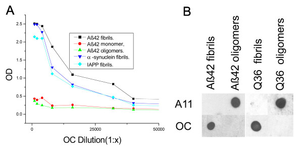
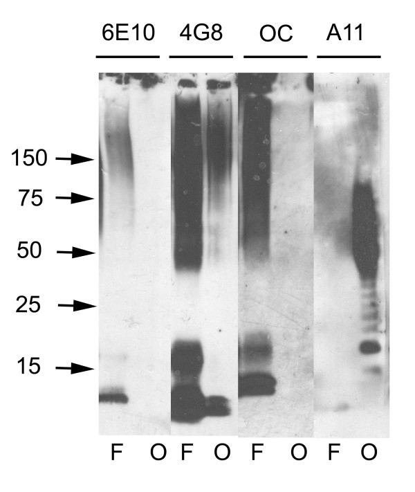
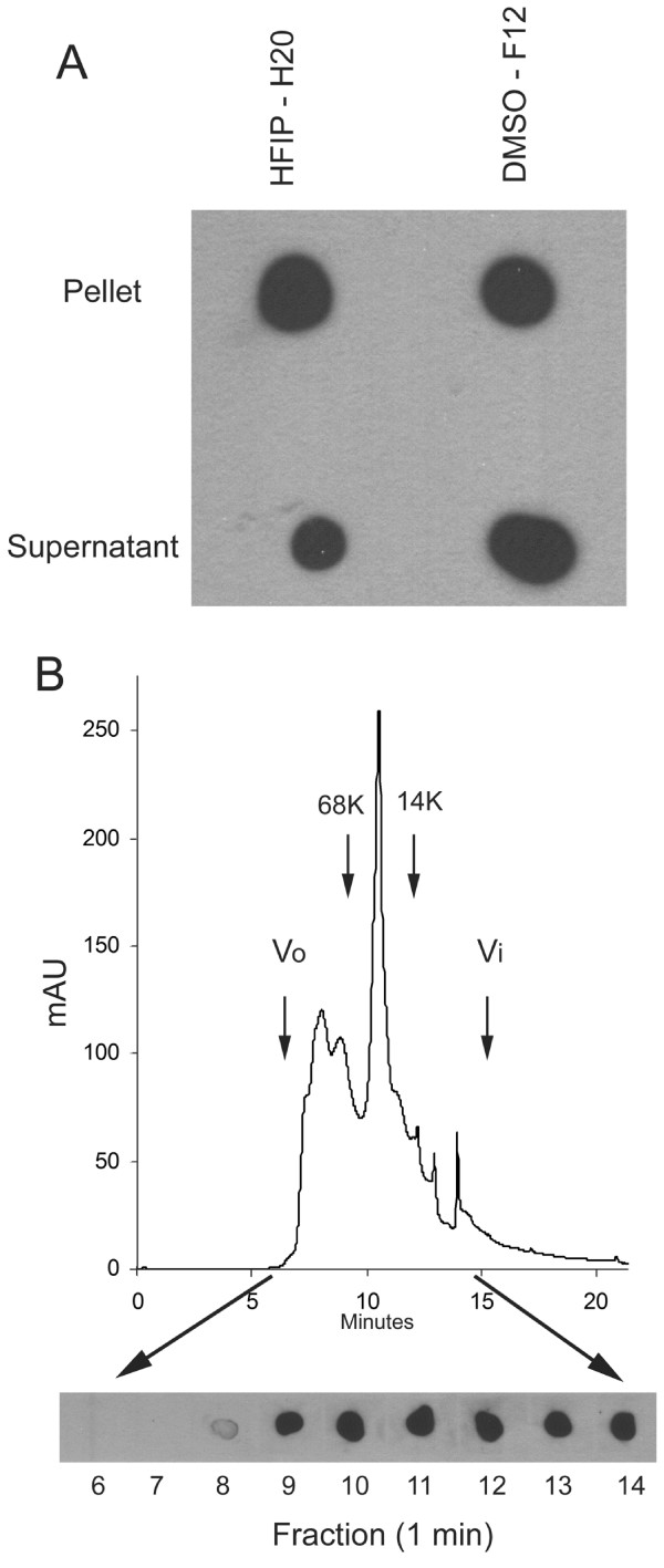
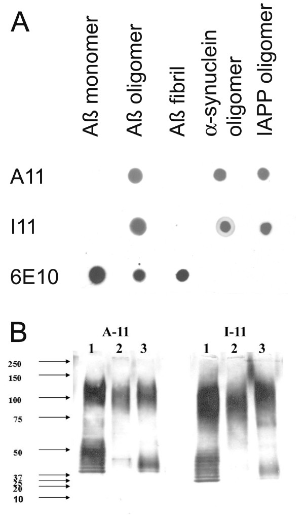
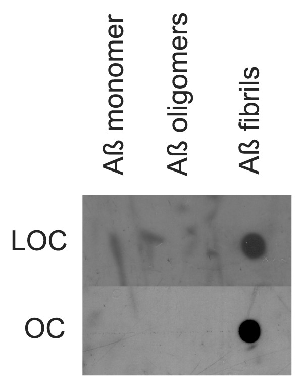
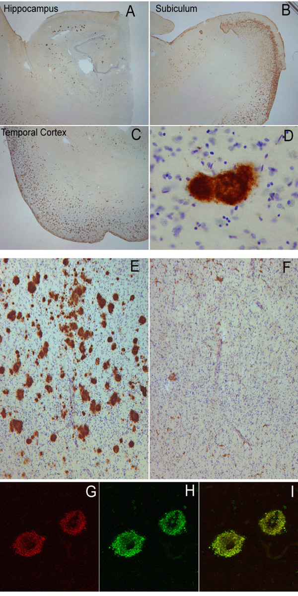
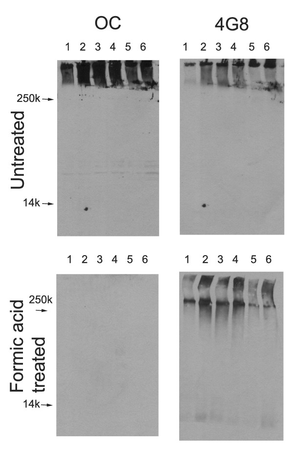
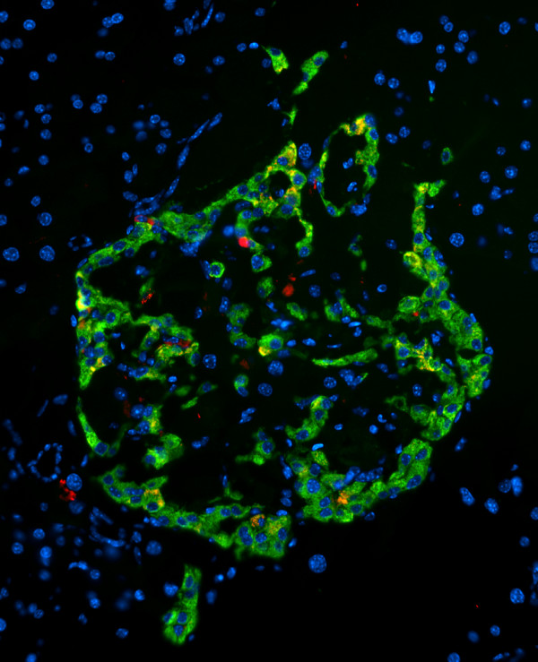
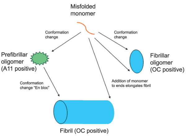
Background: Amyloid-related degenerative diseases are associated with the accumulation of misfolded proteins as amyloid fibrils in tissue. In Alzheimer disease (AD), amyloid accumulates in several distinct types of insoluble plaque deposits, intracellular Aβ and as soluble oligomers and the relationships between these deposits and their pathological significance remains unclear. Conformation dependent antibodies have been reported that specifically recognize distinct assembly states of amyloids, including prefibrillar oligomers and fibrils. Results: We immunized rabbits with a morphologically homogeneous population of Aβ42 fibrils. The resulting immune serum (OC) specifically recognizes fibrils, but not random coil monomer or prefibrillar oligomers, indicating fibrils display a distinct conformation dependent epitope that is absent in prefibrillar oligomers. The fibril epitope is also displayed by fibrils of other types of amyloids, indicating that the epitope is a generic feature of the polypeptide backbone. The fibril specific antibody also recognizes 100,000 × G soluble fibrillar oligomers ranging in size from dimer to greater than 250 kDa on western blots. The fibrillar oligomers recognized by OC are immunologically distinct from prefibrillar oligomers recognized by A11, even though their sizes overlap broadly, indicating that size is not a reliable indicator of oligomer conformation. The immune response to prefibrillar oligomers and fibrils is not sequence specific and antisera of the same specificity are produced in response to immunization with islet amyloid polypeptide prefibrillar oligomer mimics and fibrils. The fibril specific antibodies stain all types of amyloid deposits in human AD brain. Diffuse amyloid deposits stain intensely with anti-fibril antibody although they are thioflavin S negative, suggesting that they are indeed fibrillar in conformation. OC also stains islet amyloid deposits in transgenic mouse models of type II diabetes, demonstrating its generic specificity for amyloid fibrils. Conclusion: Since the fibril specific antibodies are conformation dependent, sequence-independent, and recognize epitopes that are distinct from those present in prefibrillar oligomers, they may have broad utility for detecting and characterizing the accumulation of amyloid fibrils and fibrillar type oligomers in degenerative diseases.
Overview
The claims and statistics of this paper haven’t been checked yet.
Run the check — free, no account needed. It reads the open-access text and checks the sources it cites.
Integrity signals
Registry
Cites
Statistics
Figures
Verification
Claims the paper makes
The claims and statistics of this paper haven’t been checked yet.
Run the check — free, no account needed. It reads the open-access text and checks the sources it cites.
Claims other papers make about this one
Each is checked against this paper’s own text: does it say what the citing paper says it does?
The source backs part of the claim, not all of it — or backs it for a different population, condition or comparison.
In the citing paper
“We obtained high resolution confocal images of brain slices from CRND8 mice stained with A11 and 4G8 antibodies, which detect oligomeric and other protofibrillar conformations respectively (Lesné et al., 2006; Kayed et al., 2007).”
From CX3CR1 in Microglia Regulates Brain Amyloid Deposition through Selective Protofibrillar Amyloid-β Phagocytosis (2010) · cites this paper as Kayed et al., 2007
Experimental — accuracy not yet measuredIn the citing paper
“As Aβ aggregates from monomers to oligomers and then fibrils, the conformation and antibody accessibility of peptide epitopes within Aβ can change.”
From Intraneuronal β-amyloid accumulation and synapse pathology in Alzheimer’s disease (2010) · cites this paper as [87]
Self-check
Percentages, sums and reported differences re-computed from the paper’s own numbers.
The self-check needs the paper’s full text.
Citations
Cites
Soluble protein oligomers in neurodegeneration: lessons from the Alzheimer's amyloid β-peptide
Haass, C., & Selkoe, D. J. (2007) · Nature Reviews Molecular Cell Biology · doi:10.1038/nrm2101 · Cited by 4,744
Common structure of soluble amyloid oligomers implies common mechanism of pathogenesis
Kayed, R., Head, E., Thompson, J. L., McIntire, T. M., Milton, S. C., Cotman, C. W., & Glabe, C. G. (2003) · Science · doi:10.1126/science.1079469 · Cited by 4,064
Self-citationDiffusible, nonfibrillar ligands derived from Aβ 1–42 are potent central nervous system neurotoxins
Lambert, M. P., Barlow, A. K., Chromy, B. A., Edwards, C., Freed, R., Liosatos, M., Morgan, T. E., Rozovsky, I., Trommer, B., Viola, K. L., et al. (1998) · Proceedings of the National Academy of Sciences · doi:10.1073/pnas.95.11.6448 · Cited by 3,506
Iwatsubo, T., Odaka, A., Suzuki, N., Mizusawa, H., Nukina, N., & Ihara, Y. (1994) · Neuron · doi:10.1016/0896-6273(94)90458-8 · Cited by 1,774
Soluble Amyloid β Peptide Concentration as a Predictor of Synaptic Change in Alzheimer's Disease
Lue, L.-F., Kuo, Y.-M., Roher, A. E., Brachova, L., Shen, Y., Sue, L., Beach, T., Kurth, J. H., Rydel, R. E., & Rogers, J. (1999) · The American Journal of Pathology · doi:10.1016/s0002-9440(10)65184-x · Cited by 1,530
von Bergen, M., Friedhoff, P., Biernat, J., Heberle, J., Mandelkow, E.-M., & Mandelkow, E. (2000) · Proceedings of the National Academy of Sciences · doi:10.1073/pnas.97.10.5129 · Cited by 1,021
Intraneuronal Aβ42 Accumulation in Human Brain
Gouras, G. K., Tsai, J., Naslund, J., Vincent, B., Edgar, M., Checler, F., Greenfield, J. P., Haroutunian, V., Buxbaum, J. D., Xu, H., et al. (2000) · The American Journal of Pathology · doi:10.1016/s0002-9440(10)64700-1 · Cited by 991
Some morphometric aspects of the brain in senile dementia of the alzheimer type
Terry, R. D., Peck, A., DeTeresa, R., Schechter, R., & Horoupian, D. S. (1981) · Annals of Neurology · doi:10.1002/ana.410100209 · Cited by 801
The Pathogenesis of Senile Plaques
DICKSON, D. W. (1997) · Journal of Neuropathology and Experimental Neurology · doi:10.1097/00005072-199704000-00001 · Cited by 753
Observation of metastable Aβ amyloid protofibrils by atomic force microscopy
Harper, J. D., Wong, S. S., Lieber, C. M., & Lansbury, P. T. (1997) · Chemistry & Biology · doi:10.1016/s1074-5521(97)90255-6 · Cited by 699
Research works printed but not linked: 1
- Wisniewski HM, Terry RD: Reexamination of the pathogenesis of the senile plaque. Prog Neuropathol. 1973, 333 , 2: 1-26.
Cited by
Sorted by citations
Items 601–700 of 778
Amyloid oligomer detection by immobilized molecular chaperone
Sakono, M., Zako, T., Yohda, M., & Maeda, M. (2011) · Biochemical Engineering Journal · doi:10.1016/j.bej.2011.12.005 · Cited by 6
Zhang, Y., Yu, X., Zhu, J., Liu, S., Liu, X., Dong, Q.-X., Chai, J.-Q., & Liu, R. (2017) · International Immunopharmacology · doi:10.1016/j.intimp.2017.11.012 · Cited by 6
Raimundo, A., Ferreira, S., Farrim, M. I. D. J. D., dos Santos, C. N., & Menezes, R. (2020) · Frontiers in Microbiology · doi:10.3389/fmicb.2020.02035 · Cited by 10
Optimized Ultrasonic Irradiation Finds Out Ultrastable Aβ1–40 Oligomers
Nakajima, K., So, M., Takahashi, K., Tagawa, Y., Hirao, M., Goto, Y., & Ogi, H. (2017) · The Journal of Physical Chemistry B · doi:10.1021/acs.jpcb.7b01409 · Cited by 7
Huang, Y., Bai, Y., Chang, C., Bacino, M. S., Cheng, I. C., Li, L., Habelitz, S., Li, W., & Zhang, Y. (2020) · Journal of Bone and Mineral Research · doi:10.1002/jbmr.4329 · Cited by 5
The Detection of Toxic Amyloid-β Fibril Fragments Through a Surface Plasmon Resonance Immunoassay
Beeg, M., Rocutto, B., Battocchio, E., Dacomo, L., Corbelli, A., Fiordaliso, F., Balducci, C., & Gobbi, M. (2024) · International Journal of Molecular Sciences · doi:10.3390/ijms252313020 · Cited by 5
Tahira, A. C., Gomes, M. P. B., Freire, M. H. O., Muxfeldt, M., Prosdocimi, F., Passos, Y. M., Amaral, M. S., Valadão, L. P. F., Rangel, L. P., Silva, J. L., et al. (2025) · RNA Biology · doi:10.1080/15476286.2025.2508107 · Cited by 5
Amyloid Fibrils of Pisum sativum L. Vicilin Inhibit Pathological Aggregation of Mammalian Proteins
Sulatsky, M. I., Belousov, M. V., Kosolapova, A. O., Mikhailova, E. V., Romanenko, M. N., Antonets, K. S., Константинович, Т. К., Константинович, Т. К., Nizhnikov, A. A., & Sulatskaya, A. I. (2023) · International Journal of Molecular Sciences · doi:10.3390/ijms241612932 · Cited by 5
Karunarathne, K., Kee, T. R., Jeon, H., Cazzaro, S., Gamage, Y. I., Pan, J., Woo, J. A., Kang, D. E., & Muschol, M. (2024) · Biomolecules · doi:10.3390/biom14060615 · Cited by 5
Yuan, Z., Du, M., Chen, Y., & Dou, F. (2013) · PubMed Central · doi:10.3969/j.issn.1673-5374.2013.33.004 · Cited by 6
Mapping Amyloid Regions in Gad m 1 with Peptide Arrays
Sánchez, R., Martínez, J., Montoya, L., Castellanos, M., & Gasset, M. (2018) · Methods in molecular biology · doi:10.1007/978-1-4939-7816-8_13 · Cited by 5
The Role of Inflammation in Amyloid Diseases
Azevedo, E. P., & Foguel, D. (2019) · IntechOpen eBooks · doi:10.5772/intechopen.81888 · Cited by 7
The tumor suppressor protein PTEN undergoes amyloid-like aggregation in tumor cellsPreprint
Claes, F., Maritschnegg, E., Baets, G. D., Siekierska, A., Rubio, M. S., Ramakers, M., Michiels, E., Smet, F. D., Depreeuw, J., Vergote, I. B., et al. (2020) · bioRxiv (Cold Spring Harbor Laboratory) · doi:10.1101/2020.11.30.402115 · Cited by 5
Sulatsky, M. I., Stepanenko, O. V., Stepanenko, O. V., Mikhailova, E. V., & Sulatskaya, A. I. (2025) · International Journal of Biological Macromolecules · doi:10.1016/j.ijbiomac.2025.140971 · Cited by 4
Hockaden, N., Leriger, G., Wang, J., Ray, H., Chakrabarti, S., Downing, N. F., Desmond, J., Williams, D. R., Hollenhorst, P. C., Longmore, G. D., et al. (2025) · Cell Stress and Chaperones · doi:10.1016/j.cstres.2025.03.003 · Cited by 4
Fallot, L. B., Pinc, J. R., Buselmeier, J. E., Palchak, J. C., Shroff, S. S., Zang, K., Rinauro, D. J., Bacon, K. M., Nguyen, M. P., Schleck, M. C., et al. (2025) · Proceedings of the National Academy of Sciences · doi:10.1073/pnas.2417944122 · Cited by 4
Hicks, C. W., Prokaeva, T. B., Spencer, B., Jayaraman, S., Huda, N., Wong, S., Chen, H. A., Sanchorawala, V., Lavatelli, F., & Gursky, O. (2025) · Journal of Molecular Biology · doi:10.1016/j.jmb.2025.169591 · Cited by 4
Ma, L., Reithofer, S., Pesch, V., Flores-Fernández, J. M., Sriraman, A., Duckering, C., Amidian, S., Özdüzenciler, P., Müller, L. D., Holger, W., et al. (2026) · Brain · doi:10.1093/brain/awag010 · Cited by 4
Grover, S., Pham, T., Jones, A., Pereira, C. S., Maurino, M. V. D., Garrad, E. C., Makoni, N. J., Parks, A., Domalewski, R. J., Riggio, G., et al. (2023) · Journal of Neurochemistry · doi:10.1111/jnc.15817 · Cited by 5
Wang, X., Xia, H., Li, T., Zuo, Q., Wang, Z., Yan, K., Xu, Z., Xue, W., Sun, G., Liu, Z., et al. (2024) · Advanced Healthcare Materials · doi:10.1002/adhm.202401625 · Cited by 4
Facile generation of drug-like conformational antibodies specific for amyloid fibrils
Desai, A. A., Zupancic, J. M., Trzeciakiewicz, H., Gerson, J. E., Dubois, K. N., Skinner, M. E., Sharkey, L. M., McArthur, N., Ferris, S. P., Bhatt, N. N., et al. (2025) · Nature Chemical Biology · doi:10.1038/s41589-025-01881-9 · Cited by 4
Kadu, P., Gadhe, L. G., Navalkar, A., Patel, K., Kumar, R., Sastry, M., & Maji, S. K. (2022) · Nanoscale · doi:10.1039/d2nr01942f · Cited by 4
Impaired Extracellular Proteostasis in Patients with Heart Failure
Gouveia, M., Teixeira, M., Schmidt, C., Lopes, M., Trindade, D., Magalhães, S., Henriques, A. G., Nunes, A., Santos, M., Vieira, S. I., et al. (2023) · Archives of Medical Research · doi:10.1016/j.arcmed.2023.02.001 · Cited by 4
Bhatt, N. N., Puangmalai, N., Sengupta, U., Jerez, C., Kidd, M., Gandhi, S. S., & Kayed, R. (2024) · Journal of Biological Chemistry · doi:10.1016/j.jbc.2024.105628 · Cited by 5
Self-citationDefining the Neuropathological Aggresome across in Silico, in Vitro, and ex Vivo Experiments
Gomes, G.-N. W., & Levine, Z. A. (2021) · The Journal of Physical Chemistry B · doi:10.1021/acs.jpcb.0c09193 · Cited by 4
Identification of amyloid antibodies for Alzheimer disease – immunotherapy
Huwait, E. A., Baghallab, I., Glabe, C. G., Abulnaja, K. O., Kumosani, T., & Moselhy, S. S. (2020) · Archives of Physiology and Biochemistry · doi:10.1080/13813455.2020.1767147 · Cited by 4
Self-citationPhysiological and Pathophysiological Role of Islet Amyloid Polypeptide (IAPP, Amylin)
Westermark, G. T. (2011) · Springer eBooks · doi:10.1007/978-1-4419-6956-9_15 · Cited by 4
Bemporad, F., & Chiti, F. (2013) · doi:10.1002/9783527654185.ch8 · Cited by 4
Puentes, L. N., Lengyel‐Zhand, Z., Lee, J. Y., Hsieh, C., Schneider, M. E., Edwards, K. J., Luk, K. C., Lee, V. M. ‐., Trojanowski, J. Q., & Mach, R. H. (2020) · bioRxiv (Cold Spring Harbor Laboratory) · doi:10.1101/2020.04.08.032250 · Cited by 4
Flow cytometric isolation of drug-like conformational antibodies specific for amyloid fibrilsPreprint
Desai, A. A., Zupancic, J. M., Trzeciakiewicz, H., Gerson, J. E., Dubois, K. N., Skinner, M. E., Sharkey, L. M., McArthur, N., Ferris, S. P., Bhatt, N. N., et al. (2023) · bioRxiv (Cold Spring Harbor Laboratory) · doi:10.1101/2023.07.04.547698 · Cited by 4
Panichi, V., Dolzani, P., Cattini, L., Alabiso, F., Bissoli, I., Cetrullo, S., Columbaro, M., Flamigni, F., Arciola, C. R., Filardo, G., et al. (2025) · Biochimica et Biophysica Acta (BBA) - Molecular Basis of Disease · doi:10.1016/j.bbadis.2025.167865 · Cited by 3
Latina, V., Introna, M. D., Malerba, F., Florio, R., Balzamino, B. O., Natale, G. D., Sciacca, M. F. M., Pappalardo, G., Micera, A., Pignataro, A., et al. (2025) · Acta Neuropathologica Communications · doi:10.1186/s40478-025-02022-y · Cited by 3
Lee, M. K., Barnes, J., Vermilyea, S., Meints, J., & Martinez, H. (2025) · International Journal of Molecular Sciences · doi:10.3390/ijms26136254 · Cited by 3
Moustouka, C., & Makhatadze, G. I. (2025) · Protein Science · doi:10.1002/pro.70094 · Cited by 2
Monitoring clusterin and fibrillar structures in aging and dementia
Trindade, D., Cachide, M., Martins, T. S., Guedes, S., Rosa, I. M., Silva, O. A. B. D. C. E., & Henriques, A. G. (2023) · Aging Brain · doi:10.1016/j.nbas.2023.100080 · Cited by 5
β‐Carboline Alkaloids Resist the Aggregation and Cytotoxicity of Human Islet Amyloid Polypeptide
Wang, Y., Huo, Y., Shao, W., Zheng, T., & Du, W. (2023) · ChemBioChem · doi:10.1002/cbic.202300395 · Cited by 3
Purro, S. A., Farmer, M., Noble, E., Sarell, C. J., Powell, M., Yip, D. C.-M., Giggins, L., Zakka, L., Thomas, D. X., Farrow, M. A., et al. (2023) · PLoS ONE · doi:10.1371/journal.pone.0294465 · Cited by 3
Degradation of pathogenic amyloids induced by matrix metalloproteinase-9
Stepanenko, O. V., Sulatsky, M. I., Mikhailova, E. V., Stepanenko, O. V., & Sulatskaya, A. I. (2024) · International Journal of Biological Macromolecules · doi:10.1016/j.ijbiomac.2024.136362 · Cited by 3
Reporters of Amyloid Structural Polymorphism
LeVine, H., Nilsson, K. P. R., Nilsson, R., & Hammarström, P. (2013) · Elsevier eBooks · doi:10.1016/b978-0-12-394431-3.00007-9 · Cited by 3
Гусельникова, В. В., Antipova, M. V., Fedorova, E. A., Safrai, A. E., Rukavishnikova, A. A., Mikhailova, E. V., & Korzhevsky, D. E. (2019) · Journal of Anatomy and Histopathology · doi:10.18499/2225-7357-2019-8-2-91-99 · Cited by 3
Mass spectrometric approaches for profiling protein folding and stability
Genereux, J. C. (2019) · Advances in protein chemistry and structural biology · doi:10.1016/bs.apcsb.2019.09.006 · Cited by 3
Gil, B., Rose, J. L., Demurtas, D., Mancini, G., Sordet‐Dessimoz, J., Sorrentino, V., Rudinskiy, N., Frosch, M. P., Hyman, B. T., Moniatte, M., et al. (2022) · bioRxiv (Cold Spring Harbor Laboratory) · doi:10.1101/2022.12.04.519021 · Cited by 3
Castranio, E. L., Varghese, M., Argyrousi, E. K., Tripathi, K. K., Huang, Y., Asada, A., Söderberg, L., Bresnahan, E., Lerner, D., Garretti, F., et al. (2026) · Alzheimer s & Dementia · doi:10.1002/alz.71426 · Cited by 2
Self-citationFallahBagheri, N., & Akan, Ö. B. (2025) · IEEE Transactions on Molecular Biological and Multi-Scale Communications · doi:10.1109/tmbmc.2025.3552959 · Cited by 2
Valle, M. L., Getaneh, B., William, C. M., Ghiso, J. A., & Rostagno, A. A. (2025) · Antioxidants · doi:10.3390/antiox14111375 · Cited by 2
Leech, G., Bayly, I., Talgatov, A., Barrett, P. C., Cameron, C. G., McFarland, S. A., & Storr, T. (2025) · Inorganic Chemistry · doi:10.1021/acs.inorgchem.5c04609 · Cited by 2
Yanes, K. J. O., Bui, C. T., Tomasello, J., Morsy, H., Kim, E., Lam, T., Tsourmas, K. I., Ayala, L. A., Green, K. N., Inlay, M. A., et al. (2026) · Journal of Neuroinflammation · doi:10.1186/s12974-025-03666-2 · Cited by 3
Yuan, S., Essepian, N., Roberts, R., Sherman, E., Gniadzik, W. M., Wang, Q., Erisir, A., & Jiang, L. (2026) · Acta Neuropathologica · doi:10.1007/s00401-026-03018-1 · Cited by 3
Animal Science Annual Volume 2023
Narayan, E. J. (2023) · Veterinary medicine and science · doi:10.5772/intechopen.113979 · Cited by 2
Wang, W., Wu, X., Zhang, Q., Zhang, T., Jiang, L., Qu, L., Lu, F., & Liu, F. (2024) · International Journal of Biological Macromolecules · doi:10.1016/j.ijbiomac.2024.139041 · Cited by 2
Functional amyloid protein FXR1 is recruited into neuronal stress granules
Valina, A. A., Belashova, T. A., Yuzman, A. K., Zadorsky, S. P., Sysoev, E. I., Mitkevich, V. A., Makarov, A. А., & Galkin, A. P. (2025) · Prion · doi:10.1080/19336896.2025.2505422 · Cited by 2
A Pyridyl Zn (II) Chelate for the Sensitive Detection of Aβ Fibrils
Yao, Y., Li, W., Han, Q.-Q., Lv, G., Li, C., & Sun, A. (2022) · Zeitschrift für anorganische und allgemeine Chemie · doi:10.1002/zaac.202200070 · Cited by 2
Lee, M., Liao, Y., Chen, S., & Chen, Y. (2021) · IUBMB Life · doi:10.1002/iub.2570 · Cited by 2
Siniukova, V. A., Galkina, S., & Galkin, A. P. (2022) · Biological Communications · doi:10.21638/spbu03.2022.106 · Cited by 2
Generation and study of antibodies against two triangular trimers derived from Aβ
Kreutzer, A. G., Malonis, R. J., Parrocha, C. M. T., Tong, K., Guaglianone, G., Nguyen, J. T., Diab, M. N., Lai, J. R., & Nowick, J. S. (2023) · Peptide Science · doi:10.1002/pep2.24333 · Cited by 2
Dagan, B., Oren, O., Banerjee, V., Taube, R., & Papo, N. (2019) · Proteins Structure Function and Bioinformatics · doi:10.1002/prot.25700 · Cited by 2
Amyloid fibrils are nanoparticles that target lysosomes.
Jakhria, T. (2014) · UPT. Syiah Kuala University Library (Syiah Kuala University) · Cited by 2
An anti-amyloidogenic treatment to specifically block the consolidation of traumatic events in mousePreprint
López-García, P., de Mingo, D. R., McGreevy, K. R., Pallé, A., Popiel, H. A., Santi, A., Nagai, Y., Trejo, J. L., & Carrión‐Vázquez, M. (2020) · bioRxiv (Cold Spring Harbor Laboratory) · doi:10.1101/2020.01.21.913053 · Cited by 2
Tau amyloidogenesis begins with a loss of its conformational polymorphismPreprint
Fernández‐Ramírez, M. D. C., Hervás, R., Menéndez, M., Laurents, D. V., & Carrión‐Vázquez, M. (2020) · bioRxiv (Cold Spring Harbor Laboratory) · doi:10.1101/2020.06.18.158923 · Cited by 2
Mammalian D-Cysteine is a Physiologic Down Regulator of Insulin Promoter MethylationPreprint
Roychaudhuri, R., Gadalla, M. M., Albacarys, L. K., West, T. R., Saavedra, H. G., Amzel, L. M., & Snyder, S. H. (2021) · SSRN Electronic Journal · doi:10.2139/ssrn.3908781 · Cited by 2
Fändrich, M., Wulff, M., Pedersen, J. S., & Otzen, D. E. (2013) · Wiley eBooks · doi:10.1002/9783527654185.ch15 · Cited by 2
Mukherjee, S., Sengupta, D., Sulkshane, P., Modak, A., Singh, S., Manna, S., Paul, A., Shaw, R., Devi, J., Masurkar, S. A., et al. (2025) · bioRxiv (Cold Spring Harbor Laboratory) · doi:10.64898/2025.12.05.692483 · Cited by 2
The bacterial OMP amyloids modulate α-synuclein and amyloid-β aggregation
Sulatsky, M. I., Belousov, M. V., Stepanenko, O. V., Stepanenko, O. V., Mikhailova, E. V., Fayoud, H. J., Nizhnikov, A. A., & Sulatskaya, A. I. (2025) · International Journal of Biological Macromolecules · doi:10.1016/j.ijbiomac.2025.147485 · Cited by 1
Directed evolution of drug-like Aβ conformation-specific antibodies
Desai, A. A., Smith, M. D., Zupancic, J. M., Makowski, E. K., Zhang, Y., Gerson, J. E., Moore, S. J., Sutter, A. B., Ferris, S. P., Ivanova, M. I., et al. (2025) · Frontiers in Immunology · doi:10.3389/fimmu.2025.1655893 · Cited by 1
Nanoparticle-mediated Zn delivery impacts neural protein phosphatase activity
Nadais, A., Castro, C., Martins, I. M., Trigo, D., Nunes, C., Henriques, A. G., Pereira, M. D. L., & Silva, O. A. B. D. C. E. (2025) · Biomaterials Advances · doi:10.1016/j.bioadv.2025.214675 · Cited by 2
Misfolding protein pathology detected in a chronic mouse model of multiple sclerosis
MacKeigan, T. P., Morgan, M. L., Mitchell, S., & Stys, P. K. (2025) · Neuroscience · doi:10.1016/j.neuroscience.2025.12.063 · Cited by 1
Amyloid-β fibrils accumulated in preeclamptic placentas suppress cytotrophoblast syncytialization
Nishioka, K., Ikezaki, M., Iwahashi, N., Arakawa, M., Fukushima, M., Mori, N., Mizoguchi, M., Horiuchi-Tanizaki, Y., Fujino, M., Tomiyama, T., et al. (2026) · Life Science Alliance · doi:10.26508/lsa.202503453 · Cited by 2
Nasim, N., Cheng, M.-Y., Park, H. H., Valenzuela, A., Andrew, P., Wexler, A. S., Chandrasekaran, V., Winkle, L. S. V., Bein, K. J., & Lein, P. J. (2026) · NeuroToxicology · doi:10.1016/j.neuro.2026.103472 · Cited by 1
Chang, H., Kang, H., Lee, S.-K., Lim, J., Kastanenka, K. V., Kashiwagi, S., & Jun, B. (2026) · Materials Today · doi:10.1016/j.mattod.2026.103428 · Cited by 1
Tau Oligomers as Potential Drug Target for Alzheimer Disease (AD) Treatment
Kayed, R. (2011) · InTech eBooks · doi:10.5772/16682 · Cited by 1
Self-citationStructure-Toxicity Relationships of Amyloid Peptide Oligomers
Walsh, P. J., & Sharpe, S. J. (2011) · InTech eBooks · doi:10.5772/30503 · Cited by 1
Pathogenesis of neurodegenerative diseases via templated recruitment
Guo, J. (2013) · ScholarlyCommons (University of Pennsylvania) · Cited by 1
Danial, N. N. (2011) · Cambridge University Press eBooks · doi:10.1017/cbo9780511976094.019 · Cited by 1
Conformation-Dependent Antibodies as Tools for Characterization of Amyloid Protein Aggregates
Wu, J., & Breydo, L. (2013) · Elsevier eBooks · doi:10.1016/b978-0-12-394431-3.00008-0 · Cited by 1
Self-citationМинкевич, Н. И., Rakitina, T. V., Богачук, А. П., Radchenko, V. V., Сурина, Е. А., Morozova‐Roche, L. A., Yanamandra, K., Иомдина, Е. Н., Бабиченко, И. И., Kostanyan, I. A., et al. (2012) · Russian Journal of Bioorganic Chemistry · doi:10.1134/s1068162012060118 · Cited by 1
Amyloid Hypothesis: Molecular and Cellular Aspects of Toxicity
Kayed, R., & Lasagna‐Reeves, C. A. (2013) · Modecular medicine and medicinal · doi:10.1142/9781848167551_0001 · Cited by 2
Self-citationLanning, J. D., & Meredith, S. C. (2011) · Springer eBooks · doi:10.1007/978-94-007-2774-8_14 · Cited by 1
Inhibition of Protein Fibrillation by Hydrogen Sulfide1
Rosario-Alomar, M. F., Quiñones-Ruíz, T., Kurouski, D., Sereda, V., DeBarros-Ferreira, E., Jesús-Kim, L. D., Hernández‐Rivera, S. P., Zagorevski, D. V., Cruz-Collazo, L. M., Lednev, I. K., et al. (2019) · IntechOpen eBooks · doi:10.5772/intechopen.86221 · Cited by 1
Nosova, O., Гусельникова, В. В., & Korzhevsky, D. E. (2023) · Journal of Neuroscience Methods · doi:10.1016/j.jneumeth.2023.109797 · Cited by 1
Fukui, R., Hafizal, U., Kageyama, Y., Irie, Y., Matsushima, Y., Hosoi, K., Nakayama, T., Kaneda, D., Hashizume, Y., Miki, K., et al. (2025) · Biochemical and Biophysical Research Communications · doi:10.1016/j.bbrc.2025.151655 · Cited by 1
Chang, L. Y., Li, F., Chen, X., Xu, S., Wang, C., Chen, H., & Wang, Q. (2014) · Metabolic Brain Disease · doi:10.1007/s11011-014-9521-8 · Cited by 1
Innovative Therapie der Alzheimer-Demenz
Dodel, R. C., & Bacher, M. (2008) · Der Nervenarzt · doi:10.1007/s00115-008-2499-x · Cited by 1
The Oligomer Species: Mechanistics and Biochemistry
Stefani, M. (2013) · Wiley eBooks · doi:10.1002/9783527654185.ch7 · Cited by 1
Structural and Compositional Information about Pre‐Amyloid Oligomers
Zijlstra, N., & Subramaniam, V. (2013) · Wiley eBooks · doi:10.1002/9783527654185.ch6 · Cited by 1
Mahapatra, S., Sarbahi, A., Madhu, P., Swasthi, H. M., & Mukhopadhyay, S. (2021) · bioRxiv · doi:10.1101/2021.03.08.434509 · Cited by 1
Anle138b manifests potent anti-hyperglycemic activity in type 2 diabetic mice expressing hIAPPPreprint
Albariqi, M. M. H., Baauw, S. M. G., Fens, S. J. P. J., Versteeg, S., Ryazanov, S., Leonov, A., Willemen, H. L. D. M., Stathonikos, N., Seychell, R. M., Saghir, A. E., et al. (2024) · bioRxiv (Cold Spring Harbor Laboratory) · doi:10.1101/2024.08.27.609850 · Cited by 1
Aβ*56 is present in brain extracts enriched for extracellular proteinsPreprint
Liu, P., Lapcinski, I. P., & Ashe, K. H. (2024) · bioRxiv (Cold Spring Harbor Laboratory) · doi:10.1101/2024.10.09.617424 · Cited by 1
Essepian, N., Yuan, S., Roberts, R., Sherman, E., Gniadzik, W., Wang, Q., Erişir, A., & Jiang, L. (2025) · bioRxiv (Cold Spring Harbor Laboratory) · doi:10.1101/2025.05.21.655370 · Cited by 1
Åslund, A. (2009) · KTH Publication Database DiVA (KTH Royal Institute of Technology) · Cited by 1
Yanamandra, K. (2010) · KTH Publication Database DiVA (KTH Royal Institute of Technology)
Structure and Function of Stefin B Oligomers – Important Role in Amyloidogenesis
Taler‐Verčič, A., Polajnar, M., & Žerovnik, E. (2014) · InTech eBooks · doi:10.5772/57570
Amyloid Polymorphisms: Structural Basis and Significance in Biology and Molecular Medicine
Stefani, M. (2011) · Wiley eBooks · doi:10.1002/9783527634323.ch5
Gold nanoparticles and peptoids as novel inhibitors of Abeta aggregation in Alzheimer's disease
Moore, K. A. (2013) · Scholar Commons (University of South Carolina)
Characterizing the in-vitro morphology and growth kinetics of intermediate amyloid aggregates
Hill, S. E. (2008) · Digital Commons - University of South Florida (University of South Florida)
Mutations in the Chaperone Hsp104 Differentially Propagate Yeast Prions
Dulle, J. E. (2013) · Open Scholarship Institutional Repository (Washington University in St. Louis) · doi:10.7936/k7t43r3n
Simon, D. (2011) · SZTE Repository of Dissertations (University of Szeged) · doi:10.14232/phd.1292
SPECIFIC AMYLOID-BETA OLIGOMERS IN HUMAN CEREBROSPINAL FLUID
Handoko, M. (2012) · University of Minnesota Digital Conservancy (University of Minnesota)
A Genome-Wide RNAi Screen for Modifiers of Polyglutamine-Induced Neurotoxicity in Drosophila
Voßfeldt, H. (2013) · eDiss (Georg-August-Universität Göttingen) · doi:10.53846/goediss-3767
Aβ Conformation Dependent Antibodies and Alzheimer's Disease
Sehlin, D. (2010) · KTH Publication Database DiVA (KTH Royal Institute of Technology)
Amyloid State of Proteins in Human Disease
Huang, L., & Huang, K. (2015) · Encyclopedia of Life Sciences · doi:10.1002/9780470015902.a0024459
Metadata
- Authors
- Kayed, R., Head, E., Sarsoza, F., Saing, T., Cotman, C. W., Necula, M., Margol, L., Wu, J., Breydo, L., Thompson, J. L., Rasool, S., Gurlo, T., Butler, P., & Glabe, C. G.
- Affiliations
- University of California, Irvine (US)
- University of California, Los Angeles (US)
- Corresponding author
- Glabe, Charles G
- ORCID
- Venue
- Molecular Neurodegeneration
- Published
- 2007-09-26
- Volume
- 2
- Issue
- 1
- Pages
- 18-18
- ISSN
- 1750-1326 (ISSN-L)
- PMC
- PMC2100048
- OpenAlex
- W2156193029
- Cited by
- 773
- References
- 28
- Citations by year
- 2026: 21 · 2025: 35 · 2024: 36 · 2023: 42 · 2022: 32 · 2021: 41 · 2020: 57 · 2019: 35 · 2018: 44 · 2017: 40 · 2016: 43 · 2015: 52 · 2014: 45 · 2013: 65 · 2012: 59
- FWCI
- 18.92
- Type
- journal article
- Language
- en
- Open access
- gold
- doi.org
- Licence
- cc-by
- Locations
- Molecular Neurodegeneration (publishedVersion, cc-by)
- PubMed Central (publishedVersion, cc-by)
- PubMed (publishedVersion)
- DOAJ (DOAJ: Directory of Open Access Journals) (submittedVersion, cc-by-sa)
- eScholarship (California Digital Library) (submittedVersion)
- Funders
- Larry L. Hillblom Foundation
- Cure Alzheimer's Fund
- National Institutes of Health (AG16573, AG000538)
- National Institute on Aging (P01AG000538, P50 AG 16573, AG 16573, AG-000538, P50AG016573)
- National Institute of Neurological Disorders and Stroke (R01NS031230)
- Topics
- Life Sciences › Neuroscience › Cellular and Molecular Neuroscience › Alzheimer's disease research and treatments
- Life Sciences › Biochemistry, Genetics and Molecular Biology › Molecular Biology › Prion Diseases and Protein Misfolding
- Health Sciences › Medicine › Psychiatry and Mental health › Dementia and Cognitive Impairment Research
- Keywords
- amyloid fibrils, prefibrillar oligomers, epitope mapping, amyloid-beta 42, Alzheimer's disease, amylin, type 2 diabetes, amyloid deposits, immunization, thioflavin S, degenerative diseases
About these checks
Registry, retraction, Cites and statistics results are computed, not judged, with one exception: a printed reference that no index lists is read once by a model before it counts as a citation error. Claims are extracted by a model and kept only when their quote appears verbatim in the text; each cited source is then checked, with a model’s help, for whether it says what the claim relies on. Those verdicts can be wrong — read the claim’s quote and the cited source before relying on one.
Statistics and arithmetic are recomputed, not judged.
Confidence bands are shown only for fields calibrated on our annotated test set. None is calibrated yet, so no band is shown.
What counts as a claim ›How we measure confidence ›How we check this ›
Are you an author, or do you see a problem with this page? Request a review
