

The DOI matches its Crossref record; 28 of the 28 references it cites were found in a registry or an index.
Fibril specific, conformation dependent antibodies recognize a generic epitope common to amyloid fibrils and fibrillar oligomers that is absent in prefibrillar oligomers
Open accessAbstract
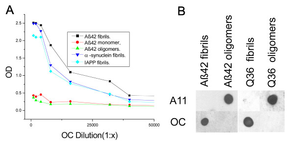
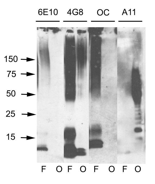
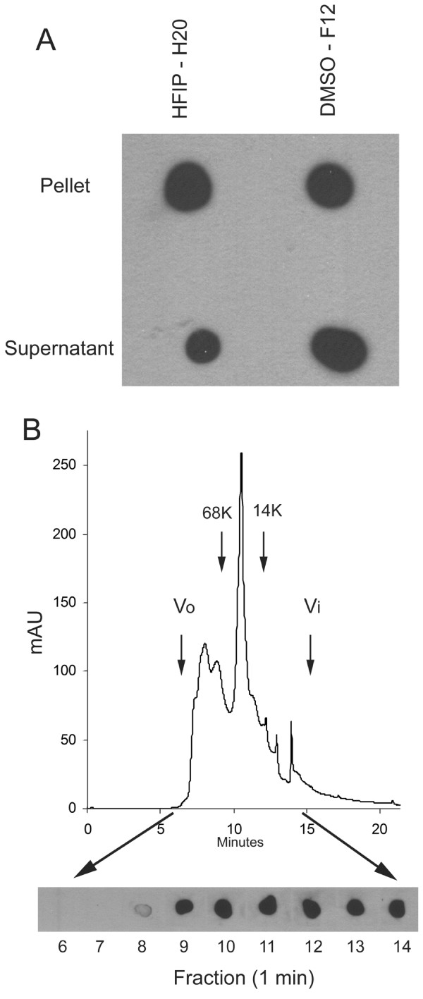
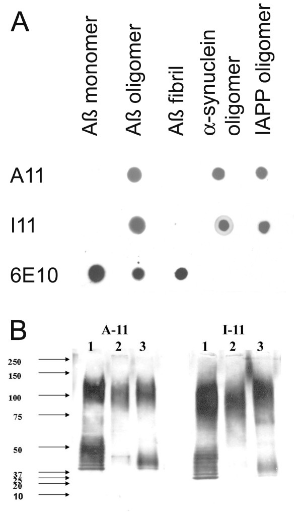
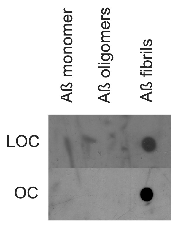
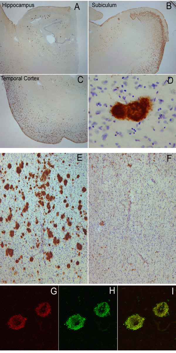
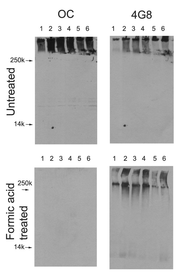
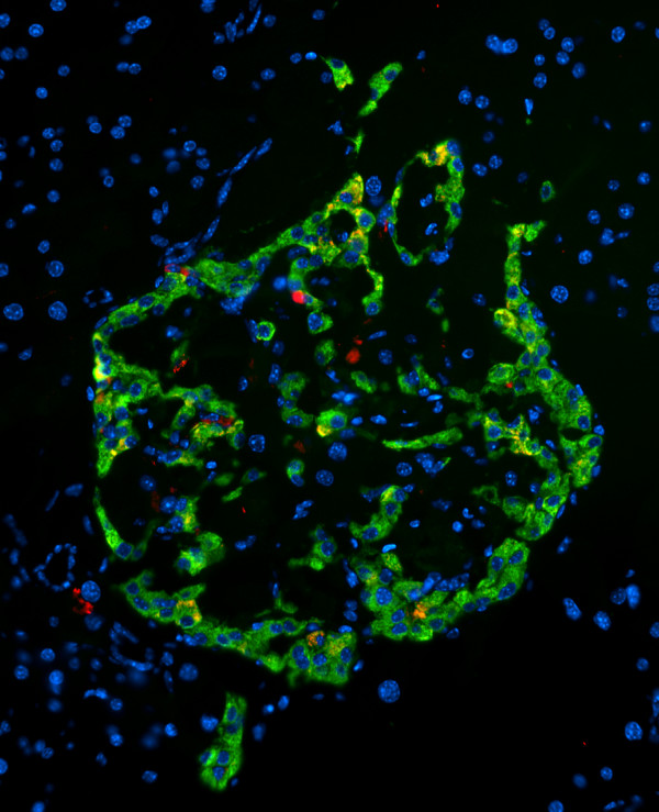
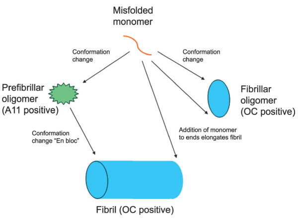
Background: Amyloid-related degenerative diseases are associated with the accumulation of misfolded proteins as amyloid fibrils in tissue. In Alzheimer disease (AD), amyloid accumulates in several distinct types of insoluble plaque deposits, intracellular Aβ and as soluble oligomers and the relationships between these deposits and their pathological significance remains unclear. Conformation dependent antibodies have been reported that specifically recognize distinct assembly states of amyloids, including prefibrillar oligomers and fibrils. Results: We immunized rabbits with a morphologically homogeneous population of Aβ42 fibrils. The resulting immune serum (OC) specifically recognizes fibrils, but not random coil monomer or prefibrillar oligomers, indicating fibrils display a distinct conformation dependent epitope that is absent in prefibrillar oligomers. The fibril epitope is also displayed by fibrils of other types of amyloids, indicating that the epitope is a generic feature of the polypeptide backbone. The fibril specific antibody also recognizes 100,000 × G soluble fibrillar oligomers ranging in size from dimer to greater than 250 kDa on western blots. The fibrillar oligomers recognized by OC are immunologically distinct from prefibrillar oligomers recognized by A11, even though their sizes overlap broadly, indicating that size is not a reliable indicator of oligomer conformation. The immune response to prefibrillar oligomers and fibrils is not sequence specific and antisera of the same specificity are produced in response to immunization with islet amyloid polypeptide prefibrillar oligomer mimics and fibrils. The fibril specific antibodies stain all types of amyloid deposits in human AD brain. Diffuse amyloid deposits stain intensely with anti-fibril antibody although they are thioflavin S negative, suggesting that they are indeed fibrillar in conformation. OC also stains islet amyloid deposits in transgenic mouse models of type II diabetes, demonstrating its generic specificity for amyloid fibrils. Conclusion: Since the fibril specific antibodies are conformation dependent, sequence-independent, and recognize epitopes that are distinct from those present in prefibrillar oligomers, they may have broad utility for detecting and characterizing the accumulation of amyloid fibrils and fibrillar type oligomers in degenerative diseases.
Overview
The claims and statistics of this paper haven’t been checked yet.
Run the check — free, no account needed. It reads the open-access text and checks the sources it cites.
Integrity signals
Registry
Cites
Statistics
Figures
Verification
Claims the paper makes
The claims and statistics of this paper haven’t been checked yet.
Run the check — free, no account needed. It reads the open-access text and checks the sources it cites.
Claims other papers make about this one
Each is checked against this paper’s own text: does it say what the citing paper says it does?
The source backs part of the claim, not all of it — or backs it for a different population, condition or comparison.
In the citing paper
“We obtained high resolution confocal images of brain slices from CRND8 mice stained with A11 and 4G8 antibodies, which detect oligomeric and other protofibrillar conformations respectively (Lesné et al., 2006; Kayed et al., 2007).”
From CX3CR1 in Microglia Regulates Brain Amyloid Deposition through Selective Protofibrillar Amyloid-β Phagocytosis (2010) · cites this paper as Kayed et al., 2007
Experimental — accuracy not yet measuredIn the citing paper
“As Aβ aggregates from monomers to oligomers and then fibrils, the conformation and antibody accessibility of peptide epitopes within Aβ can change.”
From Intraneuronal β-amyloid accumulation and synapse pathology in Alzheimer’s disease (2010) · cites this paper as [87]
Self-check
Percentages, sums and reported differences re-computed from the paper’s own numbers.
The self-check needs the paper’s full text.
Citations
Cites
Soluble protein oligomers in neurodegeneration: lessons from the Alzheimer's amyloid β-peptide
Haass, C., & Selkoe, D. J. (2007) · Nature Reviews Molecular Cell Biology · doi:10.1038/nrm2101 · Cited by 4,744
Common structure of soluble amyloid oligomers implies common mechanism of pathogenesis
Kayed, R., Head, E., Thompson, J. L., McIntire, T. M., Milton, S. C., Cotman, C. W., & Glabe, C. G. (2003) · Science · doi:10.1126/science.1079469 · Cited by 4,064
Self-citationDiffusible, nonfibrillar ligands derived from Aβ 1–42 are potent central nervous system neurotoxins
Lambert, M. P., Barlow, A. K., Chromy, B. A., Edwards, C., Freed, R., Liosatos, M., Morgan, T. E., Rozovsky, I., Trommer, B., Viola, K. L., et al. (1998) · Proceedings of the National Academy of Sciences · doi:10.1073/pnas.95.11.6448 · Cited by 3,506
Iwatsubo, T., Odaka, A., Suzuki, N., Mizusawa, H., Nukina, N., & Ihara, Y. (1994) · Neuron · doi:10.1016/0896-6273(94)90458-8 · Cited by 1,774
Soluble Amyloid β Peptide Concentration as a Predictor of Synaptic Change in Alzheimer's Disease
Lue, L.-F., Kuo, Y.-M., Roher, A. E., Brachova, L., Shen, Y., Sue, L., Beach, T., Kurth, J. H., Rydel, R. E., & Rogers, J. (1999) · The American Journal of Pathology · doi:10.1016/s0002-9440(10)65184-x · Cited by 1,530
von Bergen, M., Friedhoff, P., Biernat, J., Heberle, J., Mandelkow, E.-M., & Mandelkow, E. (2000) · Proceedings of the National Academy of Sciences · doi:10.1073/pnas.97.10.5129 · Cited by 1,021
Intraneuronal Aβ42 Accumulation in Human Brain
Gouras, G. K., Tsai, J., Naslund, J., Vincent, B., Edgar, M., Checler, F., Greenfield, J. P., Haroutunian, V., Buxbaum, J. D., Xu, H., et al. (2000) · The American Journal of Pathology · doi:10.1016/s0002-9440(10)64700-1 · Cited by 991
Some morphometric aspects of the brain in senile dementia of the alzheimer type
Terry, R. D., Peck, A., DeTeresa, R., Schechter, R., & Horoupian, D. S. (1981) · Annals of Neurology · doi:10.1002/ana.410100209 · Cited by 801
The Pathogenesis of Senile Plaques
DICKSON, D. W. (1997) · Journal of Neuropathology and Experimental Neurology · doi:10.1097/00005072-199704000-00001 · Cited by 753
Observation of metastable Aβ amyloid protofibrils by atomic force microscopy
Harper, J. D., Wong, S. S., Lieber, C. M., & Lansbury, P. T. (1997) · Chemistry & Biology · doi:10.1016/s1074-5521(97)90255-6 · Cited by 699
Research works printed but not linked: 1
- Wisniewski HM, Terry RD: Reexamination of the pathogenesis of the senile plaque. Prog Neuropathol. 1973, 333 , 2: 1-26.
Cited by
Sorted by citations
Items 101–200 of 778
Benilova, I., Gallardo, R. U., Ungureanu, A.-A., Cano, V. C., Snellinx, A., Ramakers, M., Bartic, C., Rousseau, F., Schymkowitz, J. W. H., & Strooper, B. D. (2014) · Journal of Biological Chemistry · doi:10.1074/jbc.m114.599027 · Cited by 151
Hatami, A., Monjazeb, S., Milton, S. C. F., & Glabe, C. G. (2017) · Journal of Biological Chemistry · doi:10.1074/jbc.m116.755264 · Cited by 151
Self-citationKlein, W. L. (2012) · Journal of Alzheimer s Disease · doi:10.3233/jad-2012-129039 · Cited by 145
Natural and amyloid self‐assembly of S100 proteins: structural basis of functional diversity
Fritz, G., Botelho, H. M., Morozova‐Roche, L. A., & Gomes, C. M. (2010) · FEBS Journal · doi:10.1111/j.1742-4658.2010.07887.x · Cited by 143
Esbjörner, E. K., Chan, F., Rees, E. J., Erdélyi, M., Luheshi, L. M., Bertoncini, C. W., Kaminski, C. F., Dobson, C. M., & Schierle, G. S. K. (2014) · Chemistry & Biology · doi:10.1016/j.chembiol.2014.03.014 · Cited by 138
Lee, M., Yu, W., Shih, Y., Chen, C., Guo, Z., Huang, S., Chan, J. C. C., & Chen, Y. (2018) · Scientific Reports · doi:10.1038/s41598-018-23122-x · Cited by 141
Bhaskar, K., Maphis, N. M., Xu, G., Varvel, N. H., Kokiko‐Cochran, O. N., Weick, J. P., Staugaitis, S. M., Cardona, A. E., Ransohoff, R. M., Herrup, K., et al. (2013) · Neurobiology of Disease · doi:10.1016/j.nbd.2013.10.007 · Cited by 141
Ahsan, N., Mishra, S., Jain, M. K., Surolia, A., & Gupta, S. (2015) · Scientific Reports · doi:10.1038/srep09862 · Cited by 140
Amyloid-β Annular Protofibrils Evade Fibrillar Fate in Alzheimer Disease Brain
Lasagna‐Reeves, C. A., Glabe, C. G., & Kayed, R. (2011) · Journal of Biological Chemistry · doi:10.1074/jbc.m111.236257 · Cited by 139
Self-citationp53 amyloid formation leading to its loss of function: implications in cancer pathogenesis
Ghosh, S., Salot, S., Sengupta, S., Navalkar, A., Ghosh, D., Jacob, R. S., Das, S., Kumar, R., Jha, N. N., Sahay, S., et al. (2017) · Cell Death and Differentiation · doi:10.1038/cdd.2017.105 · Cited by 136
Pagano, K., Tomaselli, S., Molinari, H., & Ragona, L. (2020) · Frontiers in Neuroscience · doi:10.3389/fnins.2020.619667 · Cited by 139
Zhang, L., Yu, X., Ji, M., Liu, S., Wu, X., Wang, Y., & Liu, R. (2018) · Food & Function · doi:10.1039/c8fo00964c · Cited by 132
Conserved features of intermediates in amyloid assembly determine their benign or toxic states
Krishnan, R., Goodman, J. L., Mukhopadhyay, S., Pacheco, C. D., Lemke, E. A., Deniz, A. A., & Lindquist, S. L. (2012) · Proceedings of the National Academy of Sciences · doi:10.1073/pnas.1209527109 · Cited by 131
Exploring the aggregation free energy landscape of the amyloid-β protein (1–40)
Zheng, W., Tsai, M., Chen, M., & Wolynes, P. G. (2016) · Proceedings of the National Academy of Sciences · doi:10.1073/pnas.1612362113 · Cited by 131
Functional Amyloid and Other Protein Fibers in the Biofilm Matrix
Erskine, E., MacPhee, C., & Stanley-Wall, N. (2018) · Journal of Molecular Biology · doi:10.1016/j.jmb.2018.07.026 · Cited by 111
Rinauro, D. J., Chiti, F., Vendruscolo, M., & Limbocker, R. (2024) · Molecular Neurodegeneration · doi:10.1186/s13024-023-00651-2 · Cited by 138
Beeg, M., Stravalaci, M., Romeo, M., Carrà, A. D., Cagnotto, A., Rossi, A., Diomede, L., Salmona, M., & Gobbi, M. (2016) · Journal of Biological Chemistry · doi:10.1074/jbc.m115.689539 · Cited by 128
P53 aggregation, interactions with tau, and impaired DNA damage response in Alzheimer’s disease
Farmer, K. M., Ghag, G., Puangmalai, N., Montalbano, M., Bhatt, N. N., & Kayed, R. (2020) · Acta Neuropathologica Communications · doi:10.1186/s40478-020-01012-6 · Cited by 130
Self-citationHsp90 Inhibits α-Synuclein Aggregation by Interacting with Soluble Oligomers
Daturpalli, S., Waudby, C. A., Meehan, S., & Jackson, S. (2013) · Journal of Molecular Biology · doi:10.1016/j.jmb.2013.08.006 · Cited by 128
Soluble Aβ Promotes Wild-Type Tau Pathology In Vivo
Chabrier, M. A., Blurton-Jones, M., Agazaryan, A. A., Nerhus, J. L., Martinez-Coria, H., & LaFerla, F. M. (2012) · The Journal of Neuroscience · doi:10.1523/jneurosci.0172-12.2012 · Cited by 123
Hatami, A., Albay, R., Monjazeb, S., Milton, S. C. F., & Glabe, C. G. (2014) · Journal of Biological Chemistry · doi:10.1074/jbc.m114.594846 · Cited by 123
Self-citationStructural Characteristics of α-Synuclein Oligomers
Cremades, N., Chen, S. W., & Dobson, C. M. (2016) · International review of cytology · doi:10.1016/bs.ircmb.2016.08.010 · Cited by 123
Targeting the Prion-like Aggregation of Mutant p53 to Combat Cancer
Silva, J. L., Cino, E. A., Soares, I. N., Ferreira, V. F., & de Oliveira, G. A. P. (2017) · Accounts of Chemical Research · doi:10.1021/acs.accounts.7b00473 · Cited by 121
Gut microbiota–driven brain Aβ amyloidosis in mice requires microglia
Dodiya, H. B., Lutz, H. L., Weigle, I. Q., Patel, P., Michalkiewicz, J., Roman-Santiago, C. J., Zhang, C. M., Liang, Y., Srinath, A., Zhang, X., et al. (2022) · Journal of Experimental Medicine · doi:10.1084/jem.20200895 · Cited by 120
Accumulation of intraneuronal Aβ correlates with ApoE4 genotype
Christensen, D. Z., Schneider-Axmann, T., Lucassen, P. J., Bayer, T. A., & Wirths, O. (2010) · Acta Neuropathologica · doi:10.1007/s00401-010-0666-1 · Cited by 120
Intracellular amyloid and the neuronal origin of Alzheimer neuritic plaques
Pensalfini, A., Albay, R., Rasool, S., Wu, J. W., Hatami, A., Arai, H., Margol, L., Milton, S., Poon, W. W., Corrada, M. M., et al. (2014) · Neurobiology of Disease · doi:10.1016/j.nbd.2014.07.011 · Cited by 120
Self-citationAmyloid-β nanotubes are associated with prion protein-dependent synaptotoxicity
Nicoll, A. J., Panico, S., Freir, D. B., Wright, D., Terry, C., Risse, E., Herron, C. E., O’Malley, T., Wadsworth, J. D. F., Farrow, M. A., et al. (2013) · Nature Communications · doi:10.1038/ncomms3416 · Cited by 119
Super‐Resolution Infrared Imaging of Polymorphic Amyloid Aggregates Directly in Neurons
Klementieva, O. L., Sandt, C., Martinsson, I., Kansiz, M., Gouras, G. K., & Borondics, F. (2020) · Advanced Science · doi:10.1002/advs.201903004 · Cited by 114
Karr, J. W., & Szalai, V. A. (2008) · Biochemistry · doi:10.1021/bi702423h · Cited by 119
Zaghi, J., Goldenson, B., Inayathullah, M., Lossinsky, A. S., Masoumi, A., Avagyan, H., Mahanian, M., Bernas, M. J., Weinand, M. E., Rosenthal, M. J., et al. (2009) · Acta Neuropathologica · doi:10.1007/s00401-008-0481-0 · Cited by 117
Amyloid-β oligomer detection by ELISA in cerebrospinal fluid and brain tissue
Bruggink, K. A., Jongbloed, W., Biemans, E. A. L. M., Veerhuis, R., Claassen, J. A. H. R., Kuiperij, H. B., & Verbeek, M. M. (2012) · Analytical Biochemistry · doi:10.1016/j.ab.2012.09.014 · Cited by 114
Davtyan, H., Ghochikyan, A., Petrushina, I., Hovakimyan, A., Davtyan, A., Poghosyan, A., Marleau, A. M., Movsesyan, N., Kiyatkin, A., Rasool, S., et al. (2013) · The Journal of Neuroscience · doi:10.1523/jneurosci.4672-12.2013 · Cited by 112
Self-citationA High Affinity Red Fluorescence and Colorimetric Probe for Amyloid β Aggregates
Rajasekhar, K., Narayanaswamy, N., Murugan, N. A., Kuang, G., Ågren, H., & Govindaraju, T. (2016) · Scientific Reports · doi:10.1038/srep23668 · Cited by 111
Kulenkampff, K., Perez, A.-M. W., Sormanni, P., Habchi, J., & Vendruscolo, M. (2021) · Nature Reviews Chemistry · doi:10.1038/s41570-021-00254-9 · Cited by 113
Pryor, N. E., Moss, M. A., & Hestekin, C. N. (2012) · International Journal of Molecular Sciences · doi:10.3390/ijms13033038 · Cited by 113
Oligomeric Aβ in the monkey brain impacts synaptic integrity and induces accelerated cortical aging
Beckman, D., Ott, S., Donis-Cox, K., Janssen, W. G., Bliss-Moreau, E., Rudebeck, P. H., Baxter, M. G., & Morrison, J. H. (2019) · Proceedings of the National Academy of Sciences · doi:10.1073/pnas.1902301116 · Cited by 111
Proteotoxicity and Cardiac Dysfunction
McLendon, P. M., & Robbins, J. M. (2015) · Circulation Research · doi:10.1161/circresaha.116.305372 · Cited by 111
Green Tea Polyphenol Epigallocatechin-Gallate in Amyloid Aggregation and Neurodegenerative Diseases
Fernandes, L., Cardim-Pires, T. R., Foguel, D., & Palhano, F. L. (2021) · Frontiers in Neuroscience · doi:10.3389/fnins.2021.718188 · Cited by 114
Soragni, A., Yousefi, S., Stoeckle, C., Soriaga, A. B., Sawaya, M. R., Kozlowski, E., Schmid, I., Radonjic‐Hoesli, S., Boutet, S., Williams, G. J., et al. (2015) · Molecular Cell · doi:10.1016/j.molcel.2015.01.026 · Cited by 115
Amyloid Formation by the Pro-Inflammatory S100A8/A9 Proteins in the Ageing Prostate
Yanamandra, K., Alexeyev, O. A., Zamotin, V., Srivastava, V., Shchukarev, A., Brorsson, A., Tartaglia, G. G., Vogl, T., Kayed, R., Wingsle, G., et al. (2009) · PLoS ONE · doi:10.1371/journal.pone.0005562 · Cited by 108
Self-citationThe role of amyloid β in the pathogenesis of Alzheimer's disease
Gilbert, B. J. (2013) · Journal of Clinical Pathology · doi:10.1136/jclinpath-2013-201515 · Cited by 109
Analysis of Protein Aggregation in Neurodegenerative Disease
Pedersen, J. T., & Heegaard, N. H. H. (2013) · Analytical Chemistry · doi:10.1021/ac400023c · Cited by 113
Kumar, S., Wirths, O., Stüber, K., Wunderlich, P., Koch, P., Theil, S., Rezaei‐Ghaleh, N., Zweckstetter, M., Bayer, T. A., Brüstle, O., et al. (2016) · Acta Neuropathologica · doi:10.1007/s00401-016-1546-0 · Cited by 107
Amyloid-β oligomers stimulate microglia through a tyrosine kinase dependent mechanism
Manocha, G. D., Floden, A. M., & Combs, C. K. (2011) · Neurobiology of Aging · doi:10.1016/j.neurobiolaging.2011.10.027 · Cited by 110
Aβ40 Oligomers Identified as a Potential Biomarker for the Diagnosis of Alzheimer's Disease
Gao, C. M., Yam, A. Y., Wang, X., Magdangal, E., Salisbury, C., Peretz, D., Zuckermann, R. N., Connolly, M. D., Hansson, O., Minthon, L., et al. (2010) · PLoS ONE · doi:10.1371/journal.pone.0015725 · Cited by 105
Rational design of potent domain antibody inhibitors of amyloid fibril assembly
Ladiwala, A. R. A., Bhattacharya, M., Perchiacca, J. M., Cao, P., Raleigh, D. P., Abedini, A., Schmidt, A. M., Varkey, J., Langen, R., & Tessier, P. M. (2012) · Proceedings of the National Academy of Sciences · doi:10.1073/pnas.1208797109 · Cited by 104
Nanoscale Dynamics of Amyloid β-42 Oligomers As Revealed by High-Speed Atomic Force Microscopy
Banerjee, S., Sun, Z., Hayden, E. Y., Teplow, D. B., & Lyubchenko, Y. L. (2017) · ACS Nano · doi:10.1021/acsnano.7b05434 · Cited by 106
Guerrero‐Munoz, M. J., Castillo‐Carranza, D. L., & Kayed, R. (2014) · Biochemical Pharmacology · doi:10.1016/j.bcp.2013.12.023 · Cited by 105
Self-citationParachikova, A. I., Vasilevko, V., Cribbs, D. H., LaFerla, F. M., & Green, K. N. (2010) · Journal of Alzheimer s Disease · doi:10.3233/jad-2010-100204 · Cited by 104
An Account of Amyloid Oligomers: Facts and Figures Obtained from Experiments and Simulations
Nagel‐Steger, L., Owen, M. C., & Strodel, B. (2016) · ChemBioChem · doi:10.1002/cbic.201500623 · Cited by 103
Chemical sensing platforms for detecting trace-level Alzheimer's core biomarkers
Kim, K., Lee, C. H., & Park, C. B. (2020) · Chemical Society Reviews · doi:10.1039/d0cs00107d · Cited by 101
Accumulation of storage proteins in plant seeds is mediated by amyloid formation
Antonets, K. S., Belousov, M. V., Sulatskaya, A. I., Belousova, M. E., Kosolapova, A. O., Sulatsky, M. I., Андреева, Е. А., Александрович, З. П., Malovichko, Y. V., Shtark, O. Y., et al. (2020) · PLoS Biology · doi:10.1371/journal.pbio.3000564 · Cited by 103
Paumier, A., Boisseau, S., Jacquier‐Sarlin, M. R., Pernet‐Gallay, K., Buisson, A., & Albrieux, M. (2021) · Brain · doi:10.1093/brain/awab281 · Cited by 114
Ryan, D. A., Narrow, W. C., Federoff, H. J., & Bowers, W. J. (2010) · Journal of Neuroscience Methods · doi:10.1016/j.jneumeth.2010.05.001 · Cited by 102
Amyloid-like ribbons of amelogenins in enamel mineralization
Carneiro, K. M. M., Zhai, H., Zhu, L., Horst, J. A., Sitlin, M., Nguyen, M., Wagner, M., Simpliciano, C., Milder, M., Chen, C., et al. (2016) · Scientific Reports · doi:10.1038/srep23105 · Cited by 98
Krotee, P., Rodríguez, J. A., Sawaya, M. R., Cascio, D., Reyes, F. E., Shi, D., Hattne, J., Nannenga, B. L., Oskarsson, M. E., Philipp, S., et al. (2017) · eLife · doi:10.7554/elife.19273 · Cited by 100
Self-citationAso, E., Martinsson, I., Appelhans, D., Effenberg, C., Benseny‐Cases, N., Cladera, J., Gouras, G. K., Ferrer, I., & Klementieva, O. L. (2019) · Nanomedicine Nanotechnology Biology and Medicine · doi:10.1016/j.nano.2019.01.010 · Cited by 101
Molecular Basis of Orb2 Amyloidogenesis and Blockade of Memory Consolidation
Hervás, R., Li, L., Majumdar, A., Fernández‐Ramírez, M. D. C., Unruh, J. R., Slaughter, B. D., Galera‐Prat, A., Santana, E., Suzuki, M., Nagai, Y., et al. (2016) · PLoS Biology · doi:10.1371/journal.pbio.1002361 · Cited by 99
Small organic probes as amyloid specific ligands – Past and recent molecular scaffolds
Nilsson, K. P. R. (2009) · FEBS Letters · doi:10.1016/j.febslet.2009.04.016 · Cited by 97
Stress-Induced Low Complexity RNA Activates Physiological Amyloidogenesis
Wang, M., Tao, X., Jacob, M. D., Bennett, C. A., Ho, J. J. D., Gonzalgo, M. L., Audas, T. E., & Lee, S. B. (2018) · Cell Reports · doi:10.1016/j.celrep.2018.07.040 · Cited by 93
Walls, K. C., Coskun, P. E., Gallegos-Pérez, J.-L., Zadourian, N., Freude, K., Rasool, S., Blurton-Jones, M. M., Green, K. N., & LaFerla, F. M. (2012) · Journal of Biological Chemistry · doi:10.1074/jbc.m112.365890 · Cited by 95
Self-citationSalahuddin, P., Fatima, M. T., Abdelhameed, A. S., Nusrat, S., & Khan, R. H. (2016) · European Journal of Medicinal Chemistry · doi:10.1016/j.ejmech.2016.02.065 · Cited by 101
Why are Functional Amyloids Non-Toxic in Humans?
Jackson, M. P., & Hewitt, E. W. (2017) · Biomolecules · doi:10.3390/biom7040071 · Cited by 98
Pre-plaque conformational changes in Alzheimer’s disease-linked Aβ and APP
Klementieva, O. L., Willén, K. I., Martinsson, I., Israelsson, B., Engdahl, A., Cladera, J., Uvdal, P., & Gouras, G. K. (2017) · Nature Communications · doi:10.1038/ncomms14726 · Cited by 98
Chadli, A., Felts, S. J., Wang, Q., Sullivan, W. P., Botuyan, M. V., Fauq, A., Ramirez-Alvarado, M., & Mer, G. (2010) · Journal of Biological Chemistry · doi:10.1074/jbc.m109.081018 · Cited by 94
Amyloid‐β impairs the phagocytosis of dystrophic synapses by astrocytes in Alzheimer's disease
Sanchez‐Mico, M. V., Jimenez, S., Gomez‐Arboledas, A., Muñoz‐Castro, C., Romero‐Molina, C., Navarro, V., Sanchez‐Mejias, E., Nuñez‐Diaz, C., Sanchez‐Varo, R., Galea, E., et al. (2021) · Glia · doi:10.1002/glia.23943 · Cited by 93
Marlatt, M. W., Potter, M. C., Bayer, T. A., van Praag, H., & Lucassen, P. J. (2013) · Current Topics in Behavioral Neurosciences · doi:10.1007/7854_2012_237 · Cited by 92
Vilella, A., Belletti, D., Sauer, A. K., Hagmeyer, S., Sarowar, T., Masoni, M., Stasiak, N., Mulvihill, J. J., Ruozi, B., Forni, F., et al. (2017) · Journal of Trace Elements in Medicine and Biology · doi:10.1016/j.jtemb.2017.12.006 · Cited by 92
Shih, Y., Tu, L., Chang, T.-Y., Ganesan, K., Chang, W.-W., Chang, P.-S., Fang, Y.-S., Lin, Y.-T., Jin, L., & Chen, Y. (2020) · Nature Communications · doi:10.1038/s41467-020-19786-7 · Cited by 92
Paciotti, S., Bellomo, G., Gatticchi, L., & Parnetti, L. (2018) · Frontiers in Neurology · doi:10.3389/fneur.2018.00415 · Cited by 93
Patten, K. T., Valenzuela, A. E., Wallis, C., Berg, E. L., Silverman, J. L., Bein, K. J., Wexler, A. S., & Lein, P. J. (2021) · Environmental Health Perspectives · doi:10.1289/ehp8905 · Cited by 92
AlphaFold and the amyloid landscape
Pinheiro, F., Santos, J., & Ventura, S. (2021) · Journal of Molecular Biology · doi:10.1016/j.jmb.2021.167059 · Cited by 88
CD45 Deficiency Drives Amyloid-β Peptide Oligomers and Neuronal Loss in Alzheimer's Disease Mice
Zhu, Y., Hou, H., Rezai-Zadeh, K. P., Giunta, B. N., Ruscin, A., Gemma, C., Jin, J., Dragicevic, N., Bradshaw, P. C., Rasool, S., et al. (2011) · Journal of Neuroscience · doi:10.1523/jneurosci.3268-10.2011 · Cited by 90
Self-citationAPP/PS1KI bigenic mice develop early synaptic deficits and hippocampus atrophy
Breyhan, H., Wirths, O., Duan, K., Marcello, A., Rettig, J., & Bayer, T. A. (2009) · Acta Neuropathologica · doi:10.1007/s00401-009-0539-7 · Cited by 87
Parachikova, A., Green, K. N., Hendrix, C., & LaFerla, F. M. (2010) · PLoS ONE · doi:10.1371/journal.pone.0014015 · Cited by 87
Non-Fibrillar Oligomeric Amyloid-β within Synapses
Pickett, E. K., Koffie, R. M., Wegmann, S., Henstridge, C. M., Herrmann, A. G., Colom-Cadena, M., Lleo, A., Kay, K. R., Vaught, M., Soberman, R., et al. (2016) · Journal of Alzheimer's Disease · doi:10.3233/jad-160007 · Cited by 90
Aβ accumulation causes MVB enlargement and is modelled by dominant negative VPS4A
Willén, K., Edgar, J. R., Hasegawa, T., Tanaka, N., Futter, C. E., & Gouras, G. K. (2017) · Molecular Neurodegeneration · doi:10.1186/s13024-017-0203-y · Cited by 86
Dual role of p53 amyloid formation in cancer; loss of function and gain of toxicity
Lasagna‐Reeves, C. A., Clos, A. L., Castillo‐Carranza, D. L., Sengupta, U., Guerrero‐Munoz, M. J., Kelly, B. C., Wagner, R. F., & Kayed, R. (2012) · Biochemical and Biophysical Research Communications · doi:10.1016/j.bbrc.2012.11.130 · Cited by 87
Self-citationJacob, R. S., Das, S., Ghosh, S., Arunagiri, A., Jha, N. N., Khan, T., Singru, P. S., Kumar, A., & Maji, S. K. (2016) · Scientific Reports · doi:10.1038/srep23370 · Cited by 85
Multiscale optical and optoacoustic imaging of amyloid-β deposits in mice
Ni, R., Chen, Z., Deán‐Ben, X. L., Voigt, F. F., Kirschenbaum, D. S., Shi, G., Villois, A., Zhou, Q., Crimi, A., Arosio, P., et al. (2022) · Nature Biomedical Engineering · doi:10.1038/s41551-022-00906-1 · Cited by 84
Quantitative imaging of protein targets in the human brain with PET
Gunn, R. N., Slifstein, M., Searle, G. E., & Price, J. C. (2015) · Physics in Medicine and Biology · doi:10.1088/0031-9155/60/22/r363 · Cited by 86
Prion Protein as a Toxic Acceptor of Amyloid-β Oligomers
Purro, S. A., Nicoll, A. J., & Collinge, J. (2017) · Biological Psychiatry · doi:10.1016/j.biopsych.2017.11.020 · Cited by 81
A Hairpin Motif in the Amyloid-β Peptide Is Important for Formation of Disease-Related Oligomers
Khaled, M., Rönnbäck, I., Ilag, L. L., Gräslund, A., Strodel, B., & Österlund, N. (2023) · Journal of the American Chemical Society · doi:10.1021/jacs.3c03980 · Cited by 87
Cross-Strand Pairing and Amyloid Assembly
Liang, Y., Pingali, S. V., Jogalekar, A. S., Snyder, J. P., Thiyagarajan, P., & Lynn, D. G. (2008) · Biochemistry · doi:10.1021/bi801081c · Cited by 84
Smith, L. M., Kostylev, M. A., Lee, S., & Strittmatter, S. M. (2019) · Journal of Biological Chemistry · doi:10.1074/jbc.ra118.006252 · Cited by 73
Selective Molecular Recognition in Amyloid Growth and Transmission and Cross-Species Barriers
Ma, B., & Nussinov, R. (2011) · Journal of Molecular Biology · doi:10.1016/j.jmb.2011.11.023 · Cited by 80
LaClair, K. D., Donde, A., Ling, J. P., Jeong, Y. H., Chhabra, R., Martin, L. J., & Wong, P. C. (2016) · Acta Neuropathologica · doi:10.1007/s00401-016-1637-y · Cited by 76
Malishev, R., Shaham‐Niv, S., Nandi, S., Kolusheva, S., Gazit, E., & Jelinek, R. (2017) · ACS Chemical Neuroscience · doi:10.1021/acschemneuro.6b00438 · Cited by 81
In Vivo Generation of Neurotoxic Prion Protein: Role for Hsp70 in Accumulation of Misfolded Isoforms
Fernández-Fúnez, P., Casas‐Tintó, S., Zhang, Y., Gómez-Velázquez, M., Morales‐Garza, M. A., Cepeda-Nieto, A. C., Castilla, J., Soto, C. A., & Rincón-Limas, D. E. (2009) · PLoS Genetics · doi:10.1371/journal.pgen.1000507 · Cited by 79
Nuclear Translocation Uncovers the Amyloid Peptide Aβ42 as a Regulator of Gene Transcription*
Barucker, C., Harmeier, A., Weiske, J., Fauler, B., Albring, K. F., Prokop, S., Hildebrand, P. W., Lurz, R., Heppner, F. L., Huber, O., et al. (2014) · Journal of Biological Chemistry · doi:10.1074/jbc.m114.564690 · Cited by 83
Polymerizing the fibre between bacteria and host cells: the biogenesis of functional amyloid fibres
Epstein, E. A., & Chapman, M. R. (2008) · Cellular Microbiology · doi:10.1111/j.1462-5822.2008.01148.x · Cited by 83
Veloso, A. J., Chow, A. M., Ganesh, H. V. S., Li, N., Dhar, D., Wu, D. C. H., Mikhaylichenko, S. N., Brown, I. R., & Kerman, K. (2014) · Analytical Chemistry · doi:10.1021/ac500424t · Cited by 79
Christensen, D. Z., Bayer, T. A., & Wirths, O. (2010) · Neurobiology of Aging · doi:10.1016/j.neurobiolaging.2008.07.022 · Cited by 77
A Luminex Assay Detects Amyloid β Oligomers in Alzheimer’s Disease Cerebrospinal Fluid
Herskovits, A. Z., Locascio, J. J., Peskind, E. R., Li, G., & Hyman, B. T. (2013) · PLoS ONE · doi:10.1371/journal.pone.0067898 · Cited by 77
Structural Evidence of Amyloid Fibril Formation in the Putative Aggregation Domain of TDP-43
Mompeán, M., Hervás, R., Xu, Y., Tran, T. H., Guarnaccia, C., Buratti, E., Baralle, F. E., Tong, L., Carrión‐Vázquez, M., McDermott, A. E., et al. (2015) · The Journal of Physical Chemistry Letters · doi:10.1021/acs.jpclett.5b00918 · Cited by 77
Picomolar Amyloid-β Peptides Enhance Spontaneous Astrocyte Calcium Transients
Lee, L., Kosuri, P., & Arancio, O. (2013) · Journal of Alzheimer s Disease · doi:10.3233/jad-130740 · Cited by 77
Rabe, M., Soragni, A., Reynolds, N. P., Verdes, D., Liverani, E., Riek, R., & Seeger, S. (2012) · ACS Chemical Neuroscience · doi:10.1021/cn3001312 · Cited by 75
Ramakrishnan, M., Kandimalla, K. K., Wengenack, T. M., Howell, K. G., & Poduslo, J. F. (2009) · Biochemistry · doi:10.1021/bi900523q · Cited by 75
Jayaraman, S., Gantz, D., Haupt, C., & Gursky, O. (2017) · Proceedings of the National Academy of Sciences · doi:10.1073/pnas.1707120114 · Cited by 74
Structural differences between amyloid beta oligomers
Breydo, L., Kurouski, D., Rasool, S., Milton, S. C. F., Wu, J., Uversky, V. N., Lednev, I. K., & Glabe, C. G. (2016) · Biochemical and Biophysical Research Communications · doi:10.1016/j.bbrc.2016.06.122 · Cited by 77
Self-citation
Metadata
- Authors
- Kayed, R., Head, E., Sarsoza, F., Saing, T., Cotman, C. W., Necula, M., Margol, L., Wu, J., Breydo, L., Thompson, J. L., Rasool, S., Gurlo, T., Butler, P., & Glabe, C. G.
- Affiliations
- University of California, Irvine (US)
- University of California, Los Angeles (US)
- Corresponding author
- Glabe, Charles G
- ORCID
- Venue
- Molecular Neurodegeneration
- Published
- 2007-09-26
- Volume
- 2
- Issue
- 1
- Pages
- 18-18
- ISSN
- 1750-1326 (ISSN-L)
- PMC
- PMC2100048
- OpenAlex
- W2156193029
- Cited by
- 773
- References
- 28
- Citations by year
- 2026: 21 · 2025: 35 · 2024: 36 · 2023: 42 · 2022: 32 · 2021: 41 · 2020: 57 · 2019: 35 · 2018: 44 · 2017: 40 · 2016: 43 · 2015: 52 · 2014: 45 · 2013: 65 · 2012: 59
- FWCI
- 18.92
- Type
- journal article
- Language
- en
- Open access
- gold
- doi.org
- Licence
- cc-by
- Locations
- Molecular Neurodegeneration (publishedVersion, cc-by)
- PubMed Central (publishedVersion, cc-by)
- PubMed (publishedVersion)
- DOAJ (DOAJ: Directory of Open Access Journals) (submittedVersion, cc-by-sa)
- eScholarship (California Digital Library) (submittedVersion)
- Funders
- Larry L. Hillblom Foundation
- Cure Alzheimer's Fund
- National Institutes of Health (AG16573, AG000538)
- National Institute on Aging (P01AG000538, P50 AG 16573, AG 16573, AG-000538, P50AG016573)
- National Institute of Neurological Disorders and Stroke (R01NS031230)
- Topics
- Life Sciences › Neuroscience › Cellular and Molecular Neuroscience › Alzheimer's disease research and treatments
- Life Sciences › Biochemistry, Genetics and Molecular Biology › Molecular Biology › Prion Diseases and Protein Misfolding
- Health Sciences › Medicine › Psychiatry and Mental health › Dementia and Cognitive Impairment Research
- Keywords
- amyloid fibrils, prefibrillar oligomers, epitope mapping, amyloid-beta 42, Alzheimer's disease, amylin, type 2 diabetes, amyloid deposits, immunization, thioflavin S, degenerative diseases
About these checks
Registry, retraction, Cites and statistics results are computed, not judged, with one exception: a printed reference that no index lists is read once by a model before it counts as a citation error. Claims are extracted by a model and kept only when their quote appears verbatim in the text; each cited source is then checked, with a model’s help, for whether it says what the claim relies on. Those verdicts can be wrong — read the claim’s quote and the cited source before relying on one.
Statistics and arithmetic are recomputed, not judged.
Confidence bands are shown only for fields calibrated on our annotated test set. None is calibrated yet, so no band is shown.
What counts as a claim ›How we measure confidence ›How we check this ›
Are you an author, or do you see a problem with this page? Request a review
