

The DOI matches its Crossref record; 3 of the 3 references it cites were found in a registry or an index.
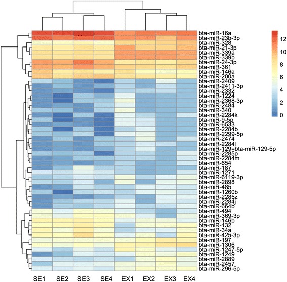
Comparative miRNAome analysis revealed different miRNA expression profiles in bovine sera and exosomes.
Overview
The claims and statistics of this paper haven’t been checked yet.
Run the check — free, no account needed. It reads the open-access text and checks the sources it cites.
Integrity signals
Registry
Cites
Statistics
Figures
Verification
Claims the paper makes
The claims and statistics of this paper haven’t been checked yet.
Run the check — free, no account needed. It reads the open-access text and checks the sources it cites.
Claims other papers make about this one
Each is checked against this paper’s own text: does it say what the citing paper says it does?
The cited source says what the claim attributes to it.
In the citing paper
“It has been reported that Ago2 complexes harbor a circulating miRNA population in human plasma”
From Red Blood Cells as Potential Repositories of MicroRNAs in the Circulatory System (2020) · cites this paper as Zhao et al., 2016
Self-check
Percentages, sums and reported differences re-computed from the paper’s own numbers.
The self-check needs the paper’s full text.
Citations
Cites
Circulating microRNAs, potential biomarkers for drug-induced liver injury
Wang, K., Zhang, S., Marzolf, B., Troisch, P., Brightman, A., Hu, Z., Hood, L. E., & Galas, D. J. (2009) · Proceedings of the National Academy of Sciences · doi:10.1073/pnas.0813371106 · Cited by 1,149
Blood Cell Origin of Circulating MicroRNAs: A Cautionary Note for Cancer Biomarker Studies
Pritchard, C. C., Kroh, E., Wood, B., Arroyo, J. D., Dougherty, K. J., Miyaji, M. M., Tait, J. F., & Tewari, M. (2012) · Cancer Prevention Research · doi:10.1158/1940-6207.capr-11-0370 · Cited by 879
Kapsimali, M., Kloosterman, W. P., de Bruijn, E., Rosa, F., Plasterk, R. H., & Wilson, S. W. (2007) · Genome Biology · doi:10.1186/gb-2007-8-8-r173 · Cited by 376
Research works printed but not linked: 62
- Seegers H, Fourichon C, Beaudeau F. Production effects related to mastitis and mastitis economics in dairy cattle herds. Vet Res. 2002;34(5):475–91. · DOI
- Schwaiger T, Beauchemin KA, Penner GB. The duration of time that beef cattle are fed a high-grain diet affects the recovery from a bout of ruminal acidosis: dry matter intake and ruminal fermentation. J Anim Sci. 2013;91(12):5729–42. · DOI
- Wierenga K, McAllister TA, Gibb DJ, Chaves AV, Okine EK, Beauchemin KA, Oba M. Evaluation of triticale dried distillers grains with solubles as a substitute for barley grain and barley silage in feedlot finishing diets. J Anim Sci. 2010;88(9):3018–29. · DOI
- Vergara CF, Döpfer D, Cook NB, Nordlund KV, McArt JAA, Nydam DV, Oetzel GR. Risk factors for postpartum problems in dairy cows: explanatory and predictive modeling. J Dairy Sci. 2014;97(7):4127–40. · DOI
- Gilad S, Meiri E, Yogev Y, Benjamin S, Lebanony D, Yerushalmi N, et al. Sera microRNAs are promising novel biomarkers. PLoS One. 2007;3(9):e3148. · DOI
- Qin X, Xu H, Gong W, Deng W. The Tumor Cytosol miRNAs, Fluid miRNAs, and Exosomes miRNAs in Lung Cancer. Front Oncol. 2013;4:357.
- Ambros V. The functions of animal microRNAs. Nature. 2004;431(7006):350–5. · DOI · Paper page
- Qin X, Yan L, Zhao X, Li C, Fu Y. microRNA-21 overexpression contributes to cell proliferation by targeting PTEN in endometrioid endometrial cancer. Oncol Lett. 2012;4(6):1290–6. · DOI
- Kim YJ, Hwang SH, Cho HH, Shin KK. MicroRNA 21 regulates the proliferation of human adipose tissue derived mesenchymal stem cells and high‐fat diet‐induced obesity alters microRNA 21 expression in white adipose tissues. J Cell Physiol. 2012;227(1):183–93. · DOI
- Zhang Z, Sun J, Bai Z, Li H, He S, Chen R. MicroRNA-153 acts as a prognostic marker in gastric cancer and its role in cell migration and invasion. Onco Targets Ther. 2015;8:357–64.
- Burgos KL, Javaherian A, Bomprezzi R, Ghaffari L, Rhodes S, Courtright A, et al. Identification of extracellular miRNA in human cerebrospinal fluid by next-generation sequencing. RNA. 2013;19(5):712–22. · DOI
- Wu F, Guo NJ, Tian H, Marohn M, Gearhart S, Bayless TM, et al. Peripheral blood microRNAs distinguish active ulcerative colitis and Crohn’s disease. Inflamm Bowel Dis. 2011;17(1):241–50. · DOI
- Hoekstra M, van der Lans CAC, Halvorsen B, Gullestad L, Kuiper J, Aukrust P, et al. The peripheral blood mononuclear cell microRNA signature of coronary artery disease. Biochem Biophys Res Commun. 2010;394(3):792–7. · DOI
- Meder B, Keller A, Vogel B, Haas J, Sedaghat-Hanmedani F, Kayvanpour E, et al. MicroRNA signatures in total peripheral blood as novel biomarkers for acute myocardial infarction. Basic Res Cardiol. 2011;106(1):13–23. · DOI
- Leidinger P, Backes C, Meder B, Meese E, Keller A. The human miRNA repertoire of different blood compounds. BMC Genomics. 2014;15:474. · DOI · Paper page
- Cheng L, Sharples RA, Scicluna BJ, Hill AF. Exosomes provide a protective and enriched source of miRNA for biomarker profiling compared to intracellular and cell-free blood. J Extracell Vesicles. 2014;3:23743. · DOI · Paper page
- Zubakov D, Boersma AW, Choi Y, van Kuijk PF, Wiemer EA, Kayser M. MicroRNA markers for forensic body fluid identification obtained from microarray screening and quantitative RT-PCR confirmation. Int Legal Med. 2010;124(3):217–26. · DOI
- Weber JA, Baxter DH, Zhang S, Huang DY, Huang KH, Lee MJ, et al. The microRNA spectrum in 12 body fluids. Clin Chem. 2010;56(11):1733–41. · DOI · Paper page
- Tzimagiorgis G, Michailidou EZ, Kritis A, Markopoulos AK, Kouidou S. Recovering circulating extracellular or cell-free RNA from bodily fluids. Cancer Epidemiol. 2011;35(6):580–9. · DOI
- Mitchell PS, Parkin RK, Kroh EM, Fritz BR, Wyman SK, Pogosova-Agadjanyan EL, et al. Circulating microRNAs as stable blood-based markers for cancer detection. Proc Natl Acad Sci U S A. 2008;105(30):10513–8. · DOI · Paper page
- Chen X, Ba Y, Ma L, Cai X, Yin Y, Wang K, et al. Characterization of microRNAs in sera: a novel class of biomarkers for diagnosis of cancer and other diseases. Cell Res. 2008;18(10):997–1006. · DOI · Paper page
- Ioannidis J, Donadeu FX. Circulating miRNA signatures of early pregnancy in cattle. BMC Genomics. 2016;17:184. · DOI
- Zheng Y, Chen KL, Zheng XM, Li HX, Wang GL. Identification and bioinformatics analysis of microRNAs associated with stress and immune response in serum of heat-stressed and normal Holstein cows. Cell Stress Chaperones. 2014;19(6):973–81. · DOI
- Han W, Zhu Y, Su Y, Li G, Qu L, Zhang H, et al. High-throughput sequencing reveals circulating miRNAs as potential biomarkers for measuring puberty onset in chicken (Gallus gallus). PLoS One. 2016;11(5):e0154958. · DOI
- Andersson P, Gidlöf O, Braun OO, Götberg M, van der Pals J, Olde B, et al. Plasma levels of liver-specific miR-122 is massively increased in a porcine cardiogenic shock model and attenuated by hypothermia. Shock. 2012;37(2):234–8. · DOI
- Ashby J, Flack K, Jimenez LA, Duan Y, Khatib A-KK, Somlo G, et al. Distribution profiling of circulating microRNAs in sera. Anal Chem. 2014;86(18):9343–9. · DOI
- Raposo G, Nijman HW, Stoorvogel W, Liejendekker R, Harding CV, Melief CJ, et al. B lymphocytes secrete antigen-presenting vesicles. J Exp Med. 1996;183(3):1161–72. · DOI · Paper page
- Denzer K, van Eijk M, Kleijmeer MJ, Jakobson E, de Groot C, Geuze HJ. Follicular dendritic cells carry MHC class II-expressing microvesicles at their surface. J Immunol. 2000;165(3):1259–65. · DOI
- Hu G, Drescher KM, Chen XM. Exosomal miRNAs: biological properties and therapeutic potential. Front Genet. 2011;3:56.
- Kourembanas S. Exosomes: vehicles of intercellular signaling, biomarkers, and vectors of cell therapy. Annu Rev Physiol. 2015;77:13–27. · DOI · Paper page
- Pritchard CC, Cheng HH, Tewari M. MicroRNA profiling: approaches and considerations. Nat Rev Gene. 2012;13(5):358–69. · DOI · Paper page
- Dhahbi JM, Spindler SR, Atamna H, Boffelli D, Mote P, Martin DI. 5’-YRNA fragments derived by processing of transcripts from specific YRNA genes and pseudogenes are abundant in human sera and plasma. Physiol Genomics. 2013;45(21):990–8. · DOI
- Dhahbi JM, Spindler SR, Atamna H, Yamakawa A, Boffelli D, Mote P, et al. 5’ tRNA halves are present as abundant complexes in sera, concentrated in blood cells, and modulated by aging and calorie restriction. BMC Genomics. 2012;14:298. · DOI
- Okamura K. Diversity of animal small RNA pathways and their biological utility. Wiley Interdiscip Rev RNA. 2012;3(3):351–68. · DOI
- Zhang C. Novel functions for small RNA molecules. Curr Opin Mol Ther. 2009;11(6):641–51.
- Esteller M. Non-coding RNAs in human disease. Nat Rev Genet. 2011;12(12):861–74. · DOI · Paper page
- Kramer BS. The science of early detection. Urol Oncol. 2004;22(4):344–7. · DOI
- Mestdagh P, Hartmann N, Baeriswyl L, Andreasen D, Bernard N, Chen C, et al. Evaluation of quantitative miRNA expression platforms in the microRNA quality control (miRQC) study. Nat Methods. 2014;11(8):809–15. · DOI · Paper page
- Rasmussen KD, Simmini S, Abreu-Goodger C, Bartonicek N, Di Giacomo M, Bilbao-Cortes D, et al. The miR-144/451 locus is required for erythroid homeostasis. J Experi Med. 2010;207(7):1351–8. · DOI · Paper page
- Spornraft M, Kirchner B, Pfaffl MW, Riedmaier I. Comparison of the miRNAome and piRNome of bovine blood and plasma by small RNA sequencing. Biotechnol Lett. 2015;37(6):1165–76. · DOI
- Vickers KC, Palmisano BT, Shoucri BM, Shamburek RD, Remaley AT. MicroRNAs are transported in plasma and delivered to recipient cells by high-density lipoproteins. Nat Cell Biol. 2011;13(4):423–33. · DOI · Paper page
- Arroyo JD, Chevillet JR, Kroh EM, Ruf IK, Pritchard CC, Gibson DF, et al. Argonaute2 complexes carry a population of circulating microRNAs independent of vesicles in human plasma. Proc Natl Acad Sci U S A. 2011;108(12):5003–8. · DOI · Paper page
- Siomi H, Siomi MC. Posttranscriptional regulation of MicroRNA biogenesis in animals. Mol Cell. 2010;38:323–32. · DOI
- Mahajan VS, Drake A, Chen J. Virus-specific host miRNAs: antiviral defenses or promoters of persistent infection? Trends Immunol. 2009;30(1):1–7. · DOI
- Hou W, Tian Q, Zheng J, Bonkovsky HL. MicroRNA‐196 represses Bach1 protein and hepatitis C virus gene expression in human hepatoma cells expressing hepatitis C viral proteins. Hepatology. 2010;51(5):1494–504. · DOI
- Pedersen IM, Cheng G, Wieland S, Volinia S, Croce CM, Chisari FV, et al. Interferon modulation of cellular microRNAs as an antiviral mechanism. Nature. 2007;449(7164):919–22. · DOI
- Ajizian SJ, English BK, Meals EA. Specific inhibitors of p38 and extracellular signal-regulated kinase mitogen-activated protein kinase pathways block inducible nitric oxide synthase and tumor necrosis factor accumulation murine macrophages stimulated with lipopolysaccharide and interferon-gamma. J Infect Dis. 1999;179(4):939–44. · DOI
- Haddad EB, Birrell M, McCluskie K, Ling A, Webber SE, Foster ML, et al. Role of p38 MAP kinase in LPS-induced airway inflammation in the rat. Br J Pharmacol. 2001;132(8):1715–24. · DOI
- Schmeck B, Zahlten J, Moog K, van Laak V, Huber S, Hocke AC, et al. Streptococcus pneumoniae-induced p38 MAPK-dependent phosphorylation of RelA at the interleukin-8 promotor. J Biol Chem. 2004;279(51):53241–7. · DOI
- Jung HJ, Coffinier C, Choe Y, Beigneux AP, Davies BS, Yang SH, et al. Regulation of prelamin A but not lamin C by miR-9, a brain-specific microRNA. Proc Natl Acad Sci U S A. 2012;109(7):E423–31. · DOI
- Nass D, Rosenwald S, Meiri E, Gilad S, Tabibian-Keissar H, Schlosberg A, et al. MiR-92b and miR‐9/9* are specifically expressed in brain primary tumors and can be used to differentiate primary from metastatic brain tumors. Brain Pathol. 2009;19(3):375–83. · DOI
- Zhao JJ, Hua YJ, Sun DG, Meng XX, Xiao HS, Ma X. Genome-wide microRNA profiling in human fetal nervous tissues by oligonucleotide microarray. Childs Nervs Syst. 2006;22(11):1419–25. · DOI
- Barturen G, Rueda A, Hamberg M. sRNAbench: profiling of small RNAs and its sequence variants in single or multi-species high-throughput experiments. Meth Next Generation Sequencing. 2014;1(1):2084–7173. · DOI
- Robinson MD, McCarthy DJ, Smyth GK. edgeR: a Bioconductor package for differential expression analysis of digital gene expression data. Bioinformatics. 2010;26(1):139–40. · DOI · Paper page
- Benjamini Y, Hochberg Y. Controlling the false discovery rate: a practical and powerful approach to multiple testing. J R Statist Soc B. 1995;57(1):289–300.
- Liang G, Malmuthuge N, McFadden TB, Bao H, Griebel PJ, Stothard P, et al. Potential regulatory role of microRNAs in the development of bovine gastrointestinal tract during early life. PLoS One. 2014;9(3):e92592. · DOI
- Enright AJ, John B, Gaul U, Tuschl T, Sander C. MicroRNA targets in Drosophila. Genome Biol. 2003;5(1):R1. · DOI · Paper page
- Livak KJ, Schmittgen TD. Analysis of relative gene expression data using real-time quantitative PCR and the 2(T)(-Delta Delta C) method. Methods. 2001;25(4):402–8. · DOI · Paper page
- Ng EK, Chong WW, Jin H, Lam EK, Shin VY, Yu J, et al. Differential expression of microRNAs in plasma of patients with colorectal cancer: a potential marker for colorectal cancer screening. Gut. 2009;58(10):1375–81. · DOI · Paper page
- Tsujiura M, Ichikawa D, Komatsu S, Shiozaki A, Takeshita H, Konishi H, et al. Circulating microRNAs in plasma of patients with gastric cancers. Br J Cancer. 2010;102(7):1174–9. · DOI
- Mi QS, Weiland M, Qi RQ, Gao XH, Poisson LM, Zhou L. Identification of mouse serum miRNA endogenous references by global gene expression profiles. PLoS One. 2012;7(2):e31278. · DOI
- Bae IS, Chung KY, Yi J, Kim TI, Choi HS, Cho YM, et al. Identification of reference genes for relative quantification of circulating microRNAs in bovine serum. PLoS One. 2015;10(3):e0122554. · DOI
Cited by
Sorted by citations
Red Blood Cells as Potential Repositories of MicroRNAs in the Circulatory System
Sun, L., Yu, Y., Niu, B., & Wang, D. (2020) · Frontiers in Genetics · doi:10.3389/fgene.2020.00442 · Cited by 83
Metadata
- Authors
- Zhao, K., Liang, G., Sun, X., & Guan, L. L.
- Venue
- BMC Genomics
- Published
- 2016-08-12
- Volume
- 17
- Issue
- 1
- ISSN
- 1471-2164
- PMC
- PMC4983018
- Cited by
- 51
- References
- 3
- Language
- en
- Funders
- Alberta Livestock and Meat Agency (CA) (ALMA 2011F129R)
About these checks
Registry, retraction, Cites and statistics results are computed, not judged, with one exception: a printed reference that no index lists is read once by a model before it counts as a citation error. Claims are extracted by a model and kept only when their quote appears verbatim in the text; each cited source is then checked, with a model’s help, for whether it says what the claim relies on. Those verdicts can be wrong — read the claim’s quote and the cited source before relying on one.
Statistics and arithmetic are recomputed, not judged.
Confidence bands are shown only for fields calibrated on our annotated test set. None is calibrated yet, so no band is shown.
What counts as a claim ›How we measure confidence ›How we check this ›
Are you an author, or do you see a problem with this page? Request a review
