

The DOI matches its Crossref record; 117 of the 117 references it cites were found in a registry or an index.
Systemic inflammation in early neonatal mice induces transient and lasting neurodegenerative effects
Open accessAbstract
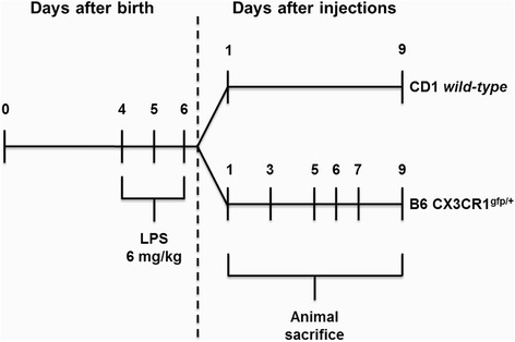
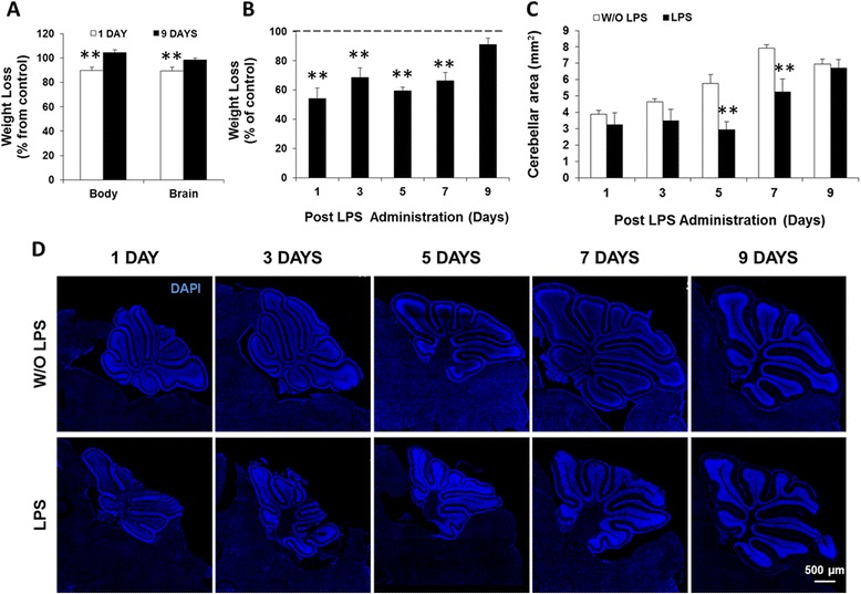
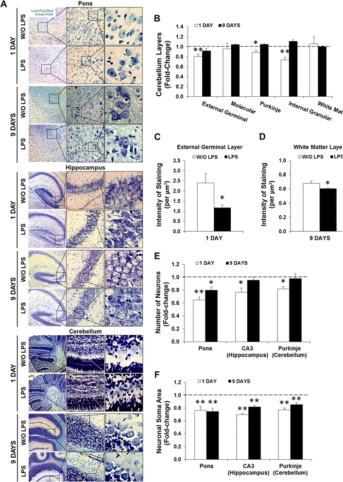
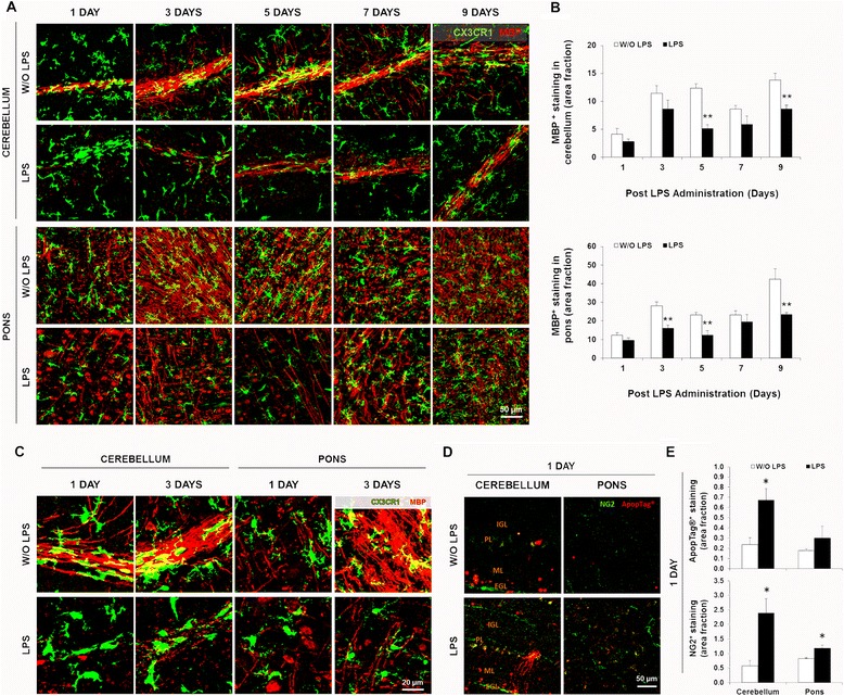
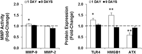
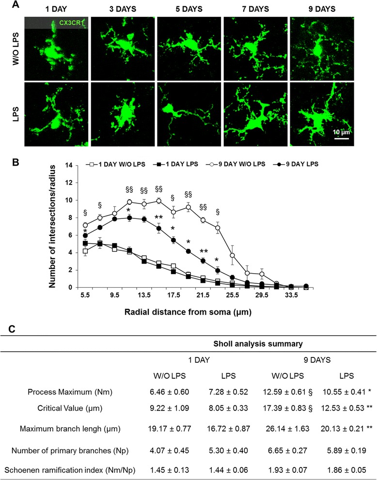
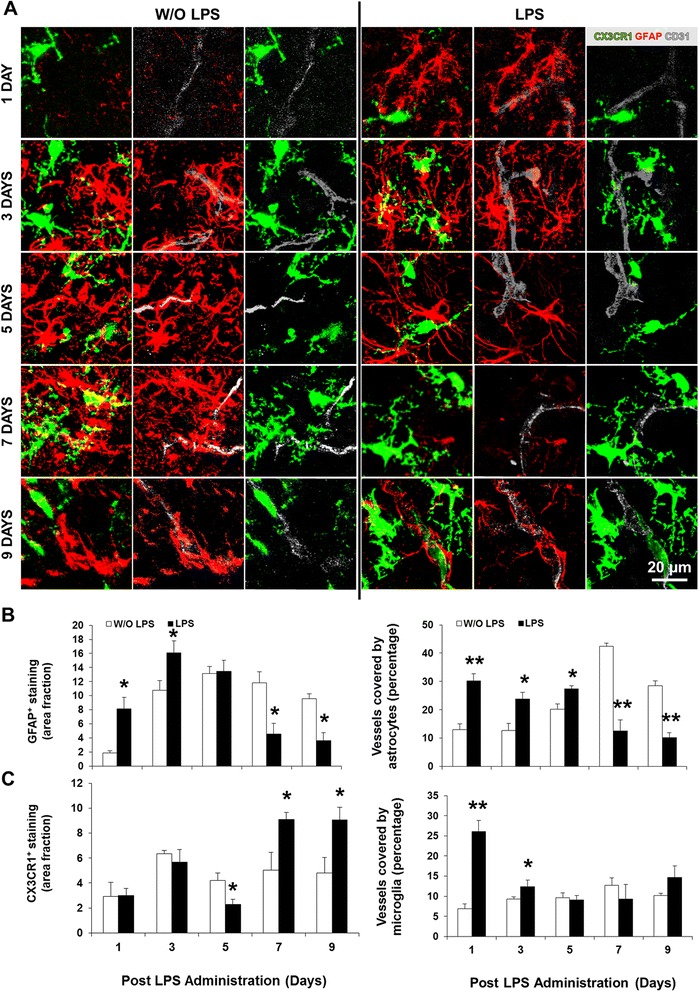
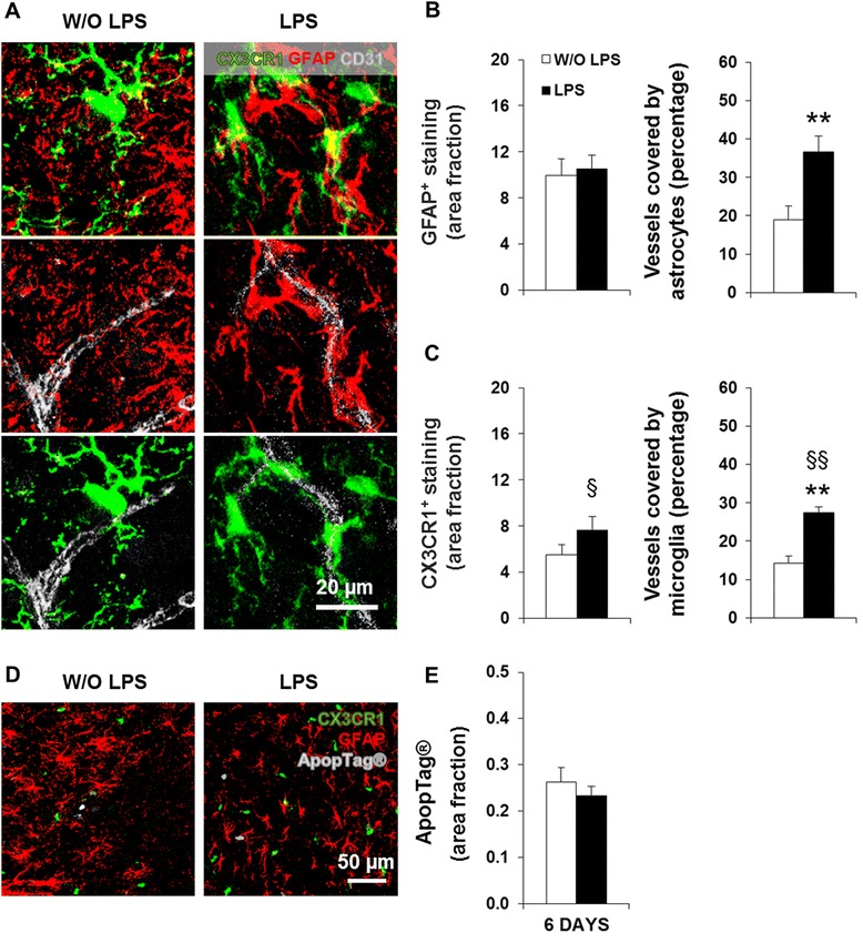
BACKGROUND: The inflammatory mediator lipopolysaccharide (LPS) has been shown to induce acute gliosis in neonatal mice. However, the progressive effects on the murine neurodevelopmental program over the week that follows systemic inflammation are not known. Thus, we investigated the effects of repeated LPS administration in the first postnatal week in mice, a condition mimicking sepsis in late preterm infants, on the developing central nervous system (CNS). METHODS: Systemic inflammation was induced by daily intraperitoneal administration (i.p.) of LPS (6 mg/kg) in newborn mice from postnatal day (PND) 4 to PND6. The effects on neurodevelopment were examined by staining the white matter and neurons with Luxol Fast Blue and Cresyl Violet, respectively. The inflammatory response was assessed by quantifying the expression/activity of matrix metalloproteinases (MMP), toll-like receptor (TLR)-4, high mobility group box (HMGB)-1, and autotaxin (ATX). In addition, B6 CX3CR1(gfp/+) mice combined with cryo-immunofluorescence were used to determine the acute, delayed, and lasting effects on myelination, microglia, and astrocytes. RESULTS: LPS administration led to acute body and brain weight loss as well as overt structural changes in the brain such as cerebellar hypoplasia, neuronal loss/shrinkage, and delayed myelination. The impaired myelination was associated with alterations in the proliferation and differentiation of NG2 progenitor cells early after LPS administration, rather than with excessive phagocytosis by CNS myeloid cells. In addition to disruptions in brain architecture, a robust inflammatory response to LPS was observed. Quantification of inflammatory biomarkers revealed decreased expression of ATX with concurrent increases in HMGB1, TLR-4, and MMP-9 expression levels. Acute astrogliosis (GFAP(+) cells) in the brain parenchyma and at the microvasculature interface together with parenchymal microgliosis (CX3CR1(+) cells) were also observed. These changes preceded the migration/proliferation of CX3CR1(+) cells around the vessels at later time points and the subsequent loss of GFAP(+) astrocytes. CONCLUSION: Collectively, our study has uncovered a complex innate inflammatory reaction and associated structural changes in the brains of neonatal mice challenged peripherally with LPS. These findings may explain some of the neurobehavioral abnormalities that develop following neonatal sepsis.
Overview
The claims and statistics of this paper haven’t been checked yet.
Run the check — free, no account needed. It reads the open-access text and checks the sources it cites.
Integrity signals
Registry
Cites
Statistics
Figures
Verification
Claims the paper makes
The claims and statistics of this paper haven’t been checked yet.
Run the check — free, no account needed. It reads the open-access text and checks the sources it cites.
Claims other papers make about this one
Each is checked against this paper’s own text: does it say what the citing paper says it does?
In the citing paper
“For instance, experimentally-induced systemic inflammation during infancy leads to neuronal loss and delayed myelination for up to a week (Cardoso et al., 2015).”
From Developmental psychoneuroendocrine and psychoneuroimmune pathways from childhood adversity to disease (2017) · cites this paper as Cardoso et al., 2015
Self-check
Percentages, sums and reported differences re-computed from the paper’s own numbers.
The self-check needs the paper’s full text.
Citations
Cites
How Matrix Metalloproteinases Regulate Cell Behavior
Sternlicht, M. D., & Werb, Z. (2001) · Annual Review of Cell and Developmental Biology · doi:10.1146/annurev.cellbio.17.1.463 · Cited by 3,959
Global, regional, and national causes of child mortality in 2008: a systematic analysis
Black, R. E., Cousens, S. N., Johnson, H. L., Lawn, J. E., Rudan, I., Bassani, D. G., Jha, P., Campbell, H. J., Walker, C. L. F., Cibulskis, R. E., et al. (2010) · The Lancet · doi:10.1016/s0140-6736(10)60549-1 · Cited by 3,062
Jung, S., Aliberti, J., Graemmel, P., Sunshine, M. J., Kreutzberg, G. W., Sher, A., & Littman, D. R. (2000) · Molecular and Cellular Biology · doi:10.1128/mcb.20.11.4106-4114.2000 · Cited by 2,632
MD-2, a Molecule that Confers Lipopolysaccharide Responsiveness on Toll-like Receptor 4
Shimazu, R., Akashi, S., Ogata, H., Nagai, Y., Fukudome, K., Miyake, K., & Kimoto, M. (1999) · The Journal of Experimental Medicine · doi:10.1084/jem.189.11.1777 · Cited by 2,187
Pericytes are required for blood–brain barrier integrity during embryogenesis
Daneman, R., Zhou, L., Kebede, A. A., & Barres, B. A. (2010) · Nature · doi:10.1038/nature09513 · Cited by 2,128
Thrombospondins are astrocyte-secreted proteins that promote CNS synaptogenesis
Christopherson, K. S., Ullian, E. M., Stokes, C. C. A., Mullowney, C. E., Hell, J. W., Agah, A., Lawler, J., Mosher, D. F., Bornstein, P., & Barres, B. A. (2005) · Cell · doi:10.1016/j.cell.2004.12.020 · Cited by 1,688
Sepsis and septic shock—A review of laboratory models and a proposal
Wichterman, K. A., Baue, A. E., & Chaudry, I. H. (1980) · Journal of Surgical Research · doi:10.1016/0022-4804(80)90037-2 · Cited by 1,466
HMGB1 is a therapeutic target for sterile inflammation and infection
Andersson, U., & Tracey, K. J. (2011) · Annual Review of Immunology · doi:10.1146/annurev-immunol-030409-101323 · Cited by 1,437
Zhan, Y., Paolicelli, R. C., Sforazzini, F., Weinhard, L., Bolasco, G., Pagani, F., Vyssotski, A. L., Bifone, A., Gozzi, A., Ragozzino, D., et al. (2014) · Nature Neuroscience · doi:10.1038/nn.3641 · Cited by 1,236
Microglia development and function
Nayak, D., Roth, T. L., & McGavern, D. B. (2014) · Annual Review of Immunology · doi:10.1146/annurev-immunol-032713-120240 · Cited by 1,154
Self-citation
Cited by
Sorted by citations
The blood-brain barrier in systemic inflammation
Varatharaj, A., & Galea, I. (2017) · Brain, Behavior, and Immunity · doi:10.1016/j.bbi.2016.03.010 · Cited by 1,172
The blood-brain barrier in aging and neurodegeneration
Knox, E. G., Aburto, M. R., Clarke, G., Cryan, J. F., & O’Driscoll, C. M. (2022) · Molecular Psychiatry · doi:10.1038/s41380-022-01511-z · Cited by 738
Disrupting the blood–brain barrier by focused ultrasound induces sterile inflammation
Kovacs, Z. I., Kim, S., Jikaria, N., Qureshi, F., Milo, B., Lewis, B. K., Bresler, M., Burks, S. R., & Frank, J. A. (2017) · Proceedings of the National Academy of Sciences · doi:10.1073/pnas.1614777114 · Cited by 458
Systemic inflammation impairs microglial Aβ clearance through NLRP3 inflammasome
Tejera, D., Mercan, D., Sanchez‐Caro, J. M., Hanan, M., Greenberg, D., Soreq, H., Latz, E., Golenbock, D., & Heneka, M. T. (2019) · The EMBO Journal · doi:10.15252/embj.2018101064 · Cited by 351
The Impact of Systemic Inflammation on Alzheimer’s Disease Pathology
Xie, J., Van Hoecke, L., & Vandenbroucke, R. E. (2022) · Frontiers in Immunology · doi:10.3389/fimmu.2021.796867 · Cited by 324
Sepsis-associated encephalopathy: A vicious cycle of immunosuppression
Ren, C., Yao, R.-Q., Zhang, H., Feng, Y.-W., & Yao, Y.-M. (2020) · Journal of Neuroinflammation · doi:10.1186/s12974-020-1701-3 · Cited by 259
Kuhlman, K. R., Chiang, J. J., Horn, S., & Bower, J. E. (2017) · Neuroscience & Biobehavioral Reviews · doi:10.1016/j.neubiorev.2017.05.020 · Cited by 208
Neuropathobiology of COVID-19: The Role for Glia
Tremblay, M.-E., Madore, C., Bordeleau, M., Tian, L., & Verkhratsky, A. (2020) · Frontiers in Cellular Neuroscience · doi:10.3389/fncel.2020.592214 · Cited by 159
Crapser, J. D., Ochaba, J., Soni, N., Reidling, J. C., Thompson, L. M., & Green, K. N. (2020) · Brain · doi:10.1093/brain/awz363 · Cited by 157
Leaky brain in neurological and psychiatric disorders: Drivers and consequences
Morris, G., Fernandes, B. S., Puri, B. K., Walker, A. J., Carvalho, A. F., & Berk, M. (2018) · Australian & New Zealand Journal of Psychiatry · doi:10.1177/0004867418796955 · Cited by 145
Cerebellar microglia are dynamically unique and survey Purkinje neurons in vivo
Stowell, R. D., Wong, E. L., Batchelor, H. N., Mendes, M. S., Lamantia, C. E., Whitelaw, B. S., & Majewska, A. K. (2018) · Developmental Neurobiology · doi:10.1002/dneu.22572 · Cited by 124
Targeting the Blood-Brain Barrier to Prevent Sepsis-Associated Cognitive Impairment
Nwafor, D. C., Brichacek, A. L., Mohammad, A. S., Griffith, J. I. G., Lucke-Wold, B. P., Benkovic, S. A., Geldenhuys, W. J., Lockman, P. R., & Brown, C. M. (2019) · Journal of Central Nervous System Disease · doi:10.1177/1179573519840652 · Cited by 126
Astroglia in Sepsis Associated Encephalopathy
Shulyatnikova, T. V., & Verkhratsky, A. (2019) · Neurochemical Research · doi:10.1007/s11064-019-02743-2 · Cited by 125
Neuroanatomy and Physiology of Brain Dysfunction in Sepsis
Mazeraud, A., Pascal, Q., Verdonk, F., Heming, N., Chrétien, F., & Sharshar, T. (2016) · Clinics in Chest Medicine · doi:10.1016/j.ccm.2016.01.013 · Cited by 107
Microglial Function in the Effects of Early-Life Stress on Brain and Behavioral Development
Catale, C., Gironda, S. C., Iacono, L. L., & Carola, V. (2020) · Journal of Clinical Medicine · doi:10.3390/jcm9020468 · Cited by 108
Air Pollution–Related Neurotoxicity Across the Life Span
Cory‐Slechta, D. A., Merrill, A. K., & Sobolewski, M. (2022) · The Annual Review of Pharmacology and Toxicology · doi:10.1146/annurev-pharmtox-051921-020812 · Cited by 89
Cunha, C., Santos, C., Gomes, C., Fernandes, A. M. A., Correia, A. M., Sebastião, A. M., Vaz, A. R., & Brites, D. M. T. O. (2017) · Molecular Neurobiology · doi:10.1007/s12035-017-0631-2 · Cited by 88
Self-citationMacrophage depletion reduced brain injury following middle cerebral artery occlusion in mice
Ma, Y., Li, Y., Jiang, L., Wang, L., Jiang, Z., Wang, Y., Zhang, Z., & Yang, G. (2016) · Journal of Neuroinflammation · doi:10.1186/s12974-016-0504-z · Cited by 80
Impact of Neuroinflammation on Hippocampal Neurogenesis: Relevance to Aging and Alzheimer’s Disease
Valero, J., Bernardino, L. I., Cardoso, F. L., Silva, A. P., Ribeiro, C., Ambrósio, A. F., & Malva, J. O. (2017) · Journal of Alzheimer s Disease · doi:10.3233/jad-170239 · Cited by 86
Self-citationExperimental necrotizing enterocolitis induces neuroinflammation in the neonatal brain
Biouss, G., Antounians, L., Li, B., O’Connell, J. S., Seo, S., Catania, V. D., Guadagno, J., Rahman, A., Zani‐Ruttenstock, E., Svergun, N., et al. (2019) · Journal of Neuroinflammation · doi:10.1186/s12974-019-1481-9 · Cited by 79
Ethanol-Induced Neurodegeneration and Glial Activation in the Developing Brain
Saito, M., Chakraborty, G., Hui, M., Masiello, K., & Saito, M. (2016) · Brain Sciences · doi:10.3390/brainsci6030031 · Cited by 72
The impact of inflammation on respiratory plasticity
Hocker, A. D., Stokes, J. A., Powell, F. L., & Huxtable, A. G. (2016) · Experimental Neurology · doi:10.1016/j.expneurol.2016.07.022 · Cited by 63
Current Understanding of Long-Term Cognitive Impairment After Sepsis
Li, Y., Ji, M., & Yang, J. (2022) · Frontiers in Immunology · doi:10.3389/fimmu.2022.855006 · Cited by 67
Salmani, H., Hosseini, M., Baghcheghi, Y., Moradi‐Marjaneh, R., & Mokhtari‐Zaer, A. (2019) · Cytokine · doi:10.1016/j.cyto.2019.154860 · Cited by 53
Adiponectin deficiency accelerates brain aging via mitochondria-associated neuroinflammation
He, K., Nie, L., Ali, T., Liu, Z., Li, W., Gao, R., Zhang, Z., Liu, J., Dai, Z., Xie, Y., et al. (2023) · Immunity & Ageing · doi:10.1186/s12979-023-00339-7 · Cited by 51
High Mobility Group Box-1: A Missing Link between Diabetes and Its Complications
Wu, H., Chen, Z., Xie, J., Kang, L., Wang, L., & Xu, B. (2016) · Mediators of Inflammation · doi:10.1155/2016/3896147 · Cited by 46
Consequences of early postnatal lipopolysaccharide exposure on developing lungs in mice
Shrestha, A. K., Bettini, M. L., Menon, R., Gopal, V. Y. N., Huang, S., Edwards, D. P., Pammi, M., Barrios, R. J., & Shivanna, B. (2018) · American Journal of Physiology-Lung Cellular and Molecular Physiology · doi:10.1152/ajplung.00560.2017 · Cited by 47
A severe mouse model of spinal muscular atrophy develops early systemic inflammation
Wan, B., Feng, P., Guan, Z., Sheng, L., Liu, Z., & Hua, Y. (2018) · Human Molecular Genetics · doi:10.1093/hmg/ddy300 · Cited by 46
Maysinger, D., Ji, J. X., Hutter, E., & COOPER, E. (2015) · Frontiers in Neuroscience · doi:10.3389/fnins.2015.00480 · Cited by 45
Stoian, A., Stoian, M., Bajkó, Z., Maier, S., Andone, S. R., Cioflinc, R. A., Moțățăianu, A., Bărcuţean, L., & Bălașa, R. (2022) · Biomedicines · doi:10.3390/biomedicines10040774 · Cited by 42
Zhang, X., Rocha‐Ferreira, E., Li, T., Vontell, R., Jabin, D., Hua, S., Zhou, K., Nazmi, A., Albertsson, A.-M., Sobotka, K. S., et al. (2017) · Journal of Neuroinflammation · doi:10.1186/s12974-017-1029-9 · Cited by 40
Salmani, H., Hosseini, M., Beheshti, F., Baghcheghi, Y., Sadeghnia, H., Soukhtanloo, M., Shafei, M., & Khazaei, M. (2018) · Life Science · doi:10.1016/j.lfs.2018.04.033 · Cited by 36
Coelho‐Santos, V., Cardoso, F. L., Leitão, R. A., Ribeiro, C., & Silva, A. P. (2017) · Brain Behavior and Immunity · doi:10.1016/j.bbi.2017.10.016 · Cited by 39
Self-citationUseinovic, N., Maksimovic, S., Liechty, C., Cabrera, O. H., Quillinan, N., & Jevtović‐Todorović, V. (2022) · British Journal of Anaesthesia · doi:10.1016/j.bja.2022.05.008 · Cited by 36
Girard, T. D., Dittus, R. S., & Ely, E. W. (2016) · Annual Review of Medicine · doi:10.1146/annurev-med-050913-015722 · Cited by 37
Masouleh, S. K., Beyer, F., Lampe, L., Loeffler, M., Luck, T., Riedel-Heller, S. G., Schroeter, M. L., Stümvoll, M., Villringer, A., & Witte, A. V. (2017) · Journal of Cerebral Blood Flow & Metabolism · doi:10.1177/0271678x17729111 · Cited by 35
Sepsis, Cytokine Storms, and Immunopathology: The Divide between Neonates and Adults
Greenfield, K. G., Badovinac, V. P., Griffith, T. S., & Knoop, K. A. (2021) · ImmunoHorizons · doi:10.4049/immunohorizons.2000104 · Cited by 32
Zhao, M., Li, C. H., & Liu, Y. L. (2016) · Genetics and Molecular Research · doi:10.4238/gmr.15026998 · Cited by 33
Enteric glial reactivity to systemic LPS administration: Changes in GFAP and S100B protein
Franceschi, R. D. C., Nardin, P., Machado, C. V., Tortorelli, L. S., Martinez‐Pereira, M. A., Zanotto, C., Gonçalves, C., & Zancan, D. M. (2017) · Neuroscience Research · doi:10.1016/j.neures.2016.12.005 · Cited by 33
Wolf, E. J., Miller, D. R., Logue, M. W., Sumner, J. A., Stoop, T. B., Leritz, E. C., Hayes, J. P., Stone, A., Schichman, S. A., McGlinchey, R. E., et al. (2017) · Brain Behavior and Immunity · doi:10.1016/j.bbi.2017.06.001 · Cited by 31
Liu, Y., Yu, Y., Liu, J., Liu, W., Cao, Y., Yan, R., & Yao, Y. (2022) · Frontiers in Neurology · doi:10.3389/fneur.2022.892480 · Cited by 32
Seman, B. G., Vance, J. K., Rawson, T. W., Witt, M. R., Huckaby, A. B., Povroznik, J. M., Bradford, S. D., Barbier, M., & Robinson, C. M. (2019) · Infection and Immunity · doi:10.1128/iai.00828-19 · Cited by 29
Advancement in biomarker based effective diagnosis of neonatal sepsis
Gopal, N., Chauhan, N., Jain, U., Dass, S. K., SHARMA, H. S., & Chandra, R. (2023) · Artificial Cells Nanomedicine and Biotechnology · doi:10.1080/21691401.2023.2252016 · Cited by 26
Pires, J. M., Foresti, M. L., Silva, C. S., Rêgo, D. B., Calió, M. L., Mosini, A. C., Nakamura, T. K. E., Leslie, A. T. F. S., & Mello, L. E. (2020) · Frontiers in Cellular Neuroscience · doi:10.3389/fncel.2020.00142 · Cited by 25
Preconditioned neurons with NaB and nicorandil, a favorable source for stroke cell therapy
Hosseini, S. M., Ziaee, S. M., Kh, H. H., Karimi, A., Tabeshmehr, P., & Abbasi, Z. (2018) · Journal of Cellular Biochemistry · doi:10.1002/jcb.27372 · Cited by 24
Denning, N., Aziz, M., Diao, L., Prince, J. M., & Wang, P. (2020) · Molecular Medicine · doi:10.1186/s10020-020-00243-6 · Cited by 24
Cerebellar development after preterm birth
Iskusnykh, I. Y., & Chizhikov, V. V. (2022) · Frontiers in Cell and Developmental Biology · doi:10.3389/fcell.2022.1068288 · Cited by 24
Neonatal white matter damage and the fetal inflammatory response
Gilles, F. H., & Leviton, A. (2020) · Seminars in Fetal and Neonatal Medicine · doi:10.1016/j.siny.2020.101111 · Cited by 23
Shrestha, A. K., Menon, R., El‐Saie, A., Barrios, R. J., Reynolds, C. L., & Shivanna, B. (2020) · American Journal of Physiology-Lung Cellular and Molecular Physiology · doi:10.1152/ajplung.00013.2020 · Cited by 25
Veerasammy, S., Steenwinckel, J. V., Charpentier, T. L., Seo, J. H., Fleiss, B., Gressèns, P., & Levison, S. W. (2020) · Brain Behavior & Immunity - Health · doi:10.1016/j.bbih.2020.100106 · Cited by 23
Shim, S., Cho, S. J., & Park, E. A. (2021) · Journal of Korean Medical Science · doi:10.3346/jkms.2021.36.e205 · Cited by 20
Zhang, D., Yang, Y., Yang, Y., Liu, J., Zhu, T., Huang, H., & Zhou, C. (2022) · BMC Medicine · doi:10.1186/s12916-022-02434-w · Cited by 19
Ureaplasma-Driven Neuroinflammation in Neonates: Assembling the Puzzle Pieces
Silwedel, C., Speer, C. P., Härtel, C., & Glaser, K. (2020) · Neonatology · doi:10.1159/000512019 · Cited by 19
Singh, K., Patro, N., Pradeepa, M., & Patro, I. K. (2017) · Annals of Neurosciences · doi:10.1159/000477152 · Cited by 19
Whitaker, E. E., Christofi, F. L., Quinn, K. M., Wiemann, B. Z., Xia, J. C., Tobias, J. D., & Bissonnette, B. (2017) · Journal of Anesthesia · doi:10.1007/s00540-016-2294-y · Cited by 18
Role of Imaging Modalities and N -Acetylcysteine Treatment in Sepsis-Associated Encephalopathy
Zhong, Y., Guan, J., Ma, Y., Xu, M., Cheng, Y., Xu, L., Lin, Y., Zhang, X., & Wu, R. (2023) · ACS Chemical Neuroscience · doi:10.1021/acschemneuro.3c00180 · Cited by 17
Fetal brain vulnerability to SARS-CoV-2 infection
McMahon, C. L., Castro, J., Silvas, J. A., Perez, A. M., Estrada, M., Carrion, R., & Hsieh, J. (2023) · Brain Behavior and Immunity · doi:10.1016/j.bbi.2023.06.015 · Cited by 17
Ijomone, O. K., Shallie, P. D., & Naicker, T. (2020) · Neurochemical Research · doi:10.1007/s11064-020-02969-5 · Cited by 16
The biological alterations of synapse/synapse formation in sepsis-associated encephalopathy
Tang, C., Ye, J., & Wang, H. (2022) · Frontiers in Synaptic Neuroscience · doi:10.3389/fnsyn.2022.1054605 · Cited by 16
Lang, G.-P., Ndongson‐Dongmo, B., Lajqi, T., Brodhun, M., Han, Y., Wetzker, R., Frasch, M. G., & Bauer, R. (2020) · Journal of Neuroinflammation · doi:10.1186/s12974-020-01954-7 · Cited by 15
Microbiome function and neurodevelopment in Black infants: vitamin B 12 emerges as a key factor
Oliphant, K., Ayala, W. C., Ilyumzhinova, R., Mbayiwa, K., Sroka, A. W., Xie, B., Andrews, B., Keenan, K. E., & Claud, E. C. (2024) · Gut Microbes · doi:10.1080/19490976.2023.2298697 · Cited by 14
Tian, L., Zhou, D., Guo, J., Liu, W., & Mu, J. (2017) · Neural Regeneration Research · doi:10.4103/1673-5374.219044 · Cited by 14
Sancho‐Balsells, A., Brito, V., Fernández, B., Pardo, M., Straccia, M., Ginés, S., Alberch, J., Hernández, I., Arranz, B., Canals, J. M., et al. (2020) · Frontiers in Cellular Neuroscience · doi:10.3389/fncel.2020.00093 · Cited by 14
Elhessy, H. M., Eltahry, H., Erfan, O. S., Mahdi, M. R., Hazem, N. M., & El-Shahat, M. A. (2020) · Acta Histochemica · doi:10.1016/j.acthis.2020.151633 · Cited by 14
Lou, J., Ding, B., Fang, M., Xie, W., Wang, X., Wang, X., Guo, X., & Zhu, J. (2024) · Molecular Neurobiology · doi:10.1007/s12035-024-04657-9 · Cited by 13
Abe, Y., Ochiai, D., Sato, Y., Kanzaki, S., Ikenoue, S., Kasuga, Y., & Tanaka, M. (2020) · International Journal of Molecular Sciences · doi:10.3390/ijms21249590 · Cited by 12
Neurodevelopment: Inflammation Matters
Davis, R. L. (2018) · Advances in neurotoxicology · doi:10.1016/bs.ant.2018.03.002 · Cited by 13
Insight Into Regulatory T Cells in Sepsis-Associated Encephalopathy
Gao, Y.-L., Liu, Y.-C., Zhang, X., Shou, S.-T., & Chai, Y.-F. (2022) · Frontiers in Neurology · doi:10.3389/fneur.2022.830784 · Cited by 11
Liao, Z., Ou, X., Zhou, C., Ma, D., Zhao, H., & Huang, H. (2022) · Frontiers in Pharmacology · doi:10.3389/fphar.2022.1002920 · Cited by 12
Hei, Y., Zhang, X., Chen, R., Zhou, Y., Gao, D., & Liu, W. (2019) · Cellular and Molecular Neurobiology · doi:10.1007/s10571-019-00702-7 · Cited by 10
Weaver, O., Gano, D. L., Zhou, Y., Kim, H., Tognatta, R., Yan, Z., Ryu, J. K., Brandt, C., Basu, T., Grana, M., et al. (2024) · Proceedings of the National Academy of Sciences · doi:10.1073/pnas.2323050121 · Cited by 9
Wu, Z., Xue, H., Zhang, Y., & Zhao, P. (2020) · Life Sciences · doi:10.1016/j.lfs.2020.118556 · Cited by 9
Glial cell responses in a murine multifactorial perinatal brain injury model
Domowicz, M. S., Wadlington, N. L., Henry, J. G., Diaz, K., Munoz, M. J., & Schwartz, N. B. (2017) · Brain Research · doi:10.1016/j.brainres.2017.12.020 · Cited by 9
Martínez‐Gopar, P. E., Pérez‐Rodríguez, M. J., Angeles-López, Q. D., Tristán-López, L. A., González‐Espinosa, C., & Pérez‐Severiano, F. (2023) · Molecular Neurobiology · doi:10.1007/s12035-023-03234-w · Cited by 7
Late Brain Involvement after Neonatal Immune Activation
Dias, P., Freiberger, V., Ventura, L., Bragagnolo, D., Dutra, M. L., Horewicz, V. V., & Comim, C. M. (2019) · BioMed Research International · doi:10.1155/2019/9573248 · Cited by 8
Balsamo, F., Li, B., Chusilp, S., Lee, D., Biouss, G., Lee, C. M., Maynes, J. T., & Pierro, A. (2023) · Pediatric Surgery International · doi:10.1007/s00383-023-05596-7 · Cited by 6
Phyllanthus amarus protects against spatial memory impairment induced by lipopolysaccharide in mice
Alagan, A. (2019) · Bioinformation · doi:10.6026/97320630015535 · Cited by 6
The Effect of Neonatal Sepsis on Risk of Autism Diagnosis
Getahun, D., Fassett, M. J., Xiang, A. H., Chiu, V. Y., Takhar, H. S., Shaw, S. F., & Peltier, M. R. (2021) · American Journal of Perinatology · doi:10.1055/s-0041-1731648 · Cited by 6
Shen, W., Wang, H., Wang, J., Yuan, Y., Luo, Y., Chen, X. G., Li, J., Liu, Y., Yin, Y., Wang, M., et al. (2026) · Cell Host & Microbe · doi:10.1016/j.chom.2026.02.004 · Cited by 5
Mujtaba, S., Patro, I. K., & Patro, N. (2023) · Brain Sciences · doi:10.3390/brainsci13101360 · Cited by 4
Neonatal Immune Activation May Provoke Long-term Depressive Attributes
Marzzani, S. H. S., Dias, P., Freiberger, V., Ventura, L., Silva, B. B., Dutra, M. L., Bobinski, F., Schlindwein, A. D., Cassol-Jr, O. J., & Comim, C. M. (2019) · Current Neurovascular Research · doi:10.2174/1567202616666191007125502 · Cited by 6
Neurodevelopment and the gut microbiome
Oliphant, K., & Lu, J. N. (2020) · Elsevier eBooks · doi:10.1016/b978-0-12-820602-7.00006-4 · Cited by 6
Rojas, D. B., Vizuete, A. F. K., Andrade, V. S., de Andrade, R. B., Gemelli, T., Kim, T. D. H., Gonçalves, C. A., Leipnitz, G., & Wannmacher, C. M. D. (2023) · International Journal of Developmental Neuroscience · doi:10.1002/jdn.10288 · Cited by 3
Neurotoxic Effects of Airborne Particulate Matter: Mechanisms and Health Implications
Thangavel, P., Park, D., & Lee, Y. (2025) · Journal of Biochemical and Molecular Toxicology · doi:10.1002/jbt.70322 · Cited by 3
Karafoulidou, E., Kesidou, E., Theotokis, P., Konstantinou, C., Nella, M., Michailidou, I., Touloumi, O., Polyzoidou, E., Salamotas, I., Einstein, O., et al. (2024) · Cells · doi:10.3390/cells13090785 · Cited by 4
Low‐dose lipopolysaccharide exposure can increase in vivo bilirubin production rates in newborn mice
Iwatani, S., Jacobsen, D. R., Wong, R. J., & Stevenson, D. K. (2019) · Acta Paediatrica · doi:10.1111/apa.15143 · Cited by 2
Zorlular, R., Değirmencioğlu, H., & Zorlular, A. (2025) · European Journal of Pediatrics · doi:10.1007/s00431-025-06571-1 · Cited by 1
Fernández-Blanco, A., Sal-Sarria, S., Conejo, N. M., & González‐Pardo, H. (2026) · Neuroscience & Biobehavioral Reviews · doi:10.1016/j.neubiorev.2026.106823 · Cited by 1
Acute seizure activity in neonatal inflammation-sensitized hypoxia-ischemia in mice
June, A. S., Matysik, W., Marlicz, M., Zucker, E., Wagley, P. K., Kuan, C.-Y., & Burnsed, J. C. (2024) · PLoS ONE · doi:10.1371/journal.pone.0295860 · Cited by 1
Hedley, K. E., Cuskelly, A., Callister, R. J., Horvat, J. C., Hodgson, D. M., & Tadros, M. A. (2024) · Journal of Neuroimmunology · doi:10.1016/j.jneuroim.2024.578316 · Cited by 2
Wang, K.-C., Ojeda, N. B., Wang, H., Chiang, H., Tucci, M. A., Lee, J. W., Wei, H.-C., Kaizaki‐Mitsumoto, A., Tanaka, S., Dankhara, N., et al. (2024) · Neurochemistry International · doi:10.1016/j.neuint.2024.105743 · Cited by 1
Therapeutic approaches for two distinct CNS pathologies
Stumpf, S. K. (2020) · eDiss (Georg-August-Universität Göttingen) · doi:10.53846/goediss-8043
APN deficiency accelerates brain aging via mitochondrial associated neuroinflammationPreprint
He, K., Nie, L., Ali, T., Liu, Z., Li, W., Gao, R., Liu, J., Dai, Z., Xie, Y., Zhang, Z., et al. (2022) · Research Square · doi:10.21203/rs.3.rs-1549257/v1
Eissa, N., Kermarrec, L., Tshikudi, D. M., Hesampour, F., & Ghia, J. (2023) · Contemporary clinical neuroscience · doi:10.1007/978-3-031-23104-9_16
Hedley, K. E., Cuskelly, A., Callister, R. J., Horvat, J. C., Hodgson, D. M., & Tadros, M. A. (2023) · bioRxiv (Cold Spring Harbor Laboratory) · doi:10.1101/2023.06.14.544893
He, K., Nie, L., Ali, T., Liu, Z., Li, W., Gao, R., Zhang, Z., Liu, J., Dai, Z., Xie, Y., et al. (2023) · Figshare · doi:10.6084/m9.figshare.22623431
Faure, F., Bokobza, C., Guenoun, D., Steenwinckel, J. V., Gressèns, P., & Demené, C. (2025) · Imaging Neuroscience · doi:10.1162/imag.a.128
He, K., Nie, L., Ali, T., Liu, Z., Li, W., Gao, R., Zhang, Z., Liu, J., Dai, Z., Yongmei, X., et al. (2023) · Figshare · doi:10.6084/m9.figshare.22623425
Lang, G.-P., Ndongson‐Dongmo, B., Lajqi, T., Brodhun, M., Han, Y., Wetzker, R., Frasch, M. G., & Bauer, R. (2020) · Figshare · doi:10.6084/m9.figshare.13065620
He, K., Nie, L., Ali, T., Liu, Z., Li, W., Gao, R., Zhang, Z., Liu, J., Dai, Z., Yongmei, X., et al. (2023) · Figshare · doi:10.6084/m9.figshare.22623428
Metadata
- Authors
- Cardoso, F. L., Herz, J., Fernandes, A., Rocha, J., Sepodes, B., Brito, M. A., McGavern, D. B., & Brites, D.
- Affiliations
- University of Lisbon (PT)
- National Institutes of Health (US)
- Corresponding author
- McGavern, Dorian B
- Brites, Dora
- ORCID
- Venue
- Journal of Neuroinflammation
- Published
- 2015-04-29
- Volume
- 12
- Issue
- 1
- Pages
- 82-82
- ISSN
- 1742-2094 (ISSN-L)
- PMC
- PMC4440597
- OpenAlex
- W2031096177
- Cited by
- 103
- References
- 117
- Citations by year
- 2026: 4 · 2025: 3 · 2024: 7 · 2023: 13 · 2022: 13 · 2021: 3 · 2020: 18 · 2019: 12 · 2018: 6 · 2017: 13 · 2016: 10 · 2015: 1
- FWCI
- 5.33
- Type
- journal article
- Language
- en
- Open access
- gold
- doi.org
- Locations
- Journal of Neuroinflammation (publishedVersion, cc-by)
- PubMed Central (publishedVersion, cc-by)
- PubMed (publishedVersion)
- Funders
- Universidade de Lisboa
- Fundação Luso-Americana para o Desenvolvimento
- National Institutes of Health
- Fundação para a Ciência e a Tecnologia (COMPETE, PTDC/SAU-FAR/118787/2010, SFRH/BD/62959/2009, /2009)
- Topics
- Life Sciences › Neuroscience › Neurology › Neuroinflammation and Neurodegeneration Mechanisms
- Health Sciences › Medicine › Pediatrics, Perinatology and Child Health › Neonatal and fetal brain pathology
- Health Sciences › Medicine › Pediatrics, Perinatology and Child Health › Neonatal and Maternal Infections
- Keywords
- systemic inflammation, lipopolysaccharide, neonatal sepsis, brain development, myelination, cerebellar hypoplasia, neuroinflammation, astrocyte activation, microglial activation, autotaxin, HMGB1, TLR4, MMP-9, CX3CR1, astrocyte loss
- MeSH terms
- CX3C Chemokine Receptor 1, Age Factors, Anethole Trithione, Animals, Animals, Newborn, Body Weight, Cerebellum, Developmental Disabilities, Demyelinating Diseases, Myelin Basic Protein, Gene Expression Regulation, Inflammation, Lipopolysaccharides, Mice, Inbred C57BL, Mice, Transgenic, Nervous System Malformations, Time Factors, Microglia, Neurodegenerative Diseases, Receptors, Chemokine, HMGB1 Protein, Green Fluorescent Proteins, Toll-Like Receptor 4, Mice
About these checks
Registry, retraction, Cites and statistics results are computed, not judged, with one exception: a printed reference that no index lists is read once by a model before it counts as a citation error. Claims are extracted by a model and kept only when their quote appears verbatim in the text; each cited source is then checked, with a model’s help, for whether it says what the claim relies on. Those verdicts can be wrong — read the claim’s quote and the cited source before relying on one.
Statistics and arithmetic are recomputed, not judged.
Confidence bands are shown only for fields calibrated on our annotated test set. None is calibrated yet, so no band is shown.
What counts as a claim ›How we measure confidence ›How we check this ›
Are you an author, or do you see a problem with this page? Request a review
