

The DOI matches its Crossref record; 3 of the 3 references it cites were found in a registry or an index.
Size dependent translocation and fetal accumulation of gold nanoparticles from maternal blood in the rat
Overview
The claims and statistics of this paper haven’t been checked yet.
Run the check — free, no account needed. It reads the open-access text and checks the sources it cites.
Integrity signals
Registry
Cites
Statistics
Figures
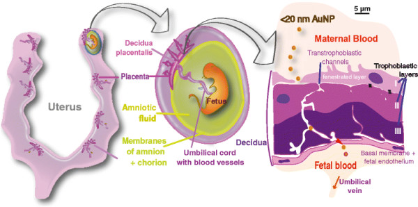
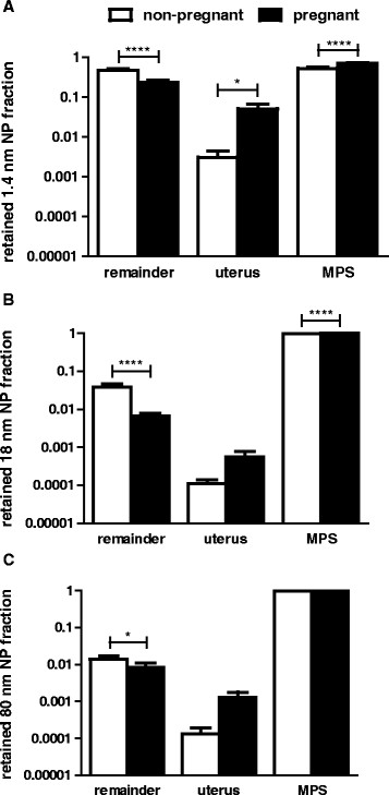
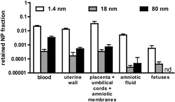
Verification
Claims the paper makes
The claims and statistics of this paper haven’t been checked yet.
Run the check — free, no account needed. It reads the open-access text and checks the sources it cites.
Claims other papers make about this one
Each is checked against this paper’s own text: does it say what the citing paper says it does?
In the citing paper
“Recently, the dose- [193] and size-dependency [194] of NP translocation from the mother to foetus was also reported in in vivo studies.”
From Particle toxicology and health - where are we? (2019) · cites this paper as [194]
Self-check
Percentages, sums and reported differences re-computed from the paper’s own numbers.
The self-check needs the paper’s full text.
Citations
Cites
Noninvasive detection of clinically occult lymph-node metastases in prostate cancer
Harisinghani, M. G., Barentsz, J., Hahn, P. F., Deserno, W. M., Tabatabaei, S., van de Kaa, C. H., de la Rosette, J., & Weissleder, R. (2003) · New England Journal of Medicine · doi:10.1056/nejmoa022749 · Cited by 1,971
Monopoli, M. P., Walczyk, D., Campbell, A., Elia, G., Lynch, I., Baldelli Bombelli, F., & Dawson, K. A. (2011) · Journal of the American Chemical Society · doi:10.1021/ja107583h · Cited by 1,835
Perspectives and opportunities for nanomedicine in the management of atherosclerosis
Lobatto, M. E., Fuster, V., Fayad, Z. A., & Mulder, W. J. M. (2011) · Nature Reviews Drug Discovery · doi:10.1038/nrd3578 · Cited by 423
Research works printed but not linked: 60
- Oberdörster G, Oberdörster E, Oberdörster J: Nanotoxicology: An Emerging Discipline Evolving from Studies of Ultrafine Particles. Environ Health Perspect 2005, 113: 823–839. 10.1289/ehp.7339 · DOI · Paper page
- Mills NL, Amin N, Robinson SD, Anand A, Davies J, Patel D, de la Fuente JM, Cassee FR, Boon NA, Macnee W, Millar AM, Donaldson K, Newby DE: Do inhaled carbon nanoparticles translocate directly into the circulation in humans? Am J Respir Crit Care Med 2006, 173: 426–431. 10.1164/rccm.200506-865OC · DOI
- Kreyling WG, Semmler-Behnke M, Seitz J, Scymczak W, Wenk A, Mayer P, Takenaka S, Oberdorster G: Size dependence of the translocation of inhaled iridium and carbon nanoparticle aggregates from the lung of rats to the blood and secondary target organs. Inhal Toxicol 2009,21(Suppl 1):55–60. 10.1080/08958370902942517 · DOI
- Lipka J, Semmler-Behnke M, Sperling RA, Wenk A, Takenaka S, Schleh C, Kissel T, Parak WJ, Kreyling WG: Biodistribution of PEG-modified gold nanoparticles following intratracheal instillation and intravenous injection. Biomaterials 2010, 31: 6574–6581. 10.1016/j.biomaterials.2010.05.009 · DOI
- Schleh C, Semmler-Behnke M, Lipka J, Wenk A, Hirn S, Schaffler M, Schmid G, Simon U, Kreyling WG: Size and surface charge of gold nanoparticles determine absorption across intestinal barriers and accumulation in secondary target organs after oral administration. Nanotoxicology 2012, 6: 36–46. 10.3109/17435390.2011.552811 · DOI · Paper page
- Hirn S, Semmler-Behnke M, Schleh C, Wenk A, Lipka J, Schaffler M, Takenaka S, Moller W, Schmid G, Simon U, Kreyling WG: Particle size-dependent and surface charge-dependent biodistribution of gold nanoparticles after intravenous administration. Eur J Pharm Biopharm 2010, 77: 407–416. 10.1016/j.ejpb.2010.12.029 · DOI
- Wiebert P, Sanchez-Crespo A, Falk R, Philipson K, Lundin A, Larsson S, Moller W, Kreyling WG, Svartengren M: No significant translocation of inhaled 35-nm carbon particles to the circulation in humans. Inhal Toxicol 2006, 18: 741–747. 10.1080/08958370600748455 · DOI
- Wiebert P, Sanchez-Crespo A, Seitz J, Falk R, Philipson K, Kreyling WG, Moller W, Sommerer K, Larsson S, Svartengren M: Negligible clearance of ultrafine particles retained in healthy and affected human lungs. Eur Respir J 2006, 28: 286–290. 10.1183/09031936.06.00103805 · DOI
- Möller W, Felten K, Sommerer K, Scheuch G, Meyer G, Meyer P, Haussinger K, Kreyling WG: Deposition, retention, and translocation of ultrafine particles from the central airways and lung periphery. Am J Respir Crit Care Med 2008, 177: 426–432. 10.1164/rccm.200602-301OC · DOI
- Semmler-Behnke M, Kreyling WG, Lipka J, Fertsch S, Wenk A, Takenaka S, Schmid G, Brandau W: Biodistribution of 1.4- and 18-nm gold particles in rats. Small 2008, 4: 2108–2111. 10.1002/smll.200800922 · DOI
- Kreyling WG, Hirn S, Möller W, Schleh C, Wenk A, Celik G, Lipka J, Schäffler M, Haberl N, Johnston BD, Sperling R, Schmid G, Simon U, Parak WJ, Semmler-Behnke M: Air¿Blood Barrier Translocation of Tracheally Instilled Gold Nanoparticles Inversely Depends on Particle Size. ACS Nano 2013, 8: 222–223. 10.1021/nn403256v · DOI · Paper page
- Rinderknecht A, Prudhomme R, Poreda R, Gelein R, Corson N, Pidruczny A, Finkelstein J, Oberdörster G, Elder A: Biokinetics of AU nanoparticles relative to size surface coating and portal of entry. 47th Annual Society of Toxicology Meeting; Seattle, WA 2008.
- Schweitzer AD, Revskaya E, Chu P, Pazo V, Friedman M, Nosanchuk JD, Cahill S, Frases S, Casadevall A, Dadachova E: Melanin-Covered Nanoparticles for Protection of Bone Marrow During Radiation Therapy of Cancer. Int J Radiat Oncol Biol Phys 2010,78(5):1494–1502. 10.1016/j.ijrobp.2010.02.020 · DOI
- Fuchs R, Ellinger I: Endocytic and transcytotic processes in villous syncytiotrophoblast: role in nutrient transport to the human fetus. Traffic 2004, 5: 725–738. 10.1111/j.1600-0854.2004.00221.x · DOI
- Takeda K, Suzuki KI, Ishihara A, Kubo-Irie M, Fujimoto R, Tabata M, Oshio S, Nihei Y, Ihara T, Sugamata M: Nanoparticles Transferred from Pregnant Mice to Their Offspring Can Damage the Genital and Cranial Nerve Systems. J Health Sci 2009, 55: 95–102. 10.1248/jhs.55.95 · DOI · Paper page
- Sugamata M, Ihara T, Sugamata M, Takeda K: Maternal exposure to diesel exhaust leads to pathological similarity to autism in newborns. J Health Sci 2006, 52: 486–488. 10.1248/jhs.52.486 · DOI
- Sugamata M, Ihara T, Takano H, Oshio S, Takeda K: Maternal diesel exhaust exposure damages newborn murine brains. J Health Sci 2006, 52: 82–84. 10.1248/jhs.52.82 · DOI
- Ritz B, Wilhelm M, Hoggatt KJ, Ghosh JK: Ambient air pollution and preterm birth in the environment and pregnancy outcomes study at the University of California, Los Angeles. Am J Epidemiol 2007, 166: 1045–1052. 10.1093/aje/kwm181 · DOI
- Coggins CRE, Ayres PH, Mosberg AT, Sagartz JW, Hayes AW: Subchronic Inhalation Study in Rats Using Aged and Diluted Sidestream Smoke from a Reference Cigarette. Inhal Toxicol 1993, 5: 77–95. 10.3109/08958379309034495 · DOI
- Witschi H, Lundgaard SM, Rajini P, Hendrickx AG, Last JA: Effects of exposure to nicotine and to sidestream smoke on pregnancy outcome in rats. Toxicol Lett 1994, 71: 279–286. 10.1016/0378-4274(94)90114-7 · DOI
- Gospe SM Jr, Zhou SS, Pinkerton KE: Effects of environmental tobacco smoke exposure in utero and/or postnatally on brain development. Pediatr Res 1996, 39: 494–498. 10.1203/00006450-199603000-00018 · DOI
- Wick P, Malek A, Manser P, Meili D, Maeder-Althaus X, Diener L, Diener PA, Zisch A, Krug HF, von Mandach U: Barrier Capacity of Human Placenta for Nanosized Materials. Environ Health Perspect 2010, 118: 432–436. 10.1289/ehp.0901200 · DOI · Paper page
- Myllynen PK, Loughran MJ, Howard CV, Sormunen R, Walsh AA, Vahakangas KH: Kinetics of gold nanoparticles in the human placenta. Reprod Toxicol 2008, 26: 130–137. 10.1016/j.reprotox.2008.06.008 · DOI · Paper page
- Yamashita K, Yoshioka Y, Higashisaka K, Mimura K, Morishita Y, Nozaki M, Yoshida T, Ogura T, Nabeshi H, Nagano K, Abe Y, Kamada H, Monobe Y, Imazawa T, Aoshima H, Shishido K, Kawai Y, Mayumi T, Tsunoda S, Itoh N, Yoshikaw T, Yanagihara I, Saito S, Tsutsumi Y: Silica and titanium dioxide nanoparticles cause pregnancy complications in mice. Nat Nanotechnol 2011, 6: 321–328. 10.1038/nnano.2011.41 · DOI
- Keelan JA: Nanotoxicology: Nanoparticles versus the placenta. Nat Nanotechnol 2011, 6: 263–264. 10.1038/nnano.2011.65 · DOI
- Rattanapinyopituk K, Shimada A, Morita T, Sakurai M, Asano A, Hasegawa T, Inoue K, Takano H: Demonstration of the Clathrin- and Caveolin-Mediated Endocytosis at the Maternal-Fetal Barrier in Mouse Placenta after Intravenous Administration of Gold Nanoparticles. J Vet Med Sci 2013, 76: 377–387. 10.1292/jvms.13-0512 · DOI
- Ockleford CD, Whyte A: Differeniated regions of human placental cell surface associated with exchange of materials between maternal and foetal blood: coated vesicles. J Cell Sci 1977, 25: 293–312. · DOI
- Ockleford CD, Whyte A, Bowyer DE: Variation in the volume of coated vesicles isoalted from human placenta. Cell Biol Int Rep 1977, 1: 137–146. 10.1016/0309-1651(77)90034-0 · DOI
- Kertschanska S, Kosanke G, Kaufmann P: Pressure dependence of so-called transtrophoblastic channels during fetal perfusion of human placental villi. Microsc Res Tech 1997, 38: 52–62. 10.1002/(SICI)1097-0029(19970701/15)38:1/2<52::AID-JEMT7>3.0.CO;2-W · DOI
- Kertschanska S, Stulcova B, Kaufmann P, Stulc J: Distensible transtrophoblastic channels in the rat placenta. Placenta 2000, 21: 670–677. 10.1053/plac.2000.0558 · DOI
- Knipp GT, Audus KL, Soares MJ: Nutrient transport across the placenta. Adv Drug Deliv Rev 1999, 38: 41–58. 10.1016/S0169-409X(99)00005-8 · DOI
- Schmid G: The relevance of shape and size of Au55 clusters. Chem Soc Rev 2008, 37: 1909–1930. 10.1039/b713631p · DOI
- King BF: A cytological study of plasma membrane modifications, intercellular junctions, and endocytic activity of amniotic epithelium. Anat Rec 1978, 190: 113–125. 10.1002/ar.1091900110 · DOI
- Modena AB, Fieni S: Amniotic fluid dynamics. Acta Biomed 2004,75(Suppl 1):11–13.
- Saunders M: Transplacental transport of nanomaterials. Wiley Interdiscip Rev Nanomed Nanobiotechnol 2009, 1: 671–684. 10.1002/wnan.53 · DOI
- Sadauskas E, Jacobsen NR, Danscher G, Stoltenberg M, Vogel U, Larsen A, Kreyling W, Wallin H: Biodistribution of gold nanoparticles in mouse lung following intratracheal instillation. Chem Central J 2009, 3: 16. 10.1186/1752-153X-3-16 · DOI
- Workshop IRSI: The relevance of the rat lung response to particle overload for human risk assessment: A workshop consensus report. Inhal Toxicol 2000, 12: 1–17.
- Conner SD, Schmid SL: Regulated portals of entry into the cell. Nature 2003, 422: 37–44. 10.1038/nature01451 · DOI
- Pan Y, Leifert A, Ruau D, Neuss S, Bornemann J, Schmid G, Brandau W, Simon U, Jahnen-Dechent W: Gold Nanoparticles of Diameter 1.4 nm Trigger Necrosis by Oxidative Stress and Mitochondrial Damage. Small 2009, 5: 2067–2076. 10.1002/smll.200900466 · DOI
- Lundqvist M, Stigler J, Elia G, Lynch I, Cedervall T, Dawson KA: Nanoparticle size and surface properties determine the protein corona with possible implications for biological impacts. Proc Natl Acad Sci U S A 2008, 105: 14265–14270. 10.1073/pnas.0805135105 · DOI · Paper page
- Lynch I, Dawson KA: Protein-nanoparticle interactions. Nano Today 2008, 3: 40–47. 10.1016/S1748-0132(08)70014-8 · DOI
- Schäffler M, Sousa F, Wenk A, Sitia L, Hirn S, Schleh C, Haberl N, Violatto M, Canovi M, Andreozzi P, Salmona M, Bigini P, Kreyling WG, Krol S: Blood protein coating of gold nanoparticles as potential tool for organ targeting. Biomaterials 2014, 35: 3455–3466. 10.1016/j.biomaterials.2013.12.100 · DOI
- Cheung CY, Brace RA: Amniotic fluid volume and composition in mouse pregnancy. J Soc Gynecol Investig 2005, 12: 558–562. 10.1016/j.jsgi.2005.08.008 · DOI
- Robertson P, Faber JJ, Brace RA, Louey S, Hohimer AR, Davis LE, Anderson DF: Responses of amniotic fluid volume and its four major flows to lung liquid diversion and amniotic infusion in the ovine fetus. Reprod Sci 2009, 16: 88–93. 10.1177/1933719108324888 · DOI
- Underwood MA, Gilbert WM, Sherman MP: Amniotic Fluid: Not Just Fetal Urine Anymore. J Perinatol 2005, 25: 341–348. 10.1038/sj.jp.7211290 · DOI
- Lutz HG: Clusters and Colloids. From Theory to Applications. VCH Verlagsgesellschaft, Weinheim/VCH Publishers, New York; 1995.
- Tominaga T, Tenma S, Watanabe H, Giebel U, Schmid G: Tracer Diffusion of a Ligand-Stabilized Two-Shell Gold Cluster. Chem Lett 1996, 25: 1033–1034. 10.1246/cl.1996.1033 · DOI
- Möller W, Gibson N, Geiser M, Pokhrel S, Wenk A, Takenaka S, Schmid O, Bulgheroni A, Simonelli F, Kozempel J, Holzwarth U, Wigge C, Eigeldinger-Berthou S, Mädler L, Kreyling W: Gold nanoparticle aerosols for rodent inhalation and translocation studies. J Nanoparticle Res 2013, 15: 1–13. 10.1007/s11051-013-1574-9 · DOI
- Semmler-Behnke M, Kreyling WG, Schulz H, Takenaka S, Butler JP, Henry FS, Tsuda A: Nanoparticle delivery in infant lungs. Proc Natl Acad Sci U S A 2012, 109: 5092–5097. 10.1073/pnas.1119339109 · DOI
- Hainfeld JF, O'Connor MJ, Dilmanian FA, Slatkin DN, Adams DJ, Smilowitz HM: Micro-CT enables microlocalisation and quantification of Her2-targeted gold nanoparticles within tumour regions. Br J Radiol 2011, 84: 526–533. 10.1259/bjr/42612922 · DOI
- Khlebtsov N, Dykman L: Biodistribution and toxicity of engineered gold nanoparticles: a review of in vitro and in vivo studies. Chem Soc Rev 2011, 40: 1647–1671. 10.1039/c0cs00018c · DOI
- Paciotti GF, Myer L, Weinreich D, Goia D, Pavel N, McLaughlin RE, Tamarkin L: Colloidal gold: a novel nanoparticle vector for tumor directed drug delivery. Drug Deliv 2004, 11: 169–183. 10.1080/10717540490433895 · DOI
- Kim Y, Lobatto ME, Kawahara T, Lee Chung B, Mieszawska AJ, Sanchez-Gaytan BL, Fay F, Senders ML, Calcagno C, Becraft J, Tun Saung M, Gordon R, Stroes ES, Ma M, Farokhzad OC, Fayad ZA, Mulder WJ, Langer R: Probing nanoparticle translocation across the permeable endothelium in experimental atherosclerosis. Proc Natl Acad Sci U S A 2014, 111: 1078–1083. 10.1073/pnas.1322725111 · DOI
- Mieszawska AJ, Kim Y, Gianella A, van Rooy I, Priem B, Labarre MP, Ozcan C, Cormode DP, Petrov A, Langer R, Farokhzad OC, Fayad ZA, Mulder WJ: Synthesis of polymer-lipid nanoparticles for image-guided delivery of dual modality therapy. Bioconjug Chem 2013, 24: 1429–1434. 10.1021/bc400166j · DOI
- McArdle HJ, Andersen HS, Jones H, Gambling L: Copper and Iron Transport Across the Placenta: Regulation and Interactions. J Neuroendocrinol 2008, 20: 427–431. 10.1111/j.1365-2826.2008.01658.x · DOI
- Turkevitch J, Stevenson PC, Hillier J: A study of the nucleation and growth processes in the synthesis of colloidal gold. Discuss Faraday Soc 1951, 11: 55–75. 10.1039/df9511100055 · DOI
- Schmid G, Lehnert A: The Complexation of Gold Colloids. Angew Chem Int Ed 1989, 28: 780–781. 10.1002/anie.198907801 · DOI
- Semmler M, Seitz J, Erbe F, Mayer P, Heyder J, Oberdorster G, Kreyling WG: Long-term clearance kinetics of inhaled ultrafine insoluble iridium particles from the rat lung, including transient translocation into secondary organs. Inhal Toxicol 2004, 16: 453–459. 10.1080/08958370490439650 · DOI · Paper page
- Semmler-Behnke M, Takenaka S, Fertsch S, Wenk A, Seitz J, Mayer P, Oberdorster G, Kreyling WG: Efficient elimination of inhaled nanoparticles from the alveolar region: evidence for interstitial uptake and subsequent reentrainment onto airways epithelium. Environ Health Perspect 2007, 115: 728–733. 10.1289/ehp.9685 · DOI · Paper page
- Kreyling WG, Semmler M, Erbe F, Mayer P, Takenaka S, Schulz H, Oberdörster G, Ziesenis A: Translocation of ultrafine insoluble iridium particles from lung epithelium to extrapulmonary organs is size dependent but very low. J Toxicol Environ Health-Part A 2002, 65: 1513–1530. 10.1080/00984100290071649 · DOI · Paper page
Cited by
Sorted by citations
Particle toxicology and health - where are we?
Riediker, M., Zink, D., Kreyling, W., Oberdörster, G., Elder, A., Graham, U., Lynch, I., Duschl, A., Ichihara, G., Ichihara, S., et al. (2019) · Particle and Fibre Toxicology · doi:10.1186/s12989-019-0302-8 · Cited by 237
Self-citationNanoparticle Transport Across the Placental Barrier: Pushing the Field Forward!
Muoth, C., Aengenheister, L., Kucki, M., Wick, P., & Buerki-Thurnherr, T. (2016) · Nanomedicine · doi:10.2217/nnm-2015-0012 · Cited by 146
Klocke, C., Allen, J. L., Sobolewski, M., Mayer-Pröschel, M., Blum, J. L., Lauterstein, D., Zelikoff, J. T., & Cory-Slechta, D. A. (2017) · Toxicological Sciences · doi:10.1093/toxsci/kfx010 · Cited by 87
Placental Drug Delivery to Treat Pre‐Eclampsia and Fetal Growth Restriction
Deepak, V., El‐Balawi, L., & Harris, L. K. (2024) · Small · doi:10.1002/smll.202311165 · Cited by 16
Metadata
- Authors
- Semmler-Behnke, M., Lipka, J., Wenk, A., Hirn, S., Schäffler, M., Tian, F., Schmid, G., Oberdörster, G., & Kreyling, W. G.
- Venue
- Particle and Fibre Toxicology
- Published
- 2014-09-10
- Volume
- 11
- Issue
- 1
- ISSN
- 1743-8977
- PMC
- PMC4445676
- Cited by
- 135
- References
- 3
- Language
- en
About these checks
Registry, retraction, Cites and statistics results are computed, not judged, with one exception: a printed reference that no index lists is read once by a model before it counts as a citation error. Claims are extracted by a model and kept only when their quote appears verbatim in the text; each cited source is then checked, with a model’s help, for whether it says what the claim relies on. Those verdicts can be wrong — read the claim’s quote and the cited source before relying on one.
Statistics and arithmetic are recomputed, not judged.
Confidence bands are shown only for fields calibrated on our annotated test set. None is calibrated yet, so no band is shown.
What counts as a claim ›How we measure confidence ›How we check this ›
Are you an author, or do you see a problem with this page? Request a review
