

The DOI matches its Crossref record; 61 of the 61 references it cites were found in a registry or an index.
Nanopolystyrene translocation and fetal deposition after acute lung exposure during late-stage pregnancy
Open accessPublished version of a preprint
Abstract
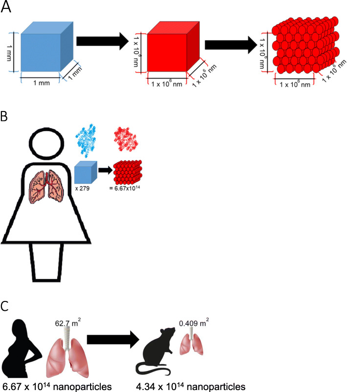
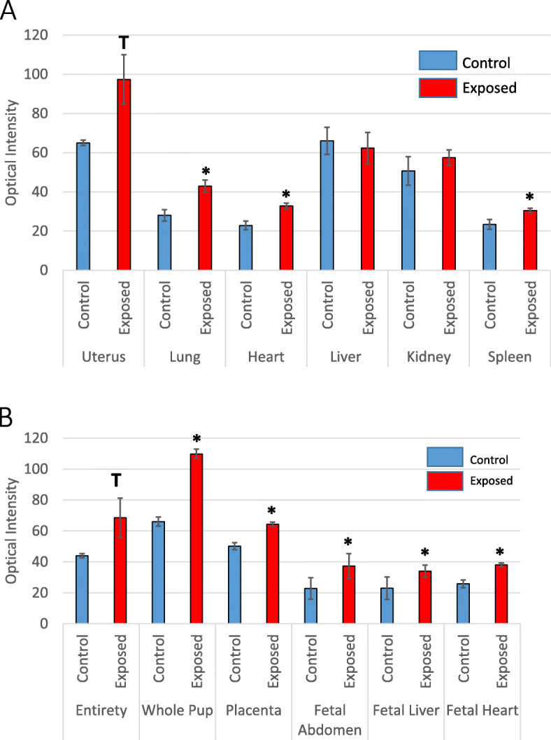
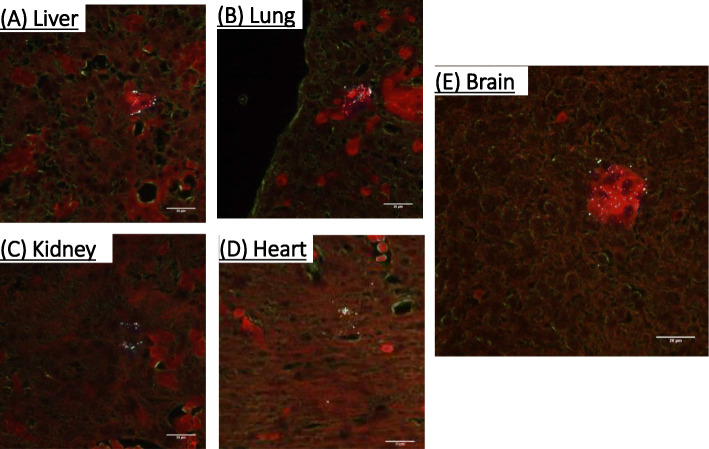
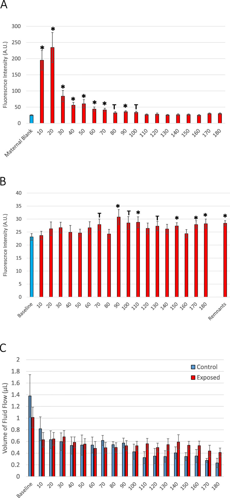
Background: Plastic is everywhere. It is used in food packaging, storage containers, electronics, furniture, clothing, and common single-use disposable items. Microplastic and nanoplastic particulates are formed from bulk fragmentation and disintegration of plastic pollution. Plastic particulates have recently been detected in indoor air and remote atmospheric fallout. Due to their small size, microplastic and nanoplastic particulate in the atmosphere can be inhaled and may pose a risk for human health, specifically in susceptible populations. When inhaled, nanosized particles have been shown to translocate across pulmonary cell barriers to secondary organs, including the placenta. However, the potential for maternal-to-fetal translocation of nanosized-plastic particles and the impact of nanoplastic deposition or accumulation on fetal health remain unknown. In this study we investigated whether nanopolystyrene particles can cross the placental barrier and deposit in fetal tissues after maternal pulmonary exposure. Results: Pregnant Sprague Dawley rats were exposed to 20 nm rhodamine-labeled nanopolystyrene beads (2.64 × 10 14 particles) via intratracheal instillation on gestational day (GD) 19. Twenty-four hours later on GD 20, maternal and fetal tissues were evaluated using fluorescent optical imaging. Fetal tissues were fixed for particle visualization with hyperspectral microscopy. Using isolated placental perfusion, a known concentration of nanopolystyrene was injected into the uterine artery. Maternal and fetal effluents were collected for 180 min and assessed for polystyrene particle concentration. Twenty-four hours after maternal exposure, fetal and placental weights were significantly lower (7 and 8%, respectively) compared with controls. Nanopolystyrene particles were detected in the maternal lung, heart, and spleen. Polystyrene nanoparticles were also observed in the placenta, fetal liver, lungs, heart, kidney, and brain suggesting maternal lung-to-fetal tissue nanoparticle translocation in late stage pregnancy. Conclusion: These studies confirm that maternal pulmonary exposure to nanopolystyrene results in the translocation of plastic particles to placental and fetal tissues and renders the fetoplacental unit vulnerable to adverse effects. These data are vital to the understanding of plastic particulate toxicology and the developmental origins of health and disease.
Overview
The claims and statistics of this paper haven’t been checked yet.
Run the check — free, no account needed. It reads the open-access text and checks the sources it cites.
Integrity signals
Registry
Cites
Statistics
Figures
Verification
Claims the paper makes
The claims and statistics of this paper haven’t been checked yet.
Run the check — free, no account needed. It reads the open-access text and checks the sources it cites.
Claims other papers make about this one
Each is checked against this paper’s own text: does it say what the citing paper says it does?
The source backs part of the claim, not all of it — or backs it for a different population, condition or comparison.
In the citing paper
“However, given the capability of these nanoplastic particles to cross the biological barrier, nanoparticles, despite the seemingly trivial contribution to the mass measurement, might play a predominant role in terms of toxicity evaluation (72, 73).”
From Rapid single-particle chemical imaging of nanoplastics by SRS microscopy (2024) · cites this paper as [72]
Experimental — accuracy not yet measured
Self-check
Percentages, sums and reported differences re-computed from the paper’s own numbers.
The self-check needs the paper’s full text.
Citations
Cites
Production, use, and fate of all plastics ever made
Geyer, R., Jambeck, J. R., & Law, K. L. (2017) · Science Advances · doi:10.1126/sciadv.1700782 · Cited by 18,606
Human Consumption of Microplastics
Cox, K. D., Covernton, G. A., Davies, H. L., Dower, J. F., Juanes, F., & Dudas, S. E. (2019) · Environmental Science & Technology · doi:10.1021/acs.est.9b01517 · Cited by 2,745
Atmospheric transport and deposition of microplastics in a remote mountain catchment
Allen, S., Allen, D., Phoenix, V. R., Roux, G. L., Durantez, P., Simonneau, A., Binet, S., & Galop, D. (2019) · Nature Geoscience · doi:10.1038/s41561-019-0335-5 · Cited by 2,584
Browne, M. A., Dissanayake, A., Galloway, T. S., Lowe, D. M., & Thompson, R. C. (2008) · Environmental Science & Technology · doi:10.1021/es800249a · Cited by 2,263
Airborne microplastics: consequences to human health?
Prata, J. C. (2018) · Environmental Pollution · doi:10.1016/j.envpol.2017.11.043 · Cited by 1,852
A first overview of textile fibers, including microplastics, in indoor and outdoor environments
Dris, R., Gaspéri, J., Mirande-Bret, C., Mandin, C., Guerrouache, M., Langlois, V., & Tassin, B. (2016) · Environmental Pollution · doi:10.1016/j.envpol.2016.12.013 · Cited by 1,635
Deng, Y., Zhang, Y., Lemos, B., & Ren, H. (2017) · Scientific Reports · doi:10.1038/srep46687 · Cited by 1,471
Pollutants bioavailability and toxicological risk from microplastics to marine mussels
Avio, C. G., Gorbi, S., Milan, M., Benedetti, M., Fattorini, D., d'Errico, G., Pauletto, M., Bargelloni, L., & Regoli, F. (2015) · Environmental Pollution · doi:10.1016/j.envpol.2014.12.021 · Cited by 1,413
Anthropogenic contamination of tap water, beer, and sea salt
Kosuth, M., Mason, S. A., & Wattenberg, E. V. (2018) · PLoS ONE · doi:10.1371/journal.pone.0194970 · Cited by 1,257
Differentially charged nanoplastics demonstrate distinct accumulation in Arabidopsis thaliana
Sun, X., Yuan, X.-Z., Jia, Y., Feng, L., Zhu, F., Dong, S., Liu, J., Kong, X., Tian, H., Duan, J.-L., et al. (2020) · Nature Nanotechnology · doi:10.1038/s41565-020-0707-4 · Cited by 1,240
Cited by
Sorted by citations
Items 101–200 of 479
Effects of nanoplastic exposure during pregnancy and lactation on neurodevelopment of rat offspring
Tian, L., Zhang, Y., Chen, J., Liu, X., Nie, H., Li, K., Liu, H., Lai, W., Shi, Y., Xi, Z., et al. (2024) · Journal of Hazardous Materials · doi:10.1016/j.jhazmat.2024.134800 · Cited by 72
Shaoyong, W., Jin, H., JIANG, X. H., Xu, B., Liu, Y., Wang, Y., & Jin, M. (2023) · Journal of Hazardous Materials · doi:10.1016/j.jhazmat.2023.131820 · Cited by 67
Bai, J. Y., Wang, Y., Deng, S., Yang, Y., Chen, S., & Wu, Z. (2024) · Particle and Fibre Toxicology · doi:10.1186/s12989-024-00595-5 · Cited by 76
“…5% goat serum in PBS for 1 h, the primary antibodies were incubated overnight at 4 °C and then washed three times in PBS. Subsequently, the sections were incubated with AlexaFluor 594 fluorescent secondary antibody in the dark for 1 h, followed by nuclear staining with nuclear dye Hoechst 33,342.”
BackgroundMethodInfluentialFan, J., Liu, L., Lu, Y., Chen, Q., Fan, S., Yang, Y., Long, Y., & Liu, X. Z. (2024) · Particle and Fibre Toxicology · doi:10.1186/s12989-024-00578-6 · Cited by 62
“To further establish their environmental or occupational relevance, we mathematically converted a typical 1 mm 3 microplastics (MPs) into NPs using a previously established method [33].”
BackgroundMethodInfluentialDeLoid, G. M., Cao, X., Coreas, R., Bitounis, D., Singh, D., Zhong, W., & Demokritou, P. (2022) · Environmental Science & Technology · doi:10.1021/acs.est.2c03195 · Cited by 61
Research progress on the cellular toxicity caused by microplastics and nanoplastics
He, T., Qu, Y., Yang, X., Liu, L., Xiong, F., Wang, D., Liu, M., & Sun, R. (2023) · Journal of Applied Toxicology · doi:10.1002/jat.4449 · Cited by 60
“…of biological samples, including lungs, intestines, liver, spleen, kidneys, placenta, breast milk, blood, urine, and feces (Braun et al., 2021; Fournier et al., 2020; Horvatits et al., 2022; Ibrahim et al., 2021; Rhodes, 2018), thereby causing hepatotoxicity, neurotoxicity, reproductive…”
BackgroundMicro(nano)plastics: an Emerging Burden for Human Health
Donisi, I., Colloca, A., Anastasio, C., Balestrieri, M. L., & D’Onofrio, N. (2024) · International Journal of Biological Sciences · doi:10.7150/ijbs.99556 · Cited by 69
“Current evidence demonstrated MNPs accumulation via the blood-placental barrier during pregnancy and trough the breast milk during nursing in different organs, leading to a dysregulated metabolism and impaired immune and reproductive function [45,46].”
BackgroundDe-la-Torre, G. E., Pizarro-Ortega, C. I., Dioses-Salinas, D. C., Ammendolia, J., & Okoffo, E. D. (2021) · Current Opinion in Toxicology · doi:10.1016/j.cotox.2021.08.002 · Cited by 62
Characteristics, Toxic Effects, and Analytical Methods of Microplastics in the Atmosphere
Yang, H., He, Y., Yan, Y., Junaid, M., & Wang, J. (2021) · Nanomaterials · doi:10.3390/nano11102747 · Cited by 67
Nanoplastic Impact on the Gut-Brain Axis: Current Knowledge and Future Directions
Grodzicki, W., Dziendzikowska, K., Gromadzka-Ostrowska, J., & Kruszewski, M. K. (2021) · International Journal of Molecular Sciences · doi:10.3390/ijms222312795 · Cited by 64
Balali, H., Morabbi, A., & Karimian, M. (2024) · Reproductive Biology and Endocrinology · doi:10.1186/s12958-024-01314-7 · Cited by 71
“The passage of NPs through the blood-placenta barrier (BPB) and their transfer via breast milk to offspring are the two main pathways through which offspring are exposed to nanoplastic particles [29, 78], and the transfer of these materials through the placenta depends on their size [164].”
BackgroundInfluentialCaputi, S., Diomede, F., Lanuti, P., Marconi, G. D., Carlo, P. D., Sinjari, B., & Trubiani, O. (2022) · International Journal of Environmental Research and Public Health · doi:10.3390/ijerph19137782 · Cited by 66
Bai, C.-L., Wang, D., Luan, Y.-L., Huang, S.-N., Liu, L., & Guo, Y. (2024) · Chemosphere · doi:10.1016/j.chemosphere.2024.142424 · Cited by 67
Polystyrene microplastic particles: In vivo and in vitro ocular surface toxicity assessment
Zhou, X., Wang, G., An, X., Wu, J., Fan, K., Xu, L., Li, C., & Xue, Y. (2022) · Environmental Pollution · doi:10.1016/j.envpol.2022.119126 · Cited by 59
Zhang, Y., Tian, L., Chen, J., Liu, X., Li, K., Liu, H., Lai, W., Shi, Y., Lin, B., & Xi, Z. (2024) · Ecotoxicology and Environmental Safety · doi:10.1016/j.ecoenv.2024.116393 · Cited by 61
Effects of micro(nano)plastics on the reproductive system: A review
Song, X., Du, L., Sima, L., Zou, D., & Qiu, X. (2023) · Chemosphere · doi:10.1016/j.chemosphere.2023.139138 · Cited by 59
Micro- and Nanoplastics’ Effects on Protein Folding and Amyloidosis
Windheim, J., Colombo, L., Battajni, N. C., Russo, L., Cagnotto, A., Diomede, L., Bigini, P., Vismara, E., Fiumara, F., Gabbrielli, S., et al. (2022) · International Journal of Molecular Sciences · doi:10.3390/ijms231810329 · Cited by 56
Ferroptosis participated in inhaled polystyrene nanoplastics-induced liver injury and fibrosis
Ge, Y., Yang, S., Zhang, T., Gong, S., Wan, X., Zhu, Y., Fang, Y., Hu, C., Yang, F., Yin, L., et al. (2024) · The Science of The Total Environment · doi:10.1016/j.scitotenv.2024.170342 · Cited by 56
Embryonic exposure of polystyrene nanoplastics affects cardiac development
Li, J., Weng, H., Liu, S., Li, F., Xu, K., Wen, S., Chen, X., Li, C., Nie, Y., Liao, B., et al. (2023) · The Science of The Total Environment · doi:10.1016/j.scitotenv.2023.167406 · Cited by 56
The bio-distribution, clearance pathways, and toxicity mechanisms of ambient ultrafine particles
Han, D., Chen, R., Kan, H., & Xu, Y. (2023) · Eco-Environment & Health · doi:10.1016/j.eehl.2023.06.001 · Cited by 52
The adverse health effects of increasing microplastic pollution on aquatic mammals
Nabi, G., Ahmad, S., Ullah, S., Zada, S., Sarfraz, M., Guo, X., Khan, M. I., & Wanghe, K. (2022) · Journal of King Saud University - Science · doi:10.1016/j.jksus.2022.102006 · Cited by 55
Shaoyong, W., Lu, S., Gan, Y., Jin, H., Wang, W., Yin, L., Wang, Y., & Jin, M. (2024) · ACS Nano · doi:10.1021/acsnano.4c08737 · Cited by 50
Huang, J., Dong, G., Liang, M., Wu, X., Xian, M., An, Y., Zhan, J., Xu, L., Xu, J., Sun, W., et al. (2022) · Chemosphere · doi:10.1016/j.chemosphere.2022.136093 · Cited by 54
A Multimedia Model for Microplastic Particles
Quik, J. T. K., Meesters, J. A. J., & Koelmans, A. A. (2022) · The Science of The Total Environment · doi:10.1016/j.scitotenv.2023.163437 · Cited by 56
Zhang, T., Yang, S., Ge, Y., Wan, X., Zhu, Y., Yang, F., Li, J., Gong, S., Cheng, Y., Hu, C., et al. (2023) · Particle and Fibre Toxicology · doi:10.1186/s12989-023-00557-3 · Cited by 52
“Due to the limitation of nanoparticle detection technology, we mathematically converted atmospheric microparticles with a diameter of 100 μm to our 40 nm polystyrene microspheres, as described previously [27, 49].”
MethodChen, C., & Lin, Z. (2024) · Environment International · doi:10.1016/j.envint.2024.108617 · Cited by 51
Cary, C. M., & Stapleton, P. A. (2023) · Archives of Toxicology · doi:10.1007/s00204-023-03528-x · Cited by 48
“2008, 2005), polystyrene (Chen et al. 2006; Fournier et al. 2020), silver (Takenaka et al.”
BackgroundSelf-citationSharma, A., Kaur, M., Sharma, K. M. T. K., Bunkar, S. K., John, P. J., & Bhatnagar, P. (2023) · NeuroToxicology · doi:10.1016/j.neuro.2023.10.009 · Cited by 47
Peng, Y., & Qi, H. (2024) · Ecotoxicology and Environmental Safety · doi:10.1016/j.ecoenv.2024.116505 · Cited by 51
Zhao, X., Sun, J., Zhou, L., Teng, M., Zhao, L., Li, Y., & Wu, F. (2023) · Environmental Science Nano · doi:10.1039/d3en00491k · Cited by 49
Jeong, B., Kim, J., Kwon, A. R., Lee, J. G., Park, S., Koo, J., Lee, W. S., Baek, J. Y., Shin, W.-H., Lee, J., et al. (2024) · Environment International · doi:10.1016/j.envint.2024.108522 · Cited by 46
Nafea, T. H., Chan, F. K. S., Xu, Y., Wang, C., Wang, X., Zhao, W., Ji, D., Xiao, H., & He, J. Y. (2024) · Earth-Science Reviews · doi:10.1016/j.earscirev.2024.104864 · Cited by 51
Mechanistic insight into potential toxic effects of microplastics and nanoplastics on human health
Thapliyal, C., Negi, S., Nagarkoti, S., & Daverey, A. (2025) · Discover Applied Sciences · doi:10.1007/s42452-025-07214-8 · Cited by 57
“The smaller size of MNPs helps them to deeply penetrate the lungs [81] and cause occupational respiratory diseases, including lung cancer, due to its prolonged exposure [62, 66, 77, 78].”
Li, M., Hou, Z., Meng, R., Hao, S., & Wang, B. (2022) · Environment International · doi:10.1016/j.envint.2022.107644 · Cited by 47
Xiong, S., He, J., Qiu, H., van Gestel, C. A. M., He, E., Qiao, Z., Cao, L., Li, J., & Chen, G. (2024) · Chemosphere · doi:10.1016/j.chemosphere.2024.141513 · Cited by 47
Polystyrene microplastics disturb maternal glucose homeostasis and induce adverse pregnancy outcomes
Zhang, R., Feng, Y., Nie, P., Wang, W., Wu, H., Wan, X., Xu, H., & Fu, F. (2024) · Ecotoxicology and Environmental Safety · doi:10.1016/j.ecoenv.2024.116492 · Cited by 46
Hu, Y. Y., Jiang, S., Zhang, Q., Zhou, W., Liang, J., Xu, Y. Q., & Su, W. (2024) · Particle and Fibre Toxicology · doi:10.1186/s12989-024-00590-w · Cited by 49
“Plastic particles invade the human body primarily through the alimentary tract [6], and also through the respiratory system [7, 8], cutaneous mucosal [9], and injection pathway [10].”
BackgroundLi, D., SUN, W. M., Jiang, X., Yu, Z., Xia, Y., Cheng, S., Mao, L., Luo, S., Tang, S., Xu, S., et al. (2022) · Ecotoxicology and Environmental Safety · doi:10.1016/j.ecoenv.2022.114104 · Cited by 45
Effects of polystyrene nanoplastic gestational exposure on mice
Wang, X., Zhao, Z., Wang, X., Hu, W., luo, C., Chu, X., Qian, M., Wang, R., Wu, Q., Wu, Q., et al. (2023) · Chemosphere · doi:10.1016/j.chemosphere.2023.138255 · Cited by 46
Ye, J., Qiu, W., Pang, X., Su, Y., Zhang, X., Huang, J., Xie, H., Liao, J., Tang, Z., Chen, Z., et al. (2024) · Environmental Pollution · doi:10.1016/j.envpol.2024.123713 · Cited by 49
Airborne Microplastics: Challenges, Prospects, and Experimental Approaches
Rednikin, A., Frank, Y. A., Rozhin, A. O., Vorobiev, D. S., & Fakhrullin, R. F. (2024) · Atmosphere · doi:10.3390/atmos15111380 · Cited by 50
Probabilistic Estimation of Airborne Micro- and Nanoplastic Intake in Humans
Chen, Y., Meng, Y., Liu, G., Huang, X., & Chai, G. (2024) · Environmental Science & Technology · doi:10.1021/acs.est.3c09189 · Cited by 47
Micro(nano)plastics in plant-derived food: Source, contamination pathways and human exposure risks
Liang, Y., Cao, X., Mo, A., Jiang, J., Zhang, Y., Gao, W., & He, D. (2023) · TrAC Trends in Analytical Chemistry · doi:10.1016/j.trac.2023.117138 · Cited by 45
D’Errico, J. N., Doherty, C., George, J. J. R., Buckley, B., & Stapleton, P. (2022) · Placenta · doi:10.1016/j.placenta.2022.03.008 · Cited by 42
Self-citationChortarea, S., Gupta, G. S., Saarimäki, L. A., Netkueakul, W., Manser, P., Aengenheister, L., Wichser, A., Fortino, V., Wick, P., Greco, D., et al. (2023) · Environment International · doi:10.1016/j.envint.2023.108015 · Cited by 44
Yun, X., Liang, L., Tian, J., Li, N., Chen, Z., Zheng, Y., Duan, S., & Zhang, L. (2024) · Journal of Hazardous Materials · doi:10.1016/j.jhazmat.2024.135271 · Cited by 44
He, F., Shi, H., Liu, R., Tian, G., Qi, Y., & Wang, T. (2023) · The Science of The Total Environment · doi:10.1016/j.scitotenv.2023.162854 · Cited by 41
Patisaul, H. B. (2021) · Advances in pharmacology · doi:10.1016/bs.apha.2021.03.007 · Cited by 44
Kļaviņš, M., Kļaviņš, L., Stabnikova, О., Stabnikov, V., Маринін, А. І., Ansone-Bērtiņa, L., Mežulis, M., & Vaseashta, A. (2022) · Microplastics · doi:10.3390/microplastics1030037 · Cited by 40
Particulate contamination of human placenta: Plastic and non-plastic
Zurub, R. E., Bainbridge, S. A., Rahman, L., Halappanavar, S. S., El‐Chaâr, D., & Wade, M. G. (2024) · Environmental Advances · doi:10.1016/j.envadv.2024.100555 · Cited by 47
A review of the toxic effects of microplastics based on studies on mammals and mammalian cell lines
Tang, K. H. D. (2024) · Environmental Science Advances · doi:10.1039/d4va00227j · Cited by 46
The biochemical mechanisms of plastic biodegradation
Gates, E. G., & Crook, N. (2024) · FEMS Microbiology Reviews · doi:10.1093/femsre/fuae027 · Cited by 46
Hong, W., Zhang, M., Wang, F., Li, Z., Li, Z., Wei, J., Song, M., Qian, Y., Pang, S., Wang, L., et al. (2024) · ACS Nano · doi:10.1021/acsnano.4c00625 · Cited by 39
Naz, S., Chatha, A. M. M., Khan, N. A., Ullah, Q., Zaman, F., Qadeer, A., Khan, I. M., Danabaş, D., Kiran, A., Skalíčková, S., et al. (2024) · Frontiers in Environmental Science · doi:10.3389/fenvs.2024.1390510 · Cited by 40
Impacts of micro- and nanoplastics on early-life health: a roadmap towards risk assessment
Christopher, E. A., Vries, Y. C., Devadoss, A., Mandemaker, L. D. B., van Boxel, J., Copsey, H., Dusza, H. M., Legler, J., Meirer, F., Muncke, J., et al. (2024) · Microplastics and Nanoplastics · doi:10.1186/s43591-024-00089-3 · Cited by 41
“MNPs have been found to accumulate in tissues and organs in rats (21 nm) [60], mice (0.1 and 2 μm) [191], zebrafish (4–40 μm) [63] and marine organisms (10 μm − 5 mm) [62].”
BackgroundInfluentialDo microplastics (MPs) and nanoplastics (NPs) directly contribute to human carcinogenesis?
Casella, C., Dondi, D., & Vadivel, D. (2025) · Environmental Pollution · doi:10.1016/j.envpol.2025.127343 · Cited by 46
Le, V., Nguyen, M., Lin, C., Nguyen, H., Nguyen, T. Q. H., Hue, N. K., Truong, Q.-M., Chang, S. W., Nguyen, X. H., & Nguyen, D. D. (2024) · Environmental Research · doi:10.1016/j.envres.2024.119345 · Cited by 39
Wan, D., Liu, Y., Chang, Q., Liu, Z., Wang, Q., Niu, R., Gao, B., Guan, Q., & Xia, Y. (2024) · Toxics · doi:10.3390/toxics12080553 · Cited by 39
Meng, X., Bao, T., Hong, L., & Wu, K. M. (2023) · Water · doi:10.3390/w15040686 · Cited by 36
Bridging the Gaps between Microplastics and Human Health
Damaj, S., Trad, F., Goevert, D., & Wilkesman, J. (2024) · Microplastics · doi:10.3390/microplastics3010004 · Cited by 43
An Overview of the Possible Exposure of Infants to Microplastics
Mišľanová, C., Valachovičová, M., & Slezáková, Z. (2024) · Life · doi:10.3390/life14030371 · Cited by 39
Li, H., Bai, L., Liang, S., Chen, X., Gu, X., Wang, C., & Gu, C. Z. (2025) · Eco-Environment & Health · doi:10.1016/j.eehl.2025.100138 · Cited by 41
Bhowmik, A., & Saha, G. (2025) · Microplastics · doi:10.3390/microplastics4020024 · Cited by 43
Microplastic pollution: a review of specific blood-tissue barrier breaches and health effects
Obeng, E. M., Hodge, C., & You, J. (2025) · Environmental Pollution · doi:10.1016/j.envpol.2025.126416 · Cited by 42
Indirect mediators of systemic health outcomes following nanoparticle inhalation exposure
Mostovenko, E., Canal, C. G., Cho, M. J., Sharma, K., Erdely, A., Campen, M. J., & Ottens, A. K. (2022) · Pharmacology & Therapeutics · doi:10.1016/j.pharmthera.2022.108120 · Cited by 39
Bhardwaj, G., Abdulkadhim, M., Joshi, K., Wankhede, L., Das, R. K., & Brar, S. K. (2025) · Applied Sciences · doi:10.3390/app15168813 · Cited by 43
Potential threats of nanoplastic accumulation in human induced pluripotent stem cells
Jeong, H., Kim, W., Choi, D., Heo, J., Han, U., Jung, S. Y., Park, H. H., Hong, S., Park, J. H., & Hong, J. (2021) · Chemical Engineering Journal · doi:10.1016/j.cej.2021.131841 · Cited by 33
Moreno, G. M., Brunson-Malone, T., Adams, S. E., Nguyen, C., Seymore, T., Cary, C. M., Polunas, M., Goedken, M. J., & Stapleton, P. A. (2024) · The Science of The Total Environment · doi:10.1016/j.scitotenv.2024.175350 · Cited by 33
Self-citationThe role of gut microbiota in MP/NP-induced toxicity
Pan, Y., Zhang, H., Zhu, L., Tan, J., Wang, B., & Li, M. (2024) · Environmental Pollution · doi:10.1016/j.envpol.2024.124742 · Cited by 35
The Impact of Maternal Nanoplastic and Microplastic Particle Exposure on Mammal’s Offspring
Yu, H., Sheen, J., & Tiao, M. (2024) · Cells · doi:10.3390/cells13161380 · Cited by 33
“Fournier et al. [37] administered 20 nm-sized PS-NPl (2.64 × 10 14 particles) via tracheal instillation to Sprague Dawley (SD) rats on gestational day 19.”
BackgroundMethodInfluentialHuang, F., You, H., Tang, X., Su, Y., Peng, H., Li, H., Wei, Z., & Hua, J. (2025) · Journal of Nanobiotechnology · doi:10.1186/s12951-025-03561-1 · Cited by 37
Plastic or plastic-free life: From formation to removal
Li, M., Pan, Y., Hou, Z., Wu, Z., Zeng, Z., & Wang, B. (2023) · The Science of The Total Environment · doi:10.1016/j.scitotenv.2023.164359 · Cited by 33
Mierzejewski, K., Kurzyńska, A., Golubska, M., Całka, J., Gałęcka, I., Szabelski, M., Paukszto, Ł., Andronowska, A., & Bogacka, I. (2023) · The Science of The Total Environment · doi:10.1016/j.scitotenv.2023.166967 · Cited by 33
Shukla, S., Khanna, S., & Khanna, K. (2024) · Toxicology Reports · doi:10.1016/j.toxrep.2024.101844 · Cited by 35
Your brain is full of microplastics: are they harming you?
Kozlov, M. (2025) · Nature · doi:10.1038/d41586-025-00405-8 · Cited by 33
Vela, L., Villacorta, A., Venus, T., Estrela‐Lopis, I., Pastor, S., García‐Rodríguez, A., Rubio, L., Marcos, R., & Hernández, A. (2023) · Environmental Pollution · doi:10.1016/j.envpol.2023.121656 · Cited by 31
Han, J., Yan, J., Li, K., Lin, B., Lai, W., Bian, L., Jia, R., Liu, X., & Xi, Z. (2023) · Toxics · doi:10.3390/toxics11050441 · Cited by 32
Rapini, C. C. S., Berardino, C. D., Peserico, A., Capacchietti, G., & Barboni, B. (2024) · International Journal of Molecular Sciences · doi:10.3390/ijms252212166 · Cited by 33
“High concentrations of PS-MNPs have been observed to accumulate in various organs, such as maternal lung, heart, spleen, and liver [140,147].”
InfluentialA Critical Review on Biological Impacts, Ecotoxicity, and Health Risks Associated with Microplastics
Ansari, I., Arora, C., Verma, A., Mahmoud, A. E. D., El‐Kady, M. M., Rajarathinam, R., Verma, D. K., & Mahish, P. K. (2025) · Water Air & Soil Pollution · doi:10.1007/s11270-024-07731-z · Cited by 31
“According to a similar study, MPs inhaled by pregnant rats penetrated their internal organs, where they were found in the placentas, fetal liver, lungs, heart, kidney, and brain (Fournier et al., 2020; Ragusa et al., 2021).”
Microplastics: A threat to Fetoplacental unit and Reproductive systems
Anifowoshe, A. T., Akhtar, M. N., Majeed, A., Singh, A. S., Ismail, T. F., & Nongthomba, U. (2025) · Toxicology Reports · doi:10.1016/j.toxrep.2025.101938 · Cited by 34
Mariani, F., Lernia, D. D., Venditti, I., Pelella, E., Muzzi, M., Giulio, A. D., & Ceschin, S. (2023) · The Science of The Total Environment · doi:10.1016/j.scitotenv.2023.164459 · Cited by 31
Bowdridge, E. C., DeVallance, E., Garner, K. L., Griffith, J. A., Schafner, K. J., Seaman, M., Engels, K. J., Wix, K., Batchelor, T., Goldsmith, W. T., et al. (2022) · Particle and Fibre Toxicology · doi:10.1186/s12989-022-00457-y · Cited by 31
“Nano-TiO2 has been shown to cross the placenta [8] during gestational exposure and potentially deposits into the pup [9], however perturbations in vasoreactivity within the uterus of the dam and/or the placenta of the pup alone are enough to result in detrimental fetal health consequences [10].”
BackgroundResultCary, C. M., Fournier, S., Adams, S., Wang, X., Yurkow, E. J., & Stapleton, P. A. (2024) · Toxicological Sciences · doi:10.1093/toxsci/kfae019 · Cited by 29
Self-citationShi, J., Yu, X., Zhao, J., et al. (2024) · Ecotoxicology and Environmental Safety · doi:10.1016/j.ecoenv.2024.116925 · Cited by 28
The microplastic-crisis: Role of bacteria in fighting microplastic-effects in the digestive system
Pacher-Deutsch, C., Schweighofer, N., Hanemaaijer, M., Marut, W., Žukauskaitė, K., Horvath, A., & Stadlbauer, V. (2024) · Environmental Pollution · doi:10.1016/j.envpol.2024.125437 · Cited by 30
Kamalasekaran, K., & Sundramoorthy, A. K. (2026) · RSC Advances · doi:10.1039/d5ra09062h · Cited by 35
“55 According to Ragusa et al. , 56 MPs are a global issue of the twenty- rst century because they have recently been identi ed inside the human placenta.”
Ahamed, M., Lateef, R., Akhtar, M. J., et al. (2022) · Molecules · doi:10.3390/molecules27217378 · Cited by 28
“A recent study observed that maternal pulmonary exposure to nanopolystyrene leads to the translocation of particles to placental and fetal tissues and renders the fetoplacental entity susceptible to hazardous effects [12].”
BackgroundChen, Z., Yin, X. H., Geng, Y., Gao, R., Zhang, Y., Ma, Y., Mu, X., Chen, X., Li, F., & He, J. (2025) · ACS Nano · doi:10.1021/acsnano.4c16786 · Cited by 28
Mass Spectrometry Imaging Reveals In Situ Behaviors of Multiple Components in Aerosol Particles
Jiang, Y., Sun, J., Xiong, C., et al. (2021) · Angewandte Chemie International Edition · doi:10.1002/anie.202103874 · Cited by 27
Muñoz, L. P., Baez, A. G., Purchase, D., Jones, H., & Garelick, H. (2021) · Environmental Science Nano · doi:10.1039/d1en00755f · Cited by 30
“In a murine model, polystyrene (PS) microplastics induced hepatic endoplasmic reticulum stress (10) and reproductive toxicity (11), while polystyrene nanoparticles caused defective neural tube morphogenesis (12) and are translocated to placental and foetal tissues (13).”
BackgroundLu, L., Zhang, R., Wang, K., Tian, J., Wu, Q., & Xu, L. (2023) · The Science of The Total Environment · doi:10.1016/j.scitotenv.2023.168834 · Cited by 29
Investigation of Microplastics (≥10 μm) in Meconium by Fourier Transform Infrared Microspectroscopy
Li, Z., Wang, J., Gao, X., Du, J., Sui, H., Jieling, W., Zhong, Y., Liang, B., Huang, Y., Ye, R., et al. (2023) · Toxics · doi:10.3390/toxics11040310 · Cited by 26
“Several studies have also confirmed that nano-sized MPs can infiltrate the fetus [12,15,50].”
BackgroundDu, T., Shao, S., Qian, L., Meng, R., Li, T., Wu, L., & Li, Y. (2023) · Water Research · doi:10.1016/j.watres.2023.120367 · Cited by 26
Yang, J.-Z., Zhang, K.-K., Hsu, C., Miao, L., Chen, L., Liu, J., Li, J.-H., Li, X., Zeng, J., Chen, L., et al. (2024) · Journal of Hazardous Materials · doi:10.1016/j.jhazmat.2024.134823 · Cited by 25
Lu, H., Xu, P., Sun, G., Chen, B., Zheng, Y., Zhang, J., & Wang, G. (2024) · The Science of The Total Environment · doi:10.1016/j.scitotenv.2024.176802 · Cited by 26
Ma, S., Li, S., Jiang, S., Wang, L., Zhan, D., Xiong, M., Jiang, Y., Huang, Q., Kui, H., & Li, X. (2025) · ACS Nano · doi:10.1021/acsnano.4c10701 · Cited by 26
Impact of microplastics and nanoplastics on human health: Mechanistic insights and exposure pathways
Rehman, M. F. U., Khan, M. M., & Khan, P. D. M. M. (2025) · Toxicology Letters · doi:10.1016/j.toxlet.2025.111769 · Cited by 31
Toxicological Effects of Ingested Microplastics on Human Health
Ayejoto, D. A., Egbueri, J. C., Onuba, L. N., Agbasi, J. C., & Nweke, N. D. (2024) · Emerging contaminants and associated treatment technologies · doi:10.1007/978-3-031-64253-1_21 · Cited by 24
Ainé, L., Jacquin, J., BREYSSE, C., Colin, C., Andanson, J., & Delor‐Jestin, F. (2025) · MethodsX · doi:10.1016/j.mex.2025.103200 · Cited by 29
Cheng, D., Zheng, D., Jiang, M., Jin, Y., Liu, R., Zhou, Y., Shen, J., Tang, J., Wang, F., Tang, J., et al. (2025) · International Immunopharmacology · doi:10.1016/j.intimp.2025.115367 · Cited by 27
Metadata
- Authors
- Fournier, S. B., D’Errico, J. N., Adler, D. S., Kollontzi, S., Goedken, M. J., Fabris, L., Yurkow, E. J., & Stapleton, P. A.
- Affiliations
- Rutgers, The State University of New Jersey (US)
- Corresponding author
- Stapleton, Phoebe A.
- Venue
- Particle and Fibre Toxicology
- Published
- 2020-10-24
- Volume
- 17
- Issue
- 1
- Pages
- 55-55
- ISSN
- 1743-8977 (ISSN-L)
- PMC
- PMC7585297
- OpenAlex
- W3094032648
- Semantic Scholar
- dcf614bf21877a0149197c66ae35c909f27165a8
- Cited by
- 482
- References
- 60
- Citations by year
- 2026: 73 · 2025: 111 · 2024: 110 · 2023: 95 · 2022: 67 · 2021: 26
- FWCI
- 11.59
- Type
- journal article
- Language
- en
- Open access
- gold
- doi.org
- Locations
- Particle and Fibre Toxicology (publishedVersion, cc-by)
- Research Square (acceptedVersion, cc-by)
- PubMed Central (publishedVersion, cc-by)
- PubMed (publishedVersion)
- PORTO@Iris (submittedVersion, cc-by)
- Rutgers University Community Repository (Rutgers University) (submittedVersion, cc-by)
- DOAJ (DOAJ: Directory of Open Access Journals) (submittedVersion, cc-by-sa)
- Funders
- National Institute of Environmental Health Sciences (R00-ES024783, P30-ES005022, T32-ES007148)
- Topics
- Physical Sciences › Environmental Science › Pollution › Microplastics and Plastic Pollution
- Physical Sciences › Materials Science › Materials Chemistry › Nanoparticles: synthesis and applications
- Physical Sciences › Environmental Science › Health, Toxicology and Mutagenesis › Air Quality and Health Impacts
- Keywords
- polystyrene nanoparticles, nanoplastics, placental barrier, fetal growth restriction, placental perfusion, intratracheal instillation, developmental origins of health and disease
- MeSH terms
- Animals, Female, Fetus, Humans, Maternal-Fetal Exchange, Particle Size, Placenta, Plastics, Polystyrenes, Pregnancy, Rats, Sprague-Dawley, Maternal Exposure, Inhalation Exposure, Rats
About these checks
Registry, retraction, Cites and statistics results are computed, not judged, with one exception: a printed reference that no index lists is read once by a model before it counts as a citation error. Claims are extracted by a model and kept only when their quote appears verbatim in the text; each cited source is then checked, with a model’s help, for whether it says what the claim relies on. Those verdicts can be wrong — read the claim’s quote and the cited source before relying on one.
Statistics and arithmetic are recomputed, not judged.
Confidence bands are shown only for fields calibrated on our annotated test set. None is calibrated yet, so no band is shown.
What counts as a claim ›How we measure confidence ›How we check this ›
Are you an author, or do you see a problem with this page? Request a review
