

The DOI matches its Crossref record; 74 of the 74 references it cites were found in a registry or an index.
Plaque associated microglia hyper-secrete extracellular vesicles and accelerate tau propagation in a humanized APP mouse model
Open accessPublished version of a preprint
Abstract
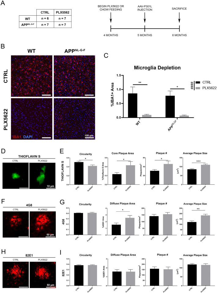
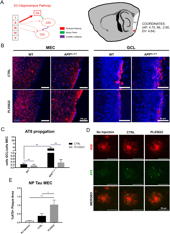
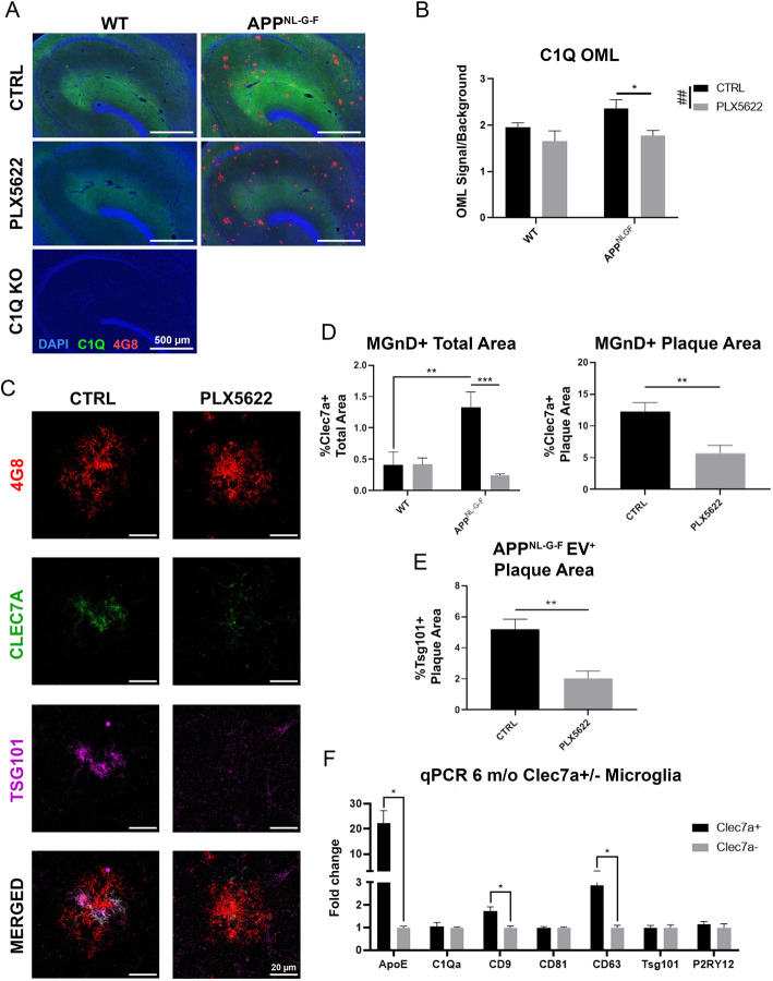
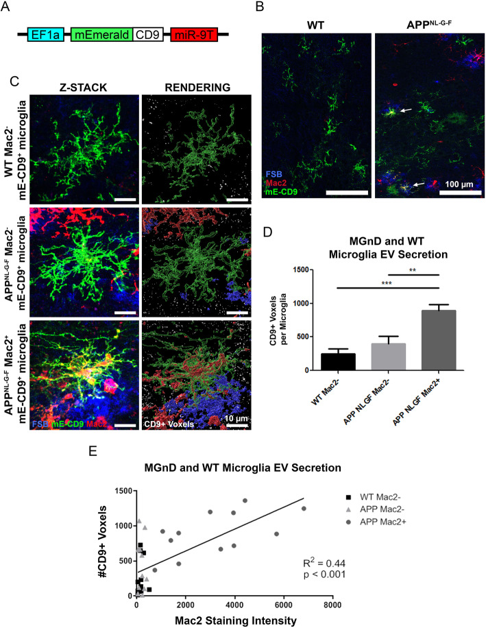
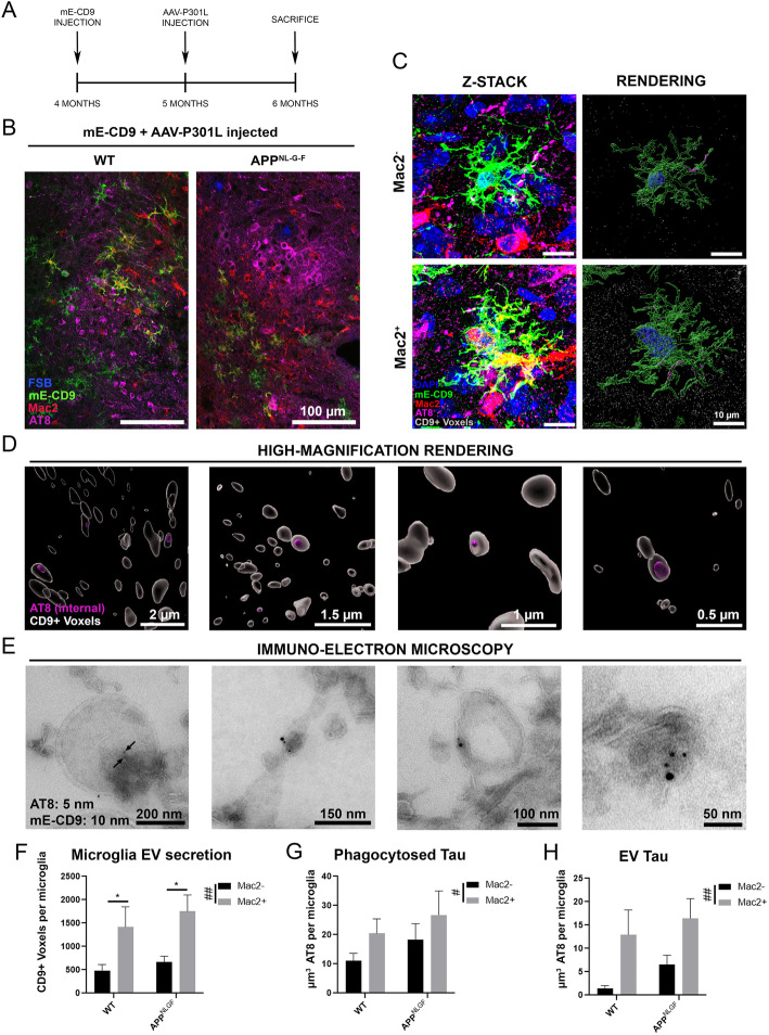
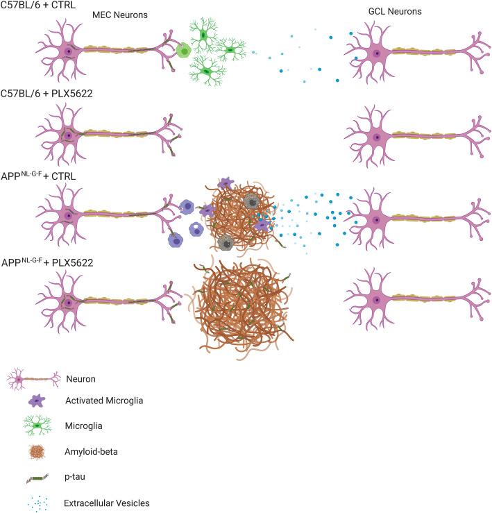
Abstract Background Recent studies suggest that microglia contribute to tau pathology progression in Alzheimer’s disease. Amyloid plaque accumulation transforms microglia, the primary innate immune cells in the brain, into neurodegenerative microglia (MGnD), which exhibit enhanced phagocytosis of plaques, apoptotic neurons and dystrophic neurites containing aggregated and phosphorylated tau (p-tau). It remains unclear how microglia promote disease progression while actively phagocytosing pathological proteins, therefore ameliorating pathology. Methods Adeno-associated virus expressing P301L tau mutant (AAV-P301L-tau) was stereotaxically injected into the medial entorhinal cortex (MEC) in C57BL/6 (WT) and humanized APP mutant knock-in homozygote ( App NL-G-F ) mice at 5 months of age. Mice were fed either chow containing a colony stimulating factor-1 receptor inhibitor (PLX5622) or control chow from 4 to 6 months of age to test the effect of microglia depletion. Animals were tested at 6 months of age for immunofluorescence, biochemistry, and FACS of microglia. In order to monitor microglial extracellular vesicle secretion in vivo, a novel lentiviral EV reporter system was engineered to express mEmerald-CD9 (mE-CD9) specifically in microglia, which was injected into the same region of MEC. Results Expressing P301L tau mutant in the MEC induced tau propagation to the granule cell layer of the hippocampal dentate gyrus, which was significantly exacerbated in App NL-G-F mice compared to WT control mice. Administration of PLX5622 depleted nearly all microglia in mouse brains and dramatically reduced propagation of p-tau in WT and to a greater extent in App NL-G-F mice, although it increased plaque burden and plaque-associated p-tau + dystrophic neurites. Plaque-associated MGnD microglia strongly expressed an EV marker, tumor susceptibility gene 101, indicative of heightened synthesis of EVs. Intracortical injection of mE-CD9 lentivirus successfully induced microglia-specific expression of mE-CD9 + EV particles, which were significantly enhanced in Mac2 + MGnD microglia compared to Mac2 − homeostatic microglia. Finally, consecutive intracortical injection of mE-CD9 lentivirus and AAV-P301L-tau into App NL-G-F mice revealed encapsulation of p-tau in microglia-specific mE-CD9 + EVs as determined by super-resolution microscopy and immuno-electron microscopy. Discussion Our findings suggest that MGnD microglia hyper-secrete p-tau + EVs while compacting Aβ plaques and clearing NP tau, which we propose as a novel mechanistic link between amyloid plaque deposition and exacerbation of tau propagation in App NL-G-F mice.
Overview
The claims and statistics of this paper haven’t been checked yet.
Run the check — free, no account needed. It reads the open-access text and checks the sources it cites.
Integrity signals
Registry
Cites
Statistics
Figures
Verification
Claims the paper makes
The claims and statistics of this paper haven’t been checked yet.
Run the check — free, no account needed. It reads the open-access text and checks the sources it cites.
Claims other papers make about this one
Each is checked against this paper’s own text: does it say what the citing paper says it does?
The source backs part of the claim, not all of it — or backs it for a different population, condition or comparison.
In the citing paper
“Amyloid-β–associated microglia were shown to secrete extracellular vesicles that contain phosphorylated tau and their depletion abrogated tau propagation (Clayton et al., 2021).”
From A novel molecular class that recruits HDAC/MECP2 complexes to PU.1 motifs reduces neuroinflammation (2023) · cites this paper as Clayton et al., 2021
Experimental — accuracy not yet measuredThe source backs part of the claim, not all of it — or backs it for a different population, condition or comparison.
In the citing paper
“demonstrating that microglia play an essential role in plaque compaction between four to six months of age in mice [61]”
From Microglia and monocytes in inflammatory CNS disease: integrating phenotype and function (2022) · cites this paper as [61]
Experimental — accuracy not yet measuredThe source backs part of the claim, not all of it — or backs it for a different population, condition or comparison.
In the citing paper
“microglia were shown to be three times as phagocytic and three times more likely to release tau-containing extracellular vesicles when situated near amyloid plaques [61]”
From Microglia and monocytes in inflammatory CNS disease: integrating phenotype and function (2022) · cites this paper as [61]
Experimental — accuracy not yet measuredThe cited source says what the claim attributes to it.
In the citing paper
“However, in the same study, an increase in plaque-associated tau in dystrophic neurites was found, indicating that microglia play a complex role in both the spread of tau and clearance of neuronal debris associated with pathological tau.”
From A novel molecular class that recruits HDAC/MECP2 complexes to PU.1 motifs reduces neuroinflammation (2023) · cites this paper as Clayton et al., 2021
The cited source says what the claim attributes to it.
In the citing paper
“microglia depletion in four-month-old humanized APP mutant knock-in homozygote (APPNL−G−F) mice resulted in reduced plaque compaction and increased plaque volume, number, and size”
From Microglia and monocytes in inflammatory CNS disease: integrating phenotype and function (2022) · cites this paper as [61]
The cited source says what the claim attributes to it.
In the citing paper
“Microglia have also been implicated in the pathological spreading of tau via the release of extracellular vesicles [12, 61, 328]”
From Microglia and monocytes in inflammatory CNS disease: integrating phenotype and function (2022) · cites this paper as [61]
The cited source says what the claim attributes to it.
In the citing paper
“This was associated with enhanced tau propagation in mice with amyloidosis, compared to wild-type mice without amyloidosis [61]”
From Microglia and monocytes in inflammatory CNS disease: integrating phenotype and function (2022) · cites this paper as [61]
In the citing paper
“in an Alzheimer’s animal model, microglia secreted abundant p-tau+ sEVs, which accelerated amyloid plaque deposition, thus exacerbating tau propagation and worsening the symptoms of the disease [41].”
From M2 microglia-derived small extracellular vesicles modulate NSC fate after ischemic stroke via miR-25-3p/miR-93-5p-TGFBR/PTEN/FOXO3 axis (2025) · cites this paper as [41]
Self-check
Percentages, sums and reported differences re-computed from the paper’s own numbers.
The self-check needs the paper’s full text.
Citations
Cites
A Unique Microglia Type Associated with Restricting Development of Alzheimer’s Disease
Keren-Shaul, H., Spinrad, A., Weiner, A., Matcovitch-Natan, O., Dvir-Szternfeld, R., Ulland, T. K., David, E., Baruch, K., Lara-Astaiso, D., Toth, B., et al. (2017) · Cell · doi:10.1016/j.cell.2017.05.018 · Cited by 5,493
Synaptic Pruning by Microglia Is Necessary for Normal Brain Development
Paolicelli, R. C., Bolasco, G., Pagani, F., Maggi, L., Scianni, M., Panzanelli, P., Giustetto, M., Ferreira, T. A., Guiducci, E., Dumas, L., et al. (2011) · Science · doi:10.1126/science.1202529 · Cited by 4,244
Microglia Sculpt Postnatal Neural Circuits in an Activity and Complement-Dependent Manner
Schafer, D. P., Lehrman, E. K., Kautzman, A. G., Koyama, R., Mardinly, A. R., Yamasaki, R., Ransohoff, R. M., Greenberg, M. E., Barres, B. A., & Stevens, B. (2012) · Neuron · doi:10.1016/j.neuron.2012.03.026 · Cited by 4,089
Complement and microglia mediate early synapse loss in Alzheimer mouse models
Hong, S., Beja-Glasser, V. F., Nfonoyim, B. M., Frouin, A., Li, S., Ramakrishnan, S., Merry, K. M., Shi, Q., Rosenthal, A., Barres, B. A., et al. (2016) · Science · doi:10.1126/science.aad8373 · Cited by 3,343 · cited 1×
“It is well known that microglia actively phagocytose synapses by synaptic tagging with complements (C1q and C3) [50–52], which also play an important role in Aβ-induced synaptic loss [53].”
BackgroundKrasemann, S., Madore, C., Cialic, R., Baufeld, C., Calcagno, N., El Fatimy, R., Beckers, L., O’Loughlin, E., Xu, Y., Fanek, Z., et al. (2017) · Immunity · doi:10.1016/j.immuni.2017.08.008 · Cited by 2,979
Self-citationIdentification of a unique TGF-β–dependent molecular and functional signature in microglia
Butovsky, O., Jedrychowski, M. P., Moore, C. S., Cialic, R., Lanser, A. J., Gabriely, G., Koeglsperger, T., Dake, B., Wu, P. M., Doykan, C. E., et al. (2014) · Nature Neuroscience · doi:10.1038/nn.3599 · Cited by 2,543
Elmore, M. R. P., Najafi, A. R., Koike, M. A., Dagher, N. N., Spangenberg, E. E., Rice, R. A., Kitazawa, M., Matusow, B., Nguyen, H., West, B. L., et al. (2014) · Neuron · doi:10.1016/j.neuron.2014.02.040 · Cited by 1,968
Depletion of microglia and inhibition of exosome synthesis halt tau propagation
Asai, H., Ikezu, S., Tsunoda, S., Medalla, M., Luebke, J., Haydar, T., Wolozin, B., Butovsky, O., Kügler, S., & Ikezu, T. (2015) · Nature Neuroscience · doi:10.1038/nn.4132 · Cited by 1,591
Self-citationThe microglial sensome revealed by direct RNA sequencing
Hickman, S. E., Kingery, N. D., Ohsumi, T. K., Borowsky, M. L., Wang, L.-C., Means, T. K., & El Khoury, J. (2013) · Nature Neuroscience · doi:10.1038/nn.3554 · Cited by 1,511
NLRP3 inflammasome activation drives tau pathology
Ising, C., Venegas, C., Zhang, S., Scheiblich, H., Schmidt, S. V., Vieira-Saecker, A., Schwartz, S., Albasset, S., McManus, R. M., Tejera, D., et al. (2019) · Nature · doi:10.1038/s41586-019-1769-z · Cited by 1,487
Disease-Associated Microglia: A Universal Immune Sensor of Neurodegeneration
Deczkowska, A., Keren-Shaul, H., Weiner, A., Colonna, M., Schwartz, M., & Amit, I. (2018) · Cell · doi:10.1016/j.cell.2018.05.003 · Cited by 1,474
Single App knock-in mouse models of Alzheimer’s disease
Saito, T., Matsuba, Y., Mihira, N., Takano, J., Nilsson, P., Itohara, S., Iwata, N., & Saido, T. C. (2014) · Nature Neuroscience · doi:10.1038/nn.3697 · Cited by 1,366
Self-citationSimard, A. R., Soulet, D., Gowing, G., Julien, J.-P., & Rivest, S. (2006) · Neuron · doi:10.1016/j.neuron.2006.01.022 · Cited by 1,265
Role of Pro-Inflammatory Cytokines Released from Microglia in Neurodegenerative Diseases
Smith, J. A., Das, A., Ray, S. K., & Banik, N. L. (2012) · Brain Research Bulletin · doi:10.1016/j.brainresbull.2011.10.004 · Cited by 1,165
Trans-Synaptic Spread of Tau Pathology In Vivo
Liu, L., Drouet, V., Wu, J., Witter, M. P., Small, S. A., Clelland, C. L. T., & Duff, K. (2012) · PLoS ONE · doi:10.1371/journal.pone.0031302 · Cited by 994
Spangenberg, E., Severson, P. L., Hohsfield, L. A., Crapser, J., Zhang, J., Burton, E. A., Zhang, Y., Spevak, W., Lin, J., Phan, N. Y., et al. (2019) · Nature Communications · doi:10.1038/s41467-019-11674-z · Cited by 939
Scavenger receptor-mediated adhesion of microglia to β-amyloid fibrils
Khoury, J. E., Hickman, S. E., Thomas, C. A., Cao, L., Silverstein, S. C., & Loike, J. D. (1996) · Nature · doi:10.1038/382716a0 · Cited by 727
The role of microglia in amyloid clearance from the AD brain
Lee, C. Y. D., & Landreth, G. E. (2010) · Journal of Neural Transmission · doi:10.1007/s00702-010-0433-4 · Cited by 701
Kitazawa, M., Oddo, S., Yamasaki, T. R., Green, K. N., & LaFerla, F. M. (2005) · The Journal of Neuroscience · doi:10.1523/jneurosci.2868-05.2005 · Cited by 697
Spangenberg, E. E., Lee, R. J., Najafi, A. R., Rice, R. A., Elmore, M. R. P., Blurton-Jones, M., West, B. L., & Green, K. N. (2016) · Brain · doi:10.1093/brain/aww016 · Cited by 695
Phagocytosis of Microglia in the Central Nervous System Diseases
Fu, R., Shen, Q., Xu, P., Luo, J. J., & Tang, Y. (2014) · Molecular Neurobiology · doi:10.1007/s12035-013-8620-6 · Cited by 687
He, Z., Guo, J. L., McBride, J. D., Narasimhan, S., Kim, H., Changolkar, L., Zhang, B., Gathagan, R. J., Yue, C., Dengler, C., et al. (2018) · Nature Medicine · doi:10.1038/nm.4443 · Cited by 645
Butowski, N. A., Colman, H., de Groot, J., Omuro, A. M. P., Nayak, L., Wen, P. Y., Cloughesy, T. F., Marimuthu, A., Haidar, S., Perry, A., et al. (2015) · Neuro-Oncology · doi:10.1093/neuonc/nov245 · Cited by 608
Ito, D., Tanaka, K., Suzuki, S., Dembo, T., & Fukuuchi, Y. (2001) · Stroke · doi:10.1161/01.str.32.5.1208 · Cited by 598
Li, Y., Liu, L., Barger, S. W., & Griffin, W. S. T. (2003) · The Journal of Neuroscience · doi:10.1523/jneurosci.23-05-01605.2003 · Cited by 563
Olmos-Alonso, A., Schetters, S. T. T., Sri, S., Askew, K., Mancuso, R., Vargas-Caballero, M., Holscher, C., Perry, V. H., & Gomez-Nicola, D. (2016) · Brain · doi:10.1093/brain/awv379 · Cited by 532
Dejanovic, B., Huntley, M. A., De Mazière, A., Meilandt, W. J., Wu, T., Srinivasan, K., Jiang, Z., Gandham, V., Friedman, B. A., Ngu, H., et al. (2018) · Neuron · doi:10.1016/j.neuron.2018.10.014 · Cited by 517
Dagher, N. N., Najafi, A. R., Kayala, K. M. N., Elmore, M. R. P., White, T. E., Medeiros, R., West, B. L., & Green, K. N. (2015) · Journal of Neuroinflammation · doi:10.1186/s12974-015-0366-9 · Cited by 505
Microglial Phagocytosis of Fibrillar β-Amyloid through a β1Integrin-Dependent Mechanism
Koenigsknecht-Talboo, J., & Landreth, G. E. (2004) · Journal of Neuroscience · doi:10.1523/jneurosci.2557-04.2004 · Cited by 457
Microglia drive APOE-dependent neurodegeneration in a tauopathy mouse model
Shi, Y., Manis, M., Long, J., Wang, K., Sullivan, P. M., Remolina Serrano, J., Hoyle, R., & Holtzman, D. M. (2019) · Journal of Experimental Medicine · doi:10.1084/jem.20190980 · Cited by 448
Local externalization of phosphatidylserine mediates developmental synaptic pruning by microglia
Scott‐Hewitt, N., Perrucci, F., Morini, R., Erreni, M., Mahoney, M., Witkowska, A., Carey, A. E., Faggiani, E., Schuetz, L. T., Mason, S., et al. (2020) · The EMBO Journal · doi:10.15252/embj.2020105380 · Cited by 436
A Dramatic Increase of C1q Protein in the CNS during Normal Aging
Stephan, A. H., Madison, D. V., Mateos, J. M., Fraser, D. A., Lovelett, E. A., Coutellier, L., Kim, L., Tsai, H.-H., Huang, E. J., Rowitch, D. H., et al. (2013) · The Journal of Neuroscience · doi:10.1523/jneurosci.1333-13.2013 · Cited by 423
Sosna, J., Philipp, S., Albay, R., Reyes-Ruiz, J. M., Baglietto-Vargas, D., LaFerla, F. M., & Glabe, C. G. (2018) · Molecular Neurodegeneration · doi:10.1186/s13024-018-0244-x · Cited by 406
Formation and maintenance of Alzheimer's disease β-amyloid plaques in the absence of microglia
Grathwohl, S. A., Kälin, R. E., Bolmont, T., Prokop, S., Winkelmann, G., Kaeser, S. A., Odenthal, J., Radde, R., Eldh, T., Gandy, S., et al. (2009) · Nature Neuroscience · doi:10.1038/nn.2432 · Cited by 401
Extracellular vesicles as mediators of neuron-glia communication
Frühbeis, C., Fröhlich, D., Kuo, W. P., & Krämer‐Albers, E. (2013) · Frontiers in Cellular Neuroscience · doi:10.3389/fncel.2013.00182 · Cited by 397
The perforant path: projections from the entorhinal cortex to the dentate gyrus
Witter, M. P. (2007) · Progress in brain research · doi:10.1016/s0079-6123(07)63003-9 · Cited by 378
Zhong, L., Xu, Y., Zhuo, R., Wang, T., Wang, K., Huang, R., Wang, D., Gao, Y., Zhu, Y., Sheng, X., et al. (2019) · Nature Communications · doi:10.1038/s41467-019-09118-9 · Cited by 371
Targeting mi R‐155 restores abnormal microglia and attenuates disease in SOD 1 mice
Butovsky, O., Jedrychowski, M. P., Cialic, R., Krasemann, S., Murugaiyan, G., Fanek, Z., Greco, D. J., Wu, P. M., Doykan, C. E., Kiner, O., et al. (2015) · Annals of Neurology · doi:10.1002/ana.24304 · Cited by 358
Boza-Serrano, A., Ruiz, R., Sanchez-Varo, R., García-Revilla, J., Yang, Y., Jimenez-Ferrer, I., Paulus, A., Wennström, M., Vilalta, A., Allendorf, D., et al. (2019) · Acta Neuropathologica · doi:10.1007/s00401-019-02013-z · Cited by 357
Microglia-Mediated Synapse Loss in Alzheimer's Disease
Rajendran, L., & Paolicelli, R. C. (2018) · The Journal of Neuroscience · doi:10.1523/jneurosci.1136-17.2017 · Cited by 355
Cell-specific deletion of C1qa identifies microglia as the dominant source of C1q in mouse brain
Fonseca, M. I., Chu, S.-H., Hernandez, M. X., Fang, M. J., Modarresi, L., Selvan, P., MacGregor, G. R., & Tenner, A. J. (2017) · Journal of Neuroinflammation · doi:10.1186/s12974-017-0814-9 · Cited by 337
Island Cells Control Temporal Association Memory
北村, 貴., Pignatelli, M., Suh, J., Kohara, K., Yoshiki, A., Abe, K., & Tonegawa, S. (2014) · Science · doi:10.1126/science.1244634 · Cited by 333
TREM2 function impedes tau seeding in neuritic plaques
Leyns, C. E. G., Gratuze, M., Narasimhan, S., Jain, N., Koscal, L. J., Jiang, H., Manis, M., Colonna, M., Lee, V. M. Y., Ulrich, J. D., et al. (2019) · Nature Neuroscience · doi:10.1038/s41593-019-0433-0 · Cited by 332
Antonucci, F., Turola, E., Riganti, L., Caleo, M., Gabrielli, M., Perrotta, C., Novellino, L., Clementi, E., Giussani, P., Viani, P., et al. (2012) · The EMBO Journal · doi:10.1038/emboj.2011.489 · Cited by 328
Tunneling nanotube (TNT)-mediated neuron-to neuron transfer of pathological Tau protein assemblies
Tardivel, M., Bégard, S., Bousset, L., Dujardin, S., Coens, A., Melki, R., Buée, L., & Colin, M. (2016) · Acta Neuropathologica Communications · doi:10.1186/s40478-016-0386-4 · Cited by 305
The role of microglia in processing and spreading of bioactive tau seeds in Alzheimer’s disease
Hopp, S. C., Lin, Y., Oakley, D., Roe, A. D., DeVos, S. L., Hanlon, D., & Hyman, B. T. (2018) · Journal of Neuroinflammation · doi:10.1186/s12974-018-1309-z · Cited by 286
Synaptic Tau Seeding Precedes Tau Pathology in Human Alzheimer’s Disease Brain
DeVos, S. L., Corjuc, B. T., Oakley, D. H., Nobuhara, C. K., Bannon, R. N., Chase, A., Commins, C., Gonzalez, J. A., Dooley, P. M., Frosch, M. P., et al. (2018) · Frontiers in Neuroscience · doi:10.3389/fnins.2018.00267 · Cited by 285
Pisanu, A., Lecca, D., Mulas, G., Wardas, J., Simbula, G., Spiga, S., & Carta, A. R. (2014) · Neurobiology of Disease · doi:10.1016/j.nbd.2014.08.011 · Cited by 273
Rice, R. A., Spangenberg, E. E., Yamate-Morgan, H., Lee, R. J., Arora, R. P. S., Hernandez, M. X., Tenner, A. J., West, B. L., & Green, K. N. (2015) · Journal of Neuroscience · doi:10.1523/jneurosci.0336-15.2015 · Cited by 270
Formation and maintenance of Alzheimer’s disease beta-amyloid plaques in the absence of microglia
Grathwohl, S. A., Kälin, R., Bolmont, T., Prokop, S., Winkelmann, G., Kaeser, S. A., Odenthal, J., Radde, R., Eldh, T., Gandy, S. E., et al. (2010) · e-Neuroforum · doi:10.1515/nf-2010-0107 · Cited by 269
Amyloid accelerates tau propagation and toxicity in a model of early Alzheimer’s disease
Pooler, A. M., Polydoro, M., Maury, E. A., Nicholls, S. B., Reddy, S. M., Wegmann, S., William, C., Saqran, L., Cagsal-Getkin, O., Pitstick, R., et al. (2015) · Acta Neuropathologica Communications · doi:10.1186/s40478-015-0199-x · Cited by 236
The Role of Microglia and the Nlrp3 Inflammasome in Alzheimer's Disease
Hanslik, K. L., & Ulland, T. K. (2020) · Frontiers in Neurology · doi:10.3389/fneur.2020.570711 · Cited by 236
CSF1R inhibitor JNJ-40346527 attenuates microglial proliferation and neurodegeneration in P301S mice
Mancuso, R., Fryatt, G., Cleal, M., Obst, J., Pipi, E., Monzón-Sandoval, J., Ribe, E., Winchester, L., Webber, C., Nevado, A., et al. (2019) · Brain · doi:10.1093/brain/awz241 · Cited by 235
Hurtado, D. E., Molina-Porcel, L., Iba, M., Aboagye, A. K., Paul, S. M., Trojanowski, J. Q., & Lee, V. M.-Y. (2010) · The American Journal of Pathology · doi:10.2353/ajpath.2010.100346 · Cited by 222
Jüttner, J., Szabo, A., Gross-Scherf, B., Morikawa, R. K., Rompani, S. B., Hantz, P., Szikra, T., Esposti, F., Cowan, C. S., Bharioke, A., et al. (2019) · Nature Neuroscience · doi:10.1038/s41593-019-0431-2 · Cited by 218
Muraoka, S., DeLeo, A. M., Sethi, M. K., Takamatsu-Yukawa, K., Yang, Z., Ko, J., Hogan, J., Ruan, Z., You, Y., Wang, Y., et al. (2020) · Alzheimer s & Dementia · doi:10.1002/alz.12089 · Cited by 195
Self-citationCasali, B. T., MacPherson, K. P., Reed-Geaghan, E. G., & Landreth, G. E. (2020) · Neurobiology of Disease · doi:10.1016/j.nbd.2020.104956 · Cited by 186
Kügler, S., Meyn, L., Holzmüller, H., Gerhardt, E., Isenmann, S., Schulz, J. B., & Bähr, M. (2001) · Molecular and Cellular Neuroscience · doi:10.1006/mcne.2000.0929 · Cited by 182
Clearance of Amyloid-β Peptides By Microglia and Macrophages: the Issue of What, When and Where
Lai, A. Y., & McLaurin, J. (2012) · Future Neurology · doi:10.2217/fnl.12.6 · Cited by 166
Microglia limit the expansion of β-amyloid plaques in a mouse model of Alzheimer’s disease
Zhao, R., Hu, W., Tsai, J., Li, W., & Gan, W.-B. (2017) · Molecular Neurodegeneration · doi:10.1186/s13024-017-0188-6 · Cited by 151
P2RX7 inhibitor suppresses exosome secretion and disease phenotype in P301S tau transgenic mice
Ruan, Z., Delpech, J.-C., Venkatesan Kalavai, S., Van Enoo, A. A., Hu, J., Ikezu, S., & Ikezu, T. (2020) · Molecular Neurodegeneration · doi:10.1186/s13024-020-00396-2 · Cited by 150
Self-citationAlzheimer's Disease: The Role of Microglia in Brain Homeostasis and Proteopathy
Clayton, K. A., Enoo, A. A. V., & Ikezu, T. (2017) · Frontiers in Neuroscience · doi:10.3389/fnins.2017.00680 · Cited by 137
Self-citationMicroglia in Infectious Diseases of the Central Nervous System
Mariani, M. M., & Kielian, T. (2009) · Journal of Neuroimmune Pharmacology · doi:10.1007/s11481-009-9170-6 · Cited by 132
Partial reduction of microglia does not affect tau pathology in aged mice
Bennett, R. E., Bryant, A., Hu, M., Robbins, A. B., Hopp, S. C., & Hyman, B. T. (2018) · Journal of Neuroinflammation · doi:10.1186/s12974-018-1348-5 · Cited by 91
Åkerblom, M., Sachdeva, R., Quintino, L., Wettergren, E. E., Chapman, K. Z., Manfre, G., Lindvall, O., Lundberg, C., & Jakobsson, J. (2013) · Nature Communications · doi:10.1038/ncomms2801 · Cited by 85
Muraoka, S., Lin, W., Chen, M., Hersh, S. W., Emili, A., Xia, W., & Ikezu, T. (2020) · Methods · doi:10.1016/j.ymeth.2020.02.002 · Cited by 69
Self-citationSpread of pathological tau proteins through communicating neurons in human Alzheimer’s diseasePreprint
Vogel, J. W., Iturria‐Medina, Y., Strandberg, O. T., Smith, R. A., Evans, A. C., Hansson, O., & for the Alzheimer’s Disease Neuroimaging Initiative, A. T. S. B. S. (2019) · bioRxiv (Cold Spring Harbor Laboratory) · doi:10.1101/555821 · Cited by 64
Tsutsumi, R., Hori, Y., Seki, T., Kurauchi, Y., Sato, M., Oshima, M., Hisatsune, A., & Katsuki, H. (2019) · Biochemical and Biophysical Research Communications · doi:10.1016/j.bbrc.2019.02.076 · Cited by 56
Son, Y., Jeong, Y. J., Shin, N., Oh, S. J., Nam, K. R., Choi, H., Choi, J. Y., & Lee, H. (2020) · International Journal of Molecular Sciences · doi:10.3390/ijms21155553 · Cited by 53
Jüttner, J., Szabó, A., Gross-Scherf, B., et al. (2018) · bioRxiv (Cold Spring Harbor Laboratory) · doi:10.1101/434720 · Cited by 26
Nelson, E. J. R., Tuschong, L. M., Hunter, M. J., Bauer, T. R., Burkholder, T. H., & Hickstein, D. D. (2010) · Gene Therapy · doi:10.1038/gt.2010.7 · Cited by 25
Zhu, K., Pieber, M., Han, J., Blomgren, K., Zhang, X., Harris, R. A., & Lund, H. (2020) · Glia · doi:10.1002/glia.23794 · Cited by 16
Exosome Release Promotes Inflammatory Resolution in Activated and Aged MicrogliaPreprint
Udeochu, J. C., Sanchez-Diaz, C., Cai, A., Jovicic, A., & Villeda, S. (2018) · bioRxiv (Cold Spring Harbor Laboratory) · doi:10.1101/423558 · Cited by 8
[Clinical signs of occlusion of the cerebral median artery].
Radijicić, B., Nikolić, M., & Lukić (1966) · PubMed · Cited by 1
Cited by
Sorted by citations
Microglia in neurodegenerative diseases: mechanism and potential therapeutic targets
Gao, C., Jiang, J., Tan, Y., & Chen, S. (2023) · Signal Transduction and Targeted Therapy · doi:10.1038/s41392-023-01588-0 · Cited by 1,616
Synaptic degeneration in Alzheimer disease
Tzioras, M., McGeachan, R. I., Durrant, C. S., & Spires-Jones, T. L. (2023) · Nature Reviews Neurology · doi:10.1038/s41582-022-00749-z · Cited by 504
The effects of microglia-associated neuroinflammation on Alzheimer’s disease
Wang, C., Zong, S., Cui, X., Wang, X., Wu, S., Wang, L., Liu, Y., & Lu, Z. (2023) · Frontiers in Immunology · doi:10.3389/fimmu.2023.1117172 · Cited by 331
The neuroimmune axis of Alzheimer’s disease
Jorfi, M., Maaser‐Hecker, A., & Tanzi, R. E. (2023) · Genome Medicine · doi:10.1186/s13073-023-01155-w · Cited by 313
The Contribution of Microglia to Neuroinflammation in Parkinson’s Disease
Badanjak, K., Fixemer, S., Smajić, S., Skupin, A., & Grünewald, A. (2021) · International Journal of Molecular Sciences · doi:10.3390/ijms22094676 · Cited by 296
Metadata
- Authors
- Clayton, K., Delpech, J. C., Herron, S., Iwahara, N., Ericsson, M., Saito, T., Saido, T. C., Ikezu, S., & Ikezu, T.
- Affiliations
- Boston University (US)
- Harvard University (US)
- RIKEN Center for Brain Science (JP)
- Corresponding author
- Ikezu, Tsuneya
- ORCID
- Venue
- Molecular Neurodegeneration
- Published
- 2021-03-22
- Volume
- 16
- Issue
- 1
- Pages
- 18-18
- ISSN
- 1750-1326 (ISSN-L)
- PMC
- PMC7986521
- OpenAlex
- W3137321433
- Cited by
- 245
- References
- 73
- Citations by year
- 2026: 35 · 2025: 42 · 2024: 49 · 2023: 43 · 2022: 49 · 2021: 26
- FWCI
- 26.24
- Type
- journal article
- Language
- en
- Update notices
- correction (2021-04-14)
- Open access
- gold
- doi.org
- Locations
- Molecular Neurodegeneration (publishedVersion, cc-by)
- bioRxiv (Cold Spring Harbor Laboratory) (acceptedVersion)
- PubMed Central (publishedVersion, cc-by)
- PubMed (publishedVersion)
- HAL (submittedVersion, cc-by)
- DOAJ (DOAJ: Directory of Open Access Journals) (submittedVersion, cc-by-sa)
- OpenBU (Boston University) (submittedVersion)
- Oskar-Bordeaux (Universite de Bordeaux) (submittedVersion, cc-by)
- Funders
- Alzheimer's Association (DVT-14-320835)
- BrightFocus Foundation (A2016551S)
- Cure Alzheimer's Fund (N/A)
- National Institute on Aging (R01 AG066429, RF1 AG054199, R56 AG057469, P30 AG0138423, F31 AG057170)
- NIGMS (T32 GM008541)
- Topics
- Life Sciences › Neuroscience › Cellular and Molecular Neuroscience › Alzheimer's disease research and treatments
- Life Sciences › Neuroscience › Neurology › Neuroinflammation and Neurodegeneration Mechanisms
- Life Sciences › Biochemistry, Genetics and Molecular Biology › Molecular Biology › Extracellular vesicles in disease
- Keywords
- microglia, extracellular vesicles, tau propagation, Alzheimer's disease, amyloid plaques, PLX5622, CSF1R inhibition, microglial depletion, CD9, TSG101, medial entorhinal cortex, dentate gyrus, dystrophic neurites, tau phosphorylation, plaque-associated microglia, disease progression
- MeSH terms
- Extracellular Vesicles, Animals, Female, Genetic Vectors, Humans, Injections, Male, Mice, Inbred C57BL, Organic Chemicals, Recombinant Proteins, Receptors, Granulocyte-Macrophage Colony-Stimulating Factor, Neurites, Amyloid beta-Protein Precursor, tau Proteins, Point Mutation, Microglia, Entorhinal Cortex, Dentate Gyrus, Mutation, Missense, Mice, Gene Knock-In Techniques, Plaque, Amyloid, Protein Aggregation, Pathological
About these checks
Registry, retraction, Cites and statistics results are computed, not judged, with one exception: a printed reference that no index lists is read once by a model before it counts as a citation error. Claims are extracted by a model and kept only when their quote appears verbatim in the text; each cited source is then checked, with a model’s help, for whether it says what the claim relies on. Those verdicts can be wrong — read the claim’s quote and the cited source before relying on one.
Statistics and arithmetic are recomputed, not judged.
Confidence bands are shown only for fields calibrated on our annotated test set. None is calibrated yet, so no band is shown.
What counts as a claim ›How we measure confidence ›How we check this ›
Are you an author, or do you see a problem with this page? Request a review
