

The DOI matches its Crossref record; 18 of the 18 references it cites were found in a registry or an index.
Tau interactome and RNA binding proteins in neurodegenerative diseases
Abstract
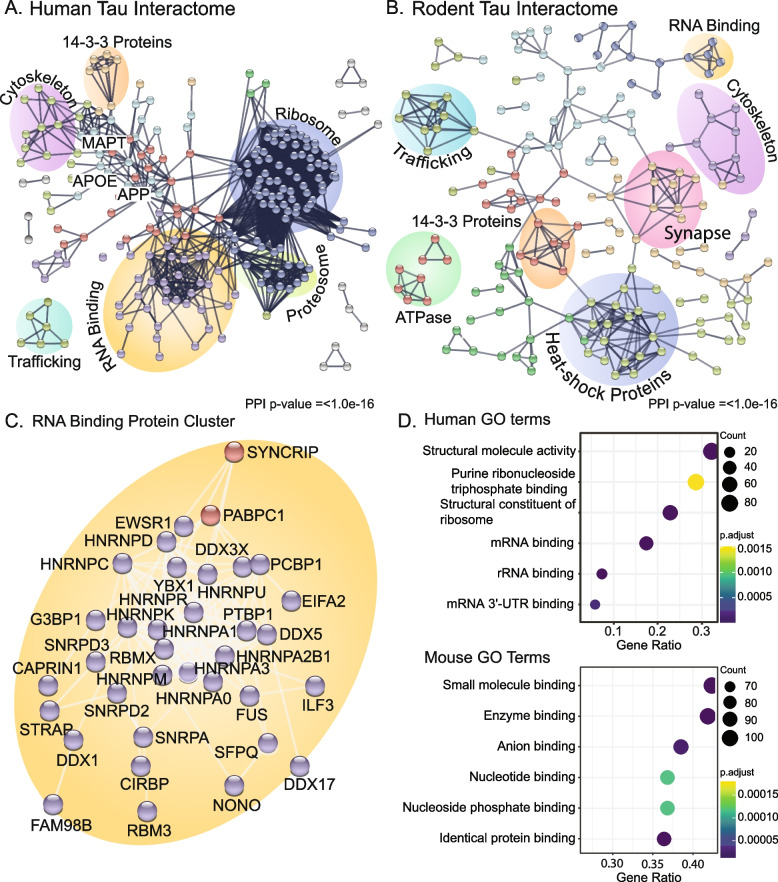
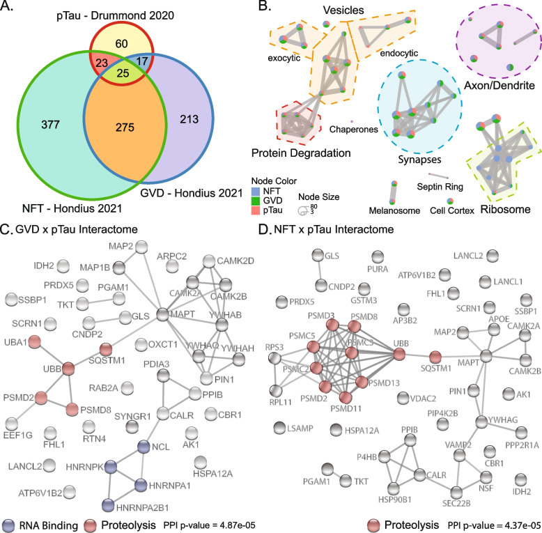
Pathological tau aggregation is a primary neuropathological feature of many neurodegenerative diseases. Intriguingly, despite the common presence of tau aggregates in these diseases the affected brain regions, clinical symptoms, and morphology, conformation, and isoform ratio present in tau aggregates varies widely. The tau-mediated disease mechanisms that drive neurodegenerative disease are still unknown. Tau interactome studies are critically important for understanding tauopathy. They reveal the interacting partners that define disease pathways, and the tau interactions present in neuropathological aggregates provide potential insight into the cellular environment and protein interactions present during pathological tau aggregation. Here we provide a combined analysis of 12 tau interactome studies of human brain tissue, human cell culture models and rodent models of disease. Together, these studies identified 2084 proteins that interact with tau in human tissue and 1152 proteins that interact with tau in rodent models of disease. Our combined analysis of the tau interactome revealed consistent enrichment of interactions between tau and proteins involved in RNA binding, ribosome, and proteasome function. Comparison of human and rodent tau interactome studies revealed substantial differences between the two species. We also performed a second analysis to identify the tau interacting proteins that are enriched in neurons containing granulovacuolar degeneration or neurofibrillary tangle pathology. These results revealed a timed dysregulation of tau interactions as pathology develops. RNA binding proteins, particularly HNRNPs, emerged as early disease-associated tau interactors and therefore may have an important role in driving tau pathology.
Overview
The claims and statistics of this paper haven’t been checked yet.
Run the check — free, no account needed. It reads the open-access text and checks the sources it cites.
Integrity signals
Registry
Cites
Statistics
Figures
Verification
Claims the paper makes
The claims and statistics of this paper haven’t been checked yet.
Run the check — free, no account needed. It reads the open-access text and checks the sources it cites.
Claims other papers make about this one
Each is checked against this paper’s own text: does it say what the citing paper says it does?
In the citing paper
“A growing body of evidence implicates interactions between pathogenic tau, RNA, and RNA-binding proteins (RBPs) in disease pathogenesis 3-13 .”
From Tau is an m 6 A-associated RNA binding protein that causes RNA dysregulation in disease (2026) · cites this paper as [11]
In the citing paper
“Physiological tau binds to many RBPs and translationassociated factors 3,11,20 .”
From Tau is an m 6 A-associated RNA binding protein that causes RNA dysregulation in disease (2026) · cites this paper as [11]
In the citing paper
“RBPs constitute a large fraction of the tau interactome 11 , and pathogenic tau can have aberrant interactions with RBPs, leading to downstream consequences in cellular and RNA-related phenotypes 3,4,9,10,14-16 .”
From Tau is an m 6 A-associated RNA binding protein that causes RNA dysregulation in disease (2026) · cites this paper as [11]
Self-check
Percentages, sums and reported differences re-computed from the paper’s own numbers.
The self-check needs the paper’s full text.
Citations
Cites
Cryo-EM structures of tau filaments from Alzheimer’s disease
Fitzpatrick, A. W. P., Falcon, B., He, S., Murzin, A. G., Murshudov, G., Garringer, H. J., Crowther, R. A., Ghetti, B., Goedert, M., & Scheres, S. H. W. (2017) · Nature · doi:10.1038/nature23002 · Cited by 2,061 · cited 1×
“For example, tau aggregates into straight or paired helical filaments in AD [3, 35].”
BackgroundTransmission and spreading of tauopathy in transgenic mouse brain
Clavaguera, F., Bolmont, T., Crowther, R. A., Abramowski, D., Frank, S., Probst, A., Fraser, G., Stalder, A. K., Beibel, M., Staufenbiel, M., et al. (2009) · Nature Cell Biology · doi:10.1038/ncb1901 · Cited by 1,803
HNRNPA2B1 Is a Mediator of m6A-Dependent Nuclear RNA Processing Events
Alarcón, C. R., Goodarzi, H., Lee, H., Liu, X., Tavazoie, S., & Tavazoie, S. F. (2015) · Cell · doi:10.1016/j.cell.2015.08.011 · Cited by 1,514
Tau PTM Profiles Identify Patient Heterogeneity and Stages of Alzheimer’s Disease
Wesseling, H., Mair, W., Kumar, M., Schlaffner, C. N., Tang, S., Beerepoot, P., Fatou, B., Guise, A. J., Cheng, L., Takeda, S., et al. (2020) · Cell · doi:10.1016/j.cell.2020.10.029 · Cited by 688 · cited 2×
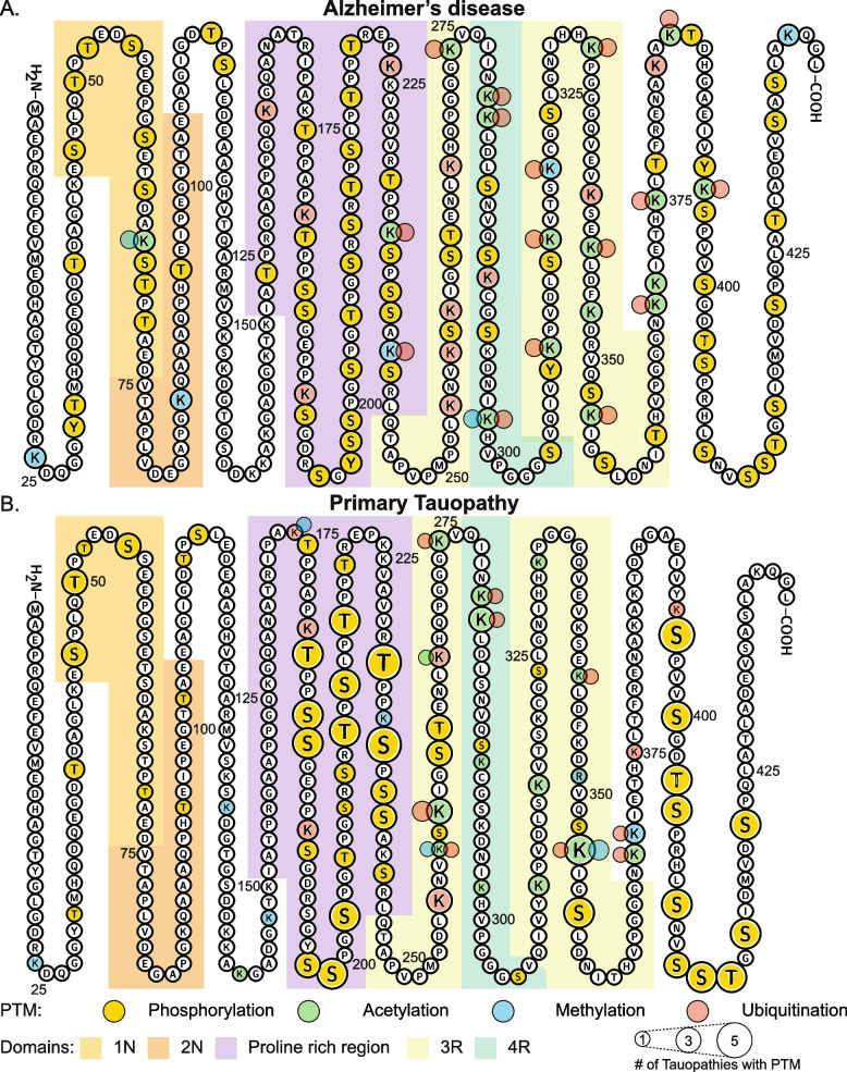
“Recent mass spectrometry studies have identified numerous additional phosphorylation sites on tau, and further characterised tau’s less abundant PTMs including acetylation, methylation, and ubiquitination [58, 113].”
BackgroundHanger, D. P., Byers, H. L., Wray, S., Leung, K.-Y., Saxton, M. J., Seereeram, A., Reynolds, C. H., Ward, M. A., & Anderton, B. H. (2007) · Journal of Biological Chemistry · doi:10.1074/jbc.m703269200 · Cited by 455
Alquezar, C., Arya, S., & Kao, A. W. (2021) · Frontiers in Neurology · doi:10.3389/fneur.2020.595532 · Cited by 367
Tau Activates Transposable Elements in Alzheimer’s Disease
Guo, C., Jeong, H.-H., Hsieh, Y.-C., Klein, H.-U., Bennett, D. A., De Jager, P. L., Liu, Z., & Shulman, J. M. (2018) · Cell Reports · doi:10.1016/j.celrep.2018.05.004 · Cited by 315
Andreadis, A. (2005) · Biochimica et Biophysica Acta (BBA) - Molecular Basis of Disease · doi:10.1016/j.bbadis.2004.08.010 · Cited by 311
Reducing the RNA binding protein TIA1 protects against tau-mediated neurodegeneration in vivo
Apicco, D. J., Ash, P. E. A., Maziuk, B., LeBlang, C., Medalla, M., Al Abdullatif, A., Ferragud, A., Botelho, E., Ballance, H. I., Dhawan, U., et al. (2018) · Nature Neuroscience · doi:10.1038/s41593-017-0022-z · Cited by 281 · cited 1×
“Reduction of TIA1 inhibits the spread and prevents toxicity of seeded tau aggregates, but interestingly, increased neurofibrillary tangle burden and neuroinflammation [8, 56, 65].”
BackgroundGaikwad, S., Puangmalai, N., Bittar, A., Montalbano, M., Garcia, S., McAllen, S., Bhatt, N., Sonawane, M., Sengupta, U., & Kayed, R. (2021) · Cell Reports · doi:10.1016/j.celrep.2021.109419 · Cited by 234
Research works printed but not linked: 103
- Abreha MH, Dammer EB, Ping L, Zhang T, Duong DM, Gearing M, Lah JJ, Levey AI, Seyfried NT. Quantitative analysis of the brain ubiquitylome in Alzheimer’s Disease. Proteomics. 2018;18(20):e1800108–e1800108. https://doi.org/10.1002/pmic.201800108. · DOI
- Ahmed Z, Cooper J, Murray TK, Garn K, McNaughton E, Clarke H, Parhizkar S, Ward MA, Cavallini A, Jackson S, Bose S, Clavaguera F, Tolnay M, Lavenir I, Goedert M, Hutton ML, O’Neill MJ. A novel in vivo model of tau propagation with rapid and progressive neurofibrillary tangle pathology: the pattern of spread is determined by connectivity, not proximity. Acta Neuropathol. 2014;127(5):667–83. https://doi.org/10.1007/s00401-014-1254-6. · DOI
- Al-Hilaly YK, Pollack SJ, Vadukul DM, Citossi F, Rickard JE, Simpson M, Storey JMD, Harrington CR, Wischik CM, Serpell LC. Alzheimer’s Disease-like paired helical filament assembly from Truncated Tau protein is independent of disulfide crosslinking. J Mol Biol. 2017;429(23):3650–65. https://doi.org/10.1016/j.jmb.2017.09.007. · DOI
- Aragão Gomes L, Uytterhoeven V, Lopez-Sanmartin D, Tomé SO, Tousseyn T, Vandenberghe R, Vandenbulcke M, von Arnim CAF, Verstreken P, Thal DR. Maturation of neuronal AD-tau pathology involves site-specific phosphorylation of cytoplasmic and synaptic tau preceding conformational change and fibril formation. Acta Neuropathol. 2021;141(2):173–92. https://doi.org/10.1007/s00401-020-02251-6. · DOI
- Arakhamia T, Lee CE, Carlomagno Y, Duong DM, Kundinger SR, Wang K, Williams D, DeTure M, Dickson DW, Cook CN, Seyfried NT, Petrucelli L, Fitzpatrick AWP. Posttranslational modifications mediate the structural diversity of tauopathy strains. Cell. 2020;180(4):633-644.e612. https://doi.org/10.1016/j.cell.2020.01.027. · DOI
- Ash PEA, Lei S, Shattuck J, Boudeau S, Carlomagno Y, Medalla M, Mashimo BL, Socorro G, Al-Mohanna LFA, Jiang L, Öztürk MM, Knobel M, Ivanov P, Petrucelli L, Wegmann S, Kanaan NM, Wolozin B. TIA1 potentiates tau phase separation and promotes generation of toxic oligomeric tau. Proc Natl Acad Sci. 2021;118(9):e2014188118. https://doi.org/10.1073/pnas.2014188118. · DOI · Paper page
- Ayyadevara S, Balasubramaniam M, Parcon PA, Barger SW, Griffin WST, Alla R, Tackett AJ, Mackintosh SG, Petricoin E, Zhou W, Shmookler Reis RJ. Proteins that mediate protein aggregation and cytotoxicity distinguish Alzheimer’s hippocampus from normal controls. Aging Cell. 2016;15(5):924–39. https://doi.org/10.1111/acel.12501. · DOI
- Bachmann S, Bell M, Klimek J, Zempel H. Differential effects of the six human TAU isoforms: somatic retention of 2N-TAU and increased microtubule number induced by 4R-TAU. Front Neurosci. 2021;15(547). https://doi.org/10.3389/fnins.2021.643115 · DOI
- Bampton A, Gatt A, Humphrey J, Cappelli S, Bhattacharya D, Foti S, Brown A-L, Asi Y, Low YH, Foiani M, Raj T, Buratti E, Fratta P, Lashley T. HnRNP K mislocalisation is a novel protein pathology of frontotemporal lobar degeneration and ageing and leads to cryptic splicing. Acta Neuropathol. 2021;142(4):609–27. https://doi.org/10.1007/s00401-021-02340-0. · DOI
- Bampton A, Gittings LM, Fratta P, Lashley T, Gatt A. The role of hnRNPs in frontotemporal dementia and amyotrophic lateral sclerosis. Acta Neuropathol. 2020;140(5):599–623. https://doi.org/10.1007/s00401-020-02203-0. · DOI
- Batlle C, Yang P, Coughlin M, Messing J, Pesarrodona M, Szulc E, Salvatella X, Kim HJ, Taylor JP, Ventura S. hnRNPDL phase separation is regulated by alternative splicing and disease-causing mutations accelerate its aggregation. Cell Rep. 2020;30(4):1117-1128.e1115. https://doi.org/10.1016/j.celrep.2019.12.080. · DOI
- Binder LI, Frankfurter A, Rebhun LI. The distribution of tau in the mammalian central nervous system. J Cell Biol. 1985;101(4):1371–8. https://doi.org/10.1083/jcb.101.4.1371. · DOI · Paper page
- Blaudin de Thé F-X, Lassus B, Schaler AW, Fowler SL, Goulbourne CN, Jeggo R, Mannoury la Cour C, Millan MJ, Duff KE. P62 accumulates through neuroanatomical circuits in response to tauopathy propagation. Acta Neuropathol Commun. 2021;9(1):177. https://doi.org/10.1186/s40478-021-01280-w. · DOI
- Boluda S, Iba M, Zhang B, Raible KM, Lee VMY, Trojanowski JQ. Differential induction and spread of tau pathology in young PS19 tau transgenic mice following intracerebral injections of pathological tau from Alzheimer’s disease or corticobasal degeneration brains. Acta Neuropathol. 2015;129(2):221–37. https://doi.org/10.1007/s00401-014-1373-0. · DOI
- Broderick J, Wang J, Andreadis A. Heterogeneous nuclear ribonucleoprotein E2 binds to tau exon 10 and moderately activates its splicing. Gene. 2004;331:107–14. https://doi.org/10.1016/j.gene.2004.02.005. · DOI
- Buée L, Delacourte A. Comparative biochemistry of Tau in progressive supranuclear palsy, corticobasal degeneration, FTDP-17 and Pick’s Disease. Brain Pathol. 1999;9(4):681–93. https://doi.org/10.1111/j.1750-3639.1999.tb00550.x. · DOI
- Bullmann T, Holzer M, Mori H, Arendt T. Pattern of tau isoforms expression during development in vivo. Int J Dev Neurosci. 2009;27(6):591–7. https://doi.org/10.1016/j.ijdevneu.2009.06.001. · DOI
- Cherry JD, Esnault CD, Baucom ZH, Tripodis Y, Huber BR, Alvarez VE, Stein TD, Dickson DW, McKee AC. Tau isoforms are differentially expressed across the hippocampus in chronic traumatic encephalopathy and Alzheimer’s disease. Acta Neuropathol Commun. 2021;9(1):86. https://doi.org/10.1186/s40478-021-01189-4. · DOI
- Chiang W-C, Lee M-H, Chen T-C, Huang J-R. Interactions between the intrinsically disordered regions of hnRNP-A2 and TDP-43 accelerate TDP-43’s conformational transition. Int J Mol Sci. 2020;21(16):5930. https://doi.org/10.3390/ijms21165930. · DOI
- Chung D-EC, Carlomagno Y, Cook CN, Jansen-West K, Daughrity L, Lewis-Tuffin LJ, Castanedes-Casey M, DeTure M, Dickson DW, Petrucelli L. Tau exhibits unique seeding properties in globular glial tauopathy. Acta Neuropathol Commun. 2019;7(1):36. https://doi.org/10.1186/s40478-019-0691-9. · DOI
- Chung D-EC, Roemer S, Petrucelli L, Dickson DW. Cellular and pathological heterogeneity of primary tauopathies. Mol Neurodegener. 2021;16(1):57. https://doi.org/10.1186/s13024-021-00476-x. · DOI
- Clavaguera F, Akatsu H, Fraser G, Crowther RA, Frank S, Hench J, Probst A, Winkler DT, Reichwald J, Staufenbiel M, Ghetti B, Goedert M, Tolnay M. Brain homogenates from human tauopathies induce tau inclusions in mouse brain. Proc Natl Acad Sci USA. 2013;110(23):9535–40. https://doi.org/10.1073/pnas.1301175110. · DOI · Paper page
- Dammer EB, Lee AK, Duong DM, Gearing M, Lah JJ, Levey AI, Seyfried NT. Quantitative phosphoproteomics of Alzheimer’s disease reveals cross-talk between kinases and small heat shock proteins. Proteomics. 2015;15(2–3):508–19. https://doi.org/10.1002/pmic.201400189. · DOI
- Drummond E, Pires G, MacMurray C, Askenazi M, Nayak S, Bourdon M, Safar J, Ueberheide B, Wisniewski T. Phosphorylated tau interactome in the human Alzheimer’s disease brain. Brain. 2020. https://doi.org/10.1093/brain/awaa223. · DOI · Paper page
- Evans HT, Benetatos J, van Roijen M, Bodea L-G, Götz J. Decreased synthesis of ribosomal proteins in tauopathy revealed by non-canonical amino acid labelling. EMBO J. 2019;38(13):e101174–e101174. https://doi.org/10.15252/embj.2018101174. · DOI · Paper page
- Evans HT, Taylor D, Kneynsberg A, Bodea L-G, Götz J. Altered ribosomal function and protein synthesis caused by tau. Acta Neuropathol Commun. 2021;9(1):110. https://doi.org/10.1186/s40478-021-01208-4. · DOI
- Falcon B, Zhang W, Murzin AG, Murshudov G, Garringer HJ, Vidal R, Crowther RA, Ghetti B, Scheres SHW, Goedert M. Structures of filaments from Pick’s disease reveal a novel tau protein fold. Nature. 2018;561(7721):137–40. https://doi.org/10.1038/s41586-018-0454-y. · DOI
- Fischer I, Baas PW. Resurrecting the mysteries of big Tau. Trends Neurosci. 2020;43(7):493–504. https://doi.org/10.1016/j.tins.2020.04.007. · DOI
- Forman MS, Zhukareva V, Bergeron C, Chin SSM, Grossman M, Clark C, Lee VMY, Trojanowski JQ. Signature Tau neuropathology in gray and white matter of corticobasal degeneration. Am J Pathol. 2002;160(6):2045–53. https://doi.org/10.1016/S0002-9440(10)61154-6. · DOI
- Forrest SL, Kril JJ, Halliday GM. Cellular and regional vulnerability in frontotemporal tauopathies. Acta Neuropathol. 2019;138(5):705–27. https://doi.org/10.1007/s00401-019-02035-7. · DOI
- Furukawa Y, Kaneko K, Nukina N. Tau protein assembles into isoform- and disulfide-dependent polymorphic fibrils with distinct structural properties. J Biol Chem. 2011;286(31):27236–46. https://doi.org/10.1074/jbc.M111.248963. · DOI
- Georgieff IS, Liem RK, Mellado W, Nunez J, Shelanski ML. High molecular weight tau: preferential localization in the peripheral nervous system. J Cell Sci. 1991;100(1):55–60. https://doi.org/10.1242/jcs.100.1.55. · DOI
- Geuens T, Bouhy D, Timmerman V. The hnRNP family: insights into their role in health and disease. Hum Genet. 2016;135(8):851–67. https://doi.org/10.1007/s00439-016-1683-5. · DOI
- Ghag G, Bhatt N, Cantu DV, Guerrero-Munoz MJ, Ellsworth A, Sengupta U, Kayed R. Soluble tau aggregates, not large fibrils, are the toxic species that display seeding and cross-seeding behavior. Protein Sci. 2018;27(11):1901–9. https://doi.org/10.1002/pro.3499. · DOI
- Gillentine MA, Wang T, Hoekzema K, Rosenfeld J, Liu P, Guo H, Kim CN, De Vries BBA, Vissers LELM, Nordenskjold M, Kvarnung M, Lindstrand A, Nordgren A, Gecz J, Iascone M, Cereda A, Scatigno A, Maitz S, Zanni G, Bertini E, Zweier C, Schuhmann S, Wiesener A, Pepper M, Panjwani H, Torti E, Abid F, Anselm I, Srivastava S, Atwal P, Bacino CA, Bhat G, Cobian K, Bird LM, Friedman J, Wright MS, Callewaert B, Petit F, Mathieu S, Afenjar A, Christensen CK, White KM, Elpeleg O, Berger I, Espineli EJ, Fagerberg C, Brasch-Andersen C, Hansen LK, Feyma T, Hughes S, Thiffault I, Sullivan B, Yan S, Keller K, Keren B, Mignot C, Kooy F, Meuwissen M, Basinger A, Kukolich M, Philips M, Ortega L, Drummond-Borg M, Lauridsen M, Sorensen K, Lehman A, Lopez-Rangel E, Levy P, Lessel D, Lotze T, Madan-Khetarpal S, Sebastian J, Vento J, Vats D, Benman LM, McKee S, Mirzaa GM, Muss C, Pappas J, Peeters H, Romano C, Elia M, Galesi O, Simon MEH, van Gassen KLI, Simpson K, Stratton R, Syed S, Thevenon J, Palafoll IV, Vitobello A, Bournez M, Faivre L, Xia K, Acampado J, Ace AJ, Amatya A, Astrovskaya I, Bashar A, Brooks E, Butler ME, Cartner LA, Chin W, Chung WK, Daniels AM, Feliciano P, Fleisch C, Ganesan S, Jensen W, Lash AE, Marini R, Myers VJ, O’Connor E, Rigby C, Robertson BE, Shah N, Shah S, Singer E, Snyder LAG, Stephens AN, Tjernagel J, Vernoia BM, Volfovsky N, White LC, Hsieh A, Shen Y, Zhou X, Turner TN, Bahl E, Thomas TR, Brueggeman L, Koomar T, Michaelson JJ, O’Roak BJ, Barnard RA, Gibbs RA, Muzny D, Sabo A, Baalman Ahmed KL, Eichler EE, Siegel M, Abbeduto L, Amaral DG, Hilscher BA, Li D, Smith K, Thompson S, Albright C, Butter EM, Eldred S, Hanna N, Jones M, Coury DL, Scherr J, Pifher T, Roby E, Dennis B, Higgins L, Brown M, Alessandri M, Gutierrez A, Hale MN, Herbert LM, Schneider HL, David G, Annett RD, Sarver DE, Arriaga I, Camba A, Gulsrud AC, Haley M, McCracken JT, Sandhu S, Tafolla M, Yang WS, Carpenter LA, Bradley CC, Gwynette F, Manning P, Shaffer R, Thomas C, Bernier RA, Fox EA, Gerdts JA, Pepper M, Ho T, Cho D, Piven J, Lechniak H, Soorya LV, Gordon R, Wainer A, Yeh L, Ochoa-Lubinoff C, Russo N, Berry-Kravis E, Booker S, Erickson CA, Prock LM, Pawlowski KG, Matthews ET, Brewster SJ, Hojlo MA, Abada E, Lamarche E, Wang T, Murali SC, Harvey WT, Kaplan HE, Pierce KL, DeMarco L, Horner S, Pandey J, Plate S, Sahin M, Riley KD, Carmody E, Constantini J, Esler A, Fatemi A, Hutter H, Landa RJ, McKenzie AP, Neely J, Singh V, Van Metre B, Wodka EL, Fombonne EJ, Huang-Storms LY, Pacheco LD, Mastel SA, Coppola LA, Francis S, Jarrett A, Jacob S, Lillie N, Gunderson J, Istephanous D, Simon L, Wasserberg O, Rachubinski AL, Rosenberg CR, Kanne SM, Shocklee AD, Takahashi N, Bridwell SL, Klimczac RL, Mahurin MA, Cotrell HE, Grant CA, Hunter SG, Martin CL, Taylor CM, Walsh LK, Dent KA, Mason A, Sziklay A, Smith CJ, Earl RK, Nowakowski T, Bernier RA, Eichler EE, Study C, Consortium S. Rare deleterious mutations of HNRNP genes result in shared neurodevelopmental disorders. Genome Med. 2021;13(1):63. https://doi.org/10.1186/s13073-021-00870-6. · DOI
- Ginsberg SD, Crino PB, Lee VMY, Eberwine JH, Trojanowski JQ. Sequestration of RNA in Alzheimer’s disease neurofibrillary tangles and senile plaques. Ann Neurol. 1997;41(2):200–9. https://doi.org/10.1002/ana.410410211. · DOI
- Ginsberg SD, Galvin JE, Chiu TS, Lee VMY, Masliah E, Trojanowski JQ. RNA sequestration to pathological lesions of neurodegenerative diseases. Acta Neuropathol. 1998;96(5):487–94. https://doi.org/10.1007/s004010050923. · DOI
- Goedert M, Spillantini MG. Ordered assembly of Tau protein and neurodegeneration. Adv Exp Med Biol. 2019;1184:3–21. https://doi.org/10.1007/978-981-32-9358-8_1. · DOI
- Grinberg LT, Wang X, Wang C, Sohn PD, Theofilas P, Sidhu M, Arevalo JB, Heinsen H, Huang EJ, Rosen H, Miller BL, Gan L, Seeley WW. Argyrophilic grain disease differs from other tauopathies by lacking tau acetylation. Acta Neuropathol. 2013;125(4):581–93. https://doi.org/10.1007/s00401-013-1080-2. · DOI
- Gunawardana CG, Mehrabian M, Wang X, Mueller I, Lubambo IB, Jonkman JEN, Wang H, Schmitt-Ulms G. The human Tau interactome: binding to the ribonucleoproteome, and impaired binding of the proline-to-leucine mutant at position 301 (P301L) to chaperones and the proteasome. Mol Cell Proteomics. 2015;14(11):3000–14. https://doi.org/10.1074/mcp.M115.050724. · DOI · Paper page
- He Z, McBride JD, Xu H, Changolkar L, Kim S-J, Zhang B, Narasimhan S, Gibbons GS, Guo JL, Kozak M, Schellenberg GD, Trojanowski JQ, Lee VMY. Transmission of tauopathy strains is independent of their isoform composition. Nat Commun. 2020;11(1):7. https://doi.org/10.1038/s41467-019-13787-x. · DOI
- Hondius DC, Koopmans F, Leistner C, Pita-Illobre D, Peferoen-Baert RM, Marbus F, Paliukhovich I, Li KW, Rozemuller AJM, Hoozemans JJM, Smit AB. The proteome of granulovacuolar degeneration and neurofibrillary tangles in Alzheimer’s disease. Acta Neuropathol. 2021;141(3):341–58. https://doi.org/10.1007/s00401-020-02261-4. · DOI
- Hoover BR, Reed MN, Su J, Penrod RD, Kotilinek LA, Grant MK, Pitstick R, Carlson GA, Lanier LM, Yuan LL, Ashe KH, Liao D. Tau mislocalization to dendritic spines mediates synaptic dysfunction independently of neurodegeneration. Neuron. 2010;68(6):1067–81. https://doi.org/10.1016/j.neuron.2010.11.030. · DOI · Paper page
- Jiang F, Tang X, Tang C, Hua Z, Ke M, Wang C, Zhao J, Gao S, Jurczyszyn A, Janz S, Beksac M, Zhan F, Gu C, Yang Y. HNRNPA2B1 promotes multiple myeloma progression by increasing AKT3 expression via m6A-dependent stabilization of ILF3 mRNA. J Hematol Oncol. 2021;14(1):54. https://doi.org/10.1186/s13045-021-01066-6. · DOI
- Jiang L, Ash PEA, Maziuk BF, Ballance HI, Boudeau S, Abdullatif AA, Orlando M, Petrucelli L, Ikezu T, Wolozin B. TIA1 regulates the generation and response to toxic tau oligomers. Acta Neuropathol. 2019;137(2):259–77. https://doi.org/10.1007/s00401-018-1937-5. · DOI · Paper page
- Kametani F, Yoshida M, Matsubara T, Murayama S, Saito Y, Kawakami I, Onaya M, Tanaka H, Kakita A, Robinson AC, Mann DMA, Hasegawa M. Comparison of common and disease-specific post-translational modifications of pathological Tau associated with a wide range of Tauopathies. Front Neurosci. 2020;14:581936. https://doi.org/10.3389/fnins.2020.581936. · DOI
- Kampers T, Friedhoff P, Biernat J, Mandelkow EM, Mandelkow E. RNA stimulates aggregation of microtubule-associated protein tau into Alzheimer-like paired helical filaments. FEBS Lett. 1996;399(3):344–9. https://doi.org/10.1016/S0014-5793(96)01386-5. · DOI · Paper page
- Keck S, Nitsch R, Grune T, Ullrich O. Proteasome inhibition by paired helical filament-tau in brains of patients with Alzheimer’s disease. J Neurochem. 2003;85(1):115–22. https://doi.org/10.1046/j.1471-4159.2003.01642.x. · DOI · Paper page
- Kim HJ, Kim NC, Wang Y-D, Scarborough EA, Moore J, Diaz Z, MacLea KS, Freibaum B, Li S, Molliex A, Kanagaraj AP, Carter R, Boylan KB, Wojtas AM, Rademakers R, Pinkus JL, Greenberg SA, Trojanowski JQ, Traynor BJ, Smith BN, Topp S, Gkazi A-S, Miller J, Shaw CE, Kottlors M, Kirschner J, Pestronk A, Li YR, Ford AF, Gitler AD, Benatar M, King OD, Kimonis VE, Ross ED, Weihl CC, Shorter J, Taylor JP. Mutations in prion-like domains in hnRNPA2B1 and hnRNPA1 cause multisystem proteinopathy and ALS. Nature. 2013;495(7442):467–73. https://doi.org/10.1038/nature11922. · DOI
- Klein H-U, McCabe C, Gjoneska E, Sullivan SE, Kaskow BJ, Tang A, Smith RV, Xu J, Pfenning AR, Bernstein BE, Meissner A, Schneider JA, Mostafavi S, Tsai L-H, Young-Pearse TL, Bennett DA, De Jager PL. Epigenome-wide study uncovers large-scale changes in histone acetylation driven by tau pathology in aging and Alzheimer’s human brains. Nat Neurosci. 2019;22(1):37–46. https://doi.org/10.1038/s41593-018-0291-1. · DOI
- Lasagna-Reeves CA, Castillo-Carranza DL, Sengupta U, Sarmiento J, Troncoso J, Jackson GR, Kayed R. Identification of oligomers at early stages of tau aggregation in Alzheimer’s disease. FASEB J. 2012;26(5):1946–59. https://doi.org/10.1096/fj.11-199851. · DOI · Paper page
- LeBlang CJ, Medalla M, Nicoletti NW, Hays EC, Zhao J, Shattuck J, Cruz AL, Wolozin B, Luebke JI. Reduction of the RNA binding protein TIA1 exacerbates neuroinflammation in tauopathy. Front Neurosci. 2020;14(285). https://doi.org/10.3389/fnins.2020.00285 · DOI
- Lefebvre T, Ferreira S, Dupont-Wallois L, Bussière T, Dupire M-J, Delacourte A, Michalski J-C, Caillet-Boudin M-L. Evidence of a balance between phosphorylation and O-GlcNAc glycosylation of Tau proteins—a role in nuclear localization. Biochim Biophys Acta. 2003;1619(2):167–76. https://doi.org/10.1016/S0304-4165(02)00477-4. · DOI
- Lester E, Ooi FK, Bakkar N, Ayers J, Woerman AL, Wheeler J, Bowser R, Carlson GA, Prusiner SB, Parker R. Tau aggregates are RNA-protein assemblies that mislocalize multiple nuclear speckle components. Neuron. 2021;109(10):1675-1691.e1679. https://doi.org/10.1016/j.neuron.2021.03.026. · DOI · Paper page
- Lippens G, Gigant B. Elucidating Tau function and dysfunction in the era of cryo-EM. J Biol Chem. 2019;294(24):9316–25. https://doi.org/10.1074/jbc.REV119.008031. · DOI
- Liu C, Götz J. Profiling murine Tau with 0N, 1N and 2N isoform-specific antibodies in brain and peripheral organs reveals distinct subcellular localization, with the 1n isoform being enriched in the nucleus. PLoS ONE. 2014;8(12):e84849. https://doi.org/10.1371/journal.pone.0084849. · DOI
- Liu C, Song X, Nisbet R, Götz J. Co-immunoprecipitation with Tau Isoform-specific Antibodies Reveals Distinct Protein Interactions and Highlights a Putative Role for 2N Tau in Disease. J Biol Chem. 2016;291(15):8173–88. https://doi.org/10.1074/jbc.M115.641902. · DOI
- Liu Y, Szaro BG. hnRNP K post-transcriptionally co-regulates multiple cytoskeletal genes needed for axonogenesis. Development. 2011;138(14):3079–90. https://doi.org/10.1242/dev.066993. · DOI
- LoPresti P, Szuchet S, Papasozomenos SC, Zinkowski RP, Binder LI. Functional implications for the microtubule-associated protein tau: localization in oligodendrocytes. Proc Natl Acad Sci USA. 1995;92(22):10369–73. https://doi.org/10.1073/pnas.92.22.10369. · DOI · Paper page
- Losev Y, Paul A, Frenkel-Pinter M, Abu-Hussein M, Khalaila I, Gazit E, Segal D. Novel model of secreted human tau protein reveals the impact of the abnormal N-glycosylation of tau on its aggregation propensity. Sci Rep. 2019;9(1):2254. https://doi.org/10.1038/s41598-019-39218-x. · DOI · Paper page
- Lu J, Li T, He R, Bartlett PF, Götz J. Visualizing the microtubule-associated protein tau in the nucleus. Sci China Life Sci. 2014;57(4):422–31. https://doi.org/10.1007/s11427-014-4635-0. · DOI
- Maina MB, Bailey LJ, Wagih S, Biasetti L, Pollack SJ, Quinn JP, Thorpe JR, Doherty AJ, Serpell LC. The involvement of tau in nucleolar transcription and the stress response. Acta Neuropathol Commun. 2018;6(1):70–70. https://doi.org/10.1186/s40478-018-0565-6. · DOI
- Martin EW, Thomasen FE, Milkovic NM, Cuneo MJ, Grace CR, Nourse A, Lindorff-Larsen K, Mittag T. Interplay of folded domains and the disordered low-complexity domain in mediating hnRNPA1 phase separation. Nucleic Acids Res. 2021;49(5):2931–45. https://doi.org/10.1093/nar/gkab063. · DOI
- Martínez-Maldonado A, Ontiveros-Torres MÁ, Harrington CR, Montiel-Sosa JF, Prandiz RG-T, Bocanegra-López P, Sorsby-Vargas AM, Bravo-Muñoz M, Florán-Garduño B, Villanueva-Fierro I, Perry G, Garcés-Ramírez L, de la Cruz F, Martínez-Robles S, Pacheco-Herrero M, Luna-Muñoz J. Molecular processing of tau protein in progressive supranuclear palsy: neuronal and glial degeneration. J Alzheimers Dis. 2021;79(4):1517–31. https://doi.org/10.3233/JAD-201139. · DOI
- McKinnon C, De Snoo ML, Gondard E, Neudorfer C, Chau H, Ngana SG, O’Hara DM, Brotchie JM, Koprich JB, Lozano AM, Kalia LV, Kalia SK. Early-onset impairment of the ubiquitin-proteasome system in dopaminergic neurons caused by α-synuclein. Acta Neuropathol Commun. 2020;8(1):17. https://doi.org/10.1186/s40478-020-0894-0. · DOI
- Meier S, Bell M, Lyons DN, Ingram A, Chen J, Gensel JC, Zhu H, Nelson PT, Abisambra JF. Identification of novel tau interactions with endoplasmic reticulum proteins in Alzheimer’s Disease brain. J Alzheimers Dis. 2015;48(3):687–702. https://doi.org/10.3233/JAD-150298. · DOI
- Montalbano M, Jaworski E, Garcia S, Ellsworth A, McAllen S, Routh A, Kayed R. Tau modulates mRNA transcription, alternative polyadenylation profiles of hnRNPs, chromatin remodeling and spliceosome complexes. Front Mol Neurosci. 2021;14:742790. https://doi.org/10.3389/fnmol.2021.742790. · DOI
- Montalbano M, McAllen S, Puangmalai N, Sengupta U, Bhatt N, Johnson OD, Kharas MG, Kayed R. RNA-binding proteins Musashi and tau soluble aggregates initiate nuclear dysfunction. Nat Commun. 2020;11(1):4305. https://doi.org/10.1038/s41467-020-18022-6. · DOI · Paper page
- Montalbano M, McAllen S, Sengupta U, Puangmalai N, Bhatt N, Ellsworth A, Kayed R. Tau oligomers mediate aggregation of RNA-binding proteins Musashi1 and Musashi2 inducing Lamin alteration. Aging Cell. 2019;18(6):e13035. https://doi.org/10.1111/acel.13035. · DOI
- Myeku N, Clelland CL, Emrani S, Kukushkin NV, Yu WH, Goldberg AL, Duff KE. Tau-driven 26S proteasome impairment and cognitive dysfunction can be prevented early in disease by activating cAMP-PKA signaling. Nat Med. 2016;22(1):46–53. https://doi.org/10.1038/nm.4011. · DOI · Paper page
- Nakano M, Riku Y, Nishioka K, Hasegawa M, Washimi Y, Arahata Y, Takeda A, Horibe K, Yamaoka A, Suzuki K, Tsujimoto M, Li Y, Yoshino H, Hattori N, Akagi A, Miyahara H, Iwasaki Y, Yoshida M. Unclassified four-repeat tauopathy associated with familial parkinsonism and progressive respiratory failure. Acta Neuropathol Commun. 2020;8(1):148. https://doi.org/10.1186/s40478-020-01025-1. · DOI
- Narasimhan S, Guo JL, Changolkar L, Stieber A, McBride JD, Silva LV, He Z, Zhang B, Gathagan RJ, Trojanowski JQ, Lee VMY. Pathological Tau Strains from human brains recapitulate the diversity of Tauopathies in nontransgenic mouse brain. J Neurosci. 2017;37(47):11406–23. https://doi.org/10.1523/JNEUROSCI.1230-17.2017. · DOI · Paper page
- Neddens J, Temmel M, Flunkert S, Kerschbaumer B, Hoeller C, Loeffler T, Niederkofler V, Daum G, Attems J, Hutter-Paier B. Phosphorylation of different tau sites during progression of Alzheimer’s disease. Acta Neuropathol Commun. 2018;6(1):52. https://doi.org/10.1186/s40478-018-0557-6. · DOI · Paper page
- Panda D, Samuel JC, Massie M, Feinstein SC, Wilson L. Differential regulation of microtubule dynamics by three- and four-repeat tau: implications for the onset of neurodegenerative disease. Proc Natl Acad Sci. 2003;100(16):9548–53. https://doi.org/10.1073/pnas.1633508100. · DOI · Paper page
- Paterno G, Bell BM, Gorion K-MM, Prokop S, Giasson BI. Reassessment of neuronal Tau distribution in adult human brain and implications for Tau pathobiology. Acta Neuropathol Commun. 2022;10(1):94. https://doi.org/10.1186/s40478-022-01394-9. · DOI
- Ping L, Kundinger SR, Duong DM, Yin L, Gearing M, Lah JJ, Levey AI, Seyfried NT. Global quantitative analysis of the human brain proteome and phosphoproteome in Alzheimer’s disease. Scientific Data. 2020;7(1):315. https://doi.org/10.1038/s41597-020-00650-8. · DOI · Paper page
- Protter DSW, Rao BS, Van Treeck B, Lin Y, Mizoue L, Rosen MK, Parker R. Intrinsically Disordered regions can contribute promiscuous interactions to RNP granule assembly. Cell Rep. 2018;22(6):1401–12. https://doi.org/10.1016/j.celrep.2018.01.036. · DOI
- Samimi N, Sharma G, Kimura T, Matsubara T, Huo A, Chiba K, Saito Y, Murayama S, Akatsu H, Hashizume Y, Hasegawa M, Farjam M, Shahpasand K, Ando K, Hisanaga S-I. Distinct phosphorylation profiles of tau in brains of patients with different tauopathies. Neurobiol Aging. 2021;108:72–9. https://doi.org/10.1016/j.neurobiolaging.2021.08.011. · DOI
- Santacruz K, Lewis J, Spires T, Paulson J, Kotilinek L, Ingelsson M, Guimaraes A, DeTure M, Ramsden M, McGowan E, Forster C, Yue M, Orne J, Janus C, Mariash A, Kuskowski M, Hyman B, Hutton M, Ashe KH. Tau suppression in a neurodegenerative mouse model improves memory function. Science. 2005;309(5733):476–81. https://doi.org/10.1126/science.1113694. · DOI · Paper page
- Scheres SH, Zhang W, Falcon B, Goedert M. Cryo-EM structures of tau filaments. Curr Opin Struct Biol. 2020;64:17–25. https://doi.org/10.1016/j.sbi.2020.05.011. · DOI
- Sealey MA, Vourkou E, Cowan CM, Bossing T, Quraishe S, Grammenoudi S, Skoulakis EMC, Mudher A. Distinct phenotypes of three-repeat and four-repeat human tau in a transgenic model of tauopathy. Neurobiol Dis. 2017;105:74–83. https://doi.org/10.1016/j.nbd.2017.05.003. · DOI
- Sengupta U, Montalbano M, McAllen S, Minuesa G, Kharas M, Kayed R. Formation of toxic oligomeric assemblies of RNA-binding protein: musashi in Alzheimer’s disease. Acta Neuropathol Commun. 2018;6(1):113. https://doi.org/10.1186/s40478-018-0615-0. · DOI · Paper page
- Sinsky J, Majerova P, Kovac A, Kotlyar M, Jurisica I, Hanes J. Physiological Tau interactome in brain and its link to Tauopathies. J Proteome Res. 2020;19(6):2429–42. https://doi.org/10.1021/acs.jproteome.0c00137. · DOI
- Swift IJ, Sogorb-Esteve A, Heller C, Synofzik M, Otto M, Graff C, Galimberti D, Todd E, Heslegrave AJ, van der Ende EL, Van Swieten JC, Zetterberg H, Rohrer JD. Fluid biomarkers in frontotemporal dementia: past, present and future. J Neurol Neurosurg Psychiatry. 2021;92(2):204. https://doi.org/10.1136/jnnp-2020-323520. · DOI · Paper page
- Tarutani A, Miyata H, Nonaka T, Hasegawa K, Yoshida M, Saito Y, Murayama S, Robinson AC, Mann DMA, Tomita T, Hasegawa M. Human tauopathy-derived tau strains determine the substrates recruited for templated amplification. Brain. 2021. https://doi.org/10.1093/brain/awab091. · DOI
- Thibaudeau TA, Anderson RT, Smith DM. A common mechanism of proteasome impairment by neurodegenerative disease-associated oligomers. Nat Commun. 2018;9(1):1097. https://doi.org/10.1038/s41467-018-03509-0. · DOI
- Togo T, Sahara N, Yen S-H, Cookson N, Ishizawa T, Hutton M, de Silva R, Lees A, Dickson DW. Argyrophilic grain disease is a sporadic 4-repeat Tauopathy. J Neuropathol Exp Neurol. 2002;61(6):547–56. https://doi.org/10.1093/jnen/61.6.547. · DOI · Paper page
- Trabzuni D, Wray S, Vandrovcova J, Ramasamy A, Walker R, Smith C, Luk C, Gibbs JR, Dillman A, Hernandez DG, Arepalli S, Singleton AB, Cookson MR, Pittman AM, de Silva R, Weale ME, Hardy J, Ryten M. MAPT expression and splicing is differentially regulated by brain region: relation to genotype and implication for tauopathies. Hum Mol Genet. 2012;21(18):4094–103. https://doi.org/10.1093/hmg/dds238. · DOI · Paper page
- Tracy TE, Madero-Pérez J, Swaney DL, Chang TS, Moritz M, Konrad C, Ward ME, Stevenson E, Hüttenhain R, Kauwe G, Mercedes M, Sweetland-Martin L, Chen X, Mok S-A, Wong MY, Telpoukhovskaia M, Min S-W, Wang C, Sohn PD, Martin J, Zhou Y, Luo W, Trojanowski JQ, Lee VMY, Gong S, Manfredi G, Coppola G, Krogan NJ, Geschwind DH, Gan L. Tau interactome maps synaptic and mitochondrial processes associated with neurodegeneration. Cell. 2022. https://doi.org/10.1016/j.cell.2021.12.041. · DOI
- Ulrich G, Salvadè A, Boersema P, Calì T, Foglieni C, Sola M, Picotti P, Papin S, Paganetti P. Phosphorylation of nuclear Tau is modulated by distinct cellular pathways. Sci Rep. 2018;8(1):17702. https://doi.org/10.1038/s41598-018-36374-4. · DOI
- Vanderweyde T, Apicco DJ, Youmans-Kidder K, Ash PEA, Cook C, Lummertz da Rocha E, Jansen-West K, Frame AA, Citro A, Leszyk JD, Ivanov P, Abisambra JF, Steffen M, Li H, Petrucelli L, Wolozin B. Interaction of tau with the RNA-binding protein TIA1 regulates tau pathophysiology and toxicity. Cell Rep. 2016;15(7):1455–66. https://doi.org/10.1016/j.celrep.2016.04.045. · DOI · Paper page
- Vanderweyde T, Yu H, Varnum M, Liu-Yesucevitz L, Citro A, Ikezu T, Duff K, Wolozin B. Contrasting pathology of the stress granule proteins TIA-1 and G3BP in Tauopathies. J Neurosci. 2012;32(24):8270. https://doi.org/10.1523/JNEUROSCI.1592-12.2012. · DOI
- Wang P, Joberty G, Buist A, Vanoosthuyse A, Stancu I-C, Vasconcelos B, Pierrot N, Faelth-Savitski M, Kienlen-Campard P, Octave J-N, Bantscheff M, Drewes G, Moechars D, Dewachter I. Tau interactome mapping based identification of Otub1 as Tau deubiquitinase involved in accumulation of pathological Tau forms in vitro and in vivo. Acta Neuropathol. 2017;133(5):731–49. https://doi.org/10.1007/s00401-016-1663-9. · DOI
- Wang X, Williams D, Müller I, Lemieux M, Dukart R, Maia IBL, Wang H, Woerman AL, Schmitt-Ulms G. Tau interactome analyses in CRISPR-Cas9 engineered neuronal cells reveal ATPase-dependent binding of wild-type but not P301L Tau to non-muscle myosins. Sci Rep. 2019;9(1):16238–16238. https://doi.org/10.1038/s41598-019-52543-5. · DOI
- Wang Y, Wang J, Gao L, Stamm S, Andreadis A. An SRp75/hnRNPG complex interacting with hnRNPE2 regulates the 5′ splice site of tau exon 10, whose misregulation causes frontotemporal dementia. Gene. 2011;485(2):130–8. https://doi.org/10.1016/j.gene.2011.06.020. · DOI
- Wang Q, Woltjer RL, Cimino PJ, Pan C, Montine KS, Zhang J, Montine TJ. Proteomic analysis of neurofibrillary tangles in Alzheimer disease identifies GAPDH as a detergent-insoluble paired helical filament tau binding protein. FASEB journal : official publication of the Federation of American Societies for Experimental Biology. 2005;19:869–71. · DOI
- Woerman AL, Aoyagi A, Patel S, Kazmi SA, Lobach I, Grinberg LT, McKee AC, Seeley WW, Olson SH, Prusiner SB. Tau prions from Alzheimer’s disease and chronic traumatic encephalopathy patients propagate in cultured cells. Proc Natl Acad Sci USA. 2016;113(50):E8187–96. https://doi.org/10.1073/pnas.1616344113. · DOI
- Wray S, Saxton M, Anderton BH, Hanger DP. Direct analysis of tau from PSP brain identifies new phosphorylation sites and a major fragment of N-terminally cleaved tau containing four microtubule-binding repeats. J Neurochem. 2008;105(6):2343–52. https://doi.org/10.1111/j.1471-4159.2008.05321.x. · DOI · Paper page
- Yu A, Fox SG, Cavallini A, Kerridge C, O’Neill MJ, Wolak J, Bose S, Morimoto RI. Tau protein aggregates inhibit the protein-folding and vesicular trafficking arms of the cellular proteostasis network. J Biol Chem. 2019;294(19):7917–30. https://doi.org/10.1074/jbc.RA119.007527. · DOI
- Zareba-Paslawska J, Patra K, Kluzer L, Revesz T, Svenningsson P. Tau isoform-driven CBD pathology transmission in oligodendrocytes in humanized tau mice. Front Neurol. 2021;11(1825). https://doi.org/10.3389/fneur.2020.589471 · DOI
- Zhang W, Falcon B, Murzin AG, Fan J, Crowther RA, Goedert M, Scheres SHW. Heparin-induced tau filaments are polymorphic and differ from those in Alzheimer’s and Pick’s diseases. Elife. 2019;8:e43584. https://doi.org/10.7554/eLife.43584. · DOI
- Zhang W, Tarutani A, Newell KL, Murzin AG, Matsubara T, Falcon B, Vidal R, Garringer HJ, Shi Y, Ikeuchi T, Murayama S, Ghetti B, Hasegawa M, Goedert M, Scheres SHW. Novel tau filament fold in corticobasal degeneration. Nature. 2020;580(7802):283–7. https://doi.org/10.1038/s41586-020-2043-0. · DOI
- Zhang X, Lin Y, Eschmann NA, Zhou H, Rauch JN, Hernandez I, Guzman E, Kosik KS, Han S. RNA stores tau reversibly in complex coacervates. PLoS Biol. 2017;15(7):e2002183. https://doi.org/10.1371/journal.pbio.2002183. · DOI · Paper page
Cited by
Compilation of reported protein changes in the brain in Alzheimer's disease
Askenazi, M., Kavanagh, T., Pires, G., Ueberheide, B., Wisniewski, T., & Drummond, E. (2023) · Nature Communications · doi:10.1038/s41467-023-40208-x · Cited by 174
Self-citationYarbro, J. M., Han, X., Dasgupta, A., Yang, K., Liu, D., Shrestha, H. K., Zaman, M., Wang, Z., Yu, K., Lee, D. G., et al. (2025) · Nature Communications · doi:10.1038/s41467-025-56853-3 · Cited by 41
Shvetcov, A., Thomson, S., Spathos, J., Cho, A.-N., Wilkins, H. M., Andrews, S. J., Delerue, F., Couttas, T. A., Issar, J. K., Isik, F., et al. (2023) · International Journal of Molecular Sciences · doi:10.3390/ijms241915011 · Cited by 12
Self-citationTijms, B. M., Vromen, E. M., Mjaavatten, O., Holstege, H., Reus, L. M., van der Lee, S., Wesenhagen, K. E., Lorenzini, L., Vermunt, L., Venkatraghavan, V., et al. (2023) · doi:10.1101/2023.05.10.23289793 · Cited by 10
Tau is an m 6 A-associated RNA binding protein that causes RNA dysregulation in disease
Dixon, G., Keys, A., Rothamel, K., McKetney, J., Jiang, Y. X., Gordon, L. A., Page, M. L., Liu, C., Hou, K., Flanagan, K., et al. (2026) · doi:10.64898/2026.10.26.762095
Metadata
- Authors
- Kavanagh, T., Halder, A., & Drummond, E.
- Venue
- Molecular Neurodegeneration
- Published
- 2022-10-17
- Volume
- 17
- Issue
- 1
- ISSN
- 1750-1326
- PMC
- PMC9575286
- Cited by
- 112
- References
- 18
- Language
- en
- Funders
- Bluesand Foundation
- Alzheimer's Association (AARG-21-852072)
- NIH (AG060882)
- National Health and Medical Research Council (1132524)
About these checks
Registry, retraction, Cites and statistics results are computed, not judged, with one exception: a printed reference that no index lists is read once by a model before it counts as a citation error. Claims are extracted by a model and kept only when their quote appears verbatim in the text; each cited source is then checked, with a model’s help, for whether it says what the claim relies on. Those verdicts can be wrong — read the claim’s quote and the cited source before relying on one.
Statistics and arithmetic are recomputed, not judged.
Confidence bands are shown only for fields calibrated on our annotated test set. None is calibrated yet, so no band is shown.
What counts as a claim ›How we measure confidence ›How we check this ›
Are you an author, or do you see a problem with this page? Request a review
Data sources
Bibliographic data: Semantic Scholar, OpenAlex, Crossref.
Contains information from the Semantic Scholar Academic Graph Datasets, made available under the ODC Attribution License (ODC-BY 1.0).
Semantic Scholar’s data is described in: Kinney, R., et al. (2023). The Semantic Scholar Open Data Platform. arXiv:2301.10140.
