

The DOI matches its Crossref record; 61 of the 61 references it cites were found in a registry or an index.
Diffusion kurtosis imaging allows the early detection and longitudinal follow‐up of amyloid‐β‐induced pathology
Open accessAbstract
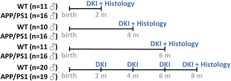
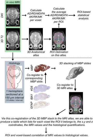
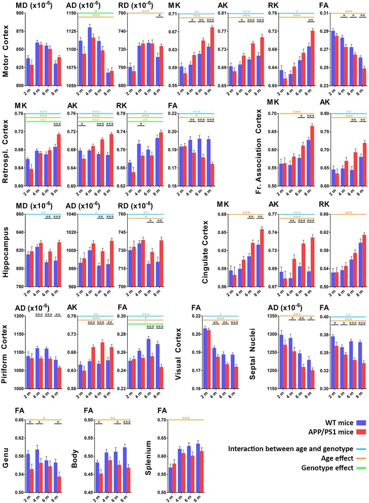
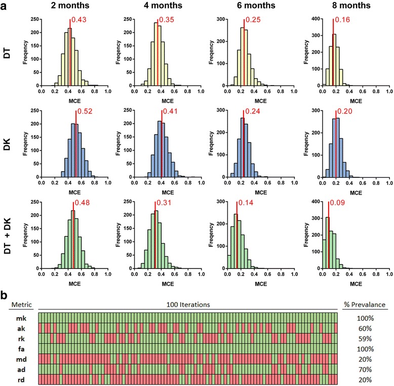
BACKGROUND: Alzheimer's disease (AD) is a progressive neurodegenerative disorder and the most common cause of dementia in the elderly population. In this study, we used the APP/PS1 transgenic mouse model to explore the feasibility of using diffusion kurtosis imaging (DKI) as a tool for the early detection of microstructural changes in the brain due to amyloid-β (Aβ) plaque deposition. METHODS: We longitudinally acquired DKI data of wild-type (WT) and APP/PS1 mice at 2, 4, 6 and 8 months of age, after which these mice were sacrificed for histological examination. Three additional cohorts of mice were also included at 2, 4 and 6 months of age to allow voxel-based co-registration between diffusion tensor and diffusion kurtosis metrics and immunohistochemistry. RESULTS: Changes were observed in diffusion tensor (DT) and diffusion kurtosis (DK) metrics in many of the 23 regions of interest that were analysed. Mean and axial kurtosis were greatly increased owing to Aβ-induced pathological changes in the motor cortex of APP/PS1 mice at 4, 6 and 8 months of age. Additionally, fractional anisotropy (FA) was decreased in APP/PS1 mice at these respective ages. Linear discriminant analysis of the motor cortex data indicated that combining diffusion tensor and diffusion kurtosis metrics permits improved separation of WT from APP/PS1 mice compared with either diffusion tensor or diffusion kurtosis metrics alone. We observed that mean kurtosis and FA are the critical metrics for a correct genotype classification. Furthermore, using a newly developed platform to co-register the in vivo diffusion-weighted magnetic resonance imaging with multiple 3D histological stacks, we found high correlations between DK metrics and anti-Aβ (clone 4G8) antibody, glial fibrillary acidic protein, ionised calcium-binding adapter molecule 1 and myelin basic protein immunohistochemistry. Finally, we observed reduced FA in the septal nuclei of APP/PS1 mice at all ages investigated. The latter was at least partially also observed by voxel-based statistical parametric mapping, which showed significantly reduced FA in the septal nuclei, as well as in the corpus callosum, of 8-month-old APP/PS1 mice compared with WT mice. CONCLUSIONS: Our results indicate that DKI metrics hold tremendous potential for the early detection and longitudinal follow-up of Aβ-induced pathology.
Overview
The claims and statistics of this paper haven’t been checked yet.
Run the check — free, no account needed. It reads the open-access text and checks the sources it cites.
Integrity signals
Registry
Cites
Statistics
Figures
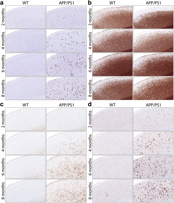
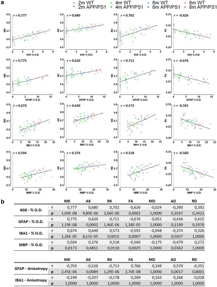
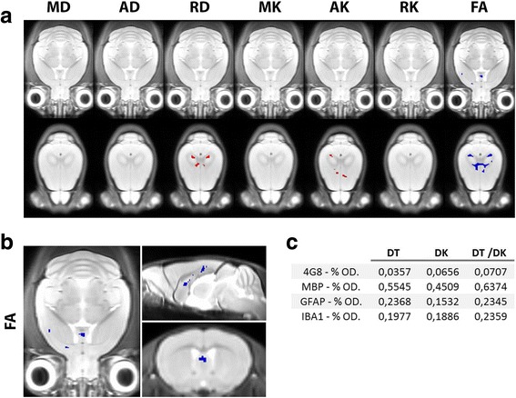
Verification
Claims the paper makes
The claims and statistics of this paper haven’t been checked yet.
Run the check — free, no account needed. It reads the open-access text and checks the sources it cites.
Claims other papers make about this one
Each is checked against this paper’s own text: does it say what the citing paper says it does?
The source backs part of the claim, not all of it — or backs it for a different population, condition or comparison.
In the citing paper
“Although Alzheimer’s disease (AD) is a leading cause, other forms such as vascular dementia (VD), Lewy body dementia (LBD) and frontotemporal dementia (FTD) are also prevalent9–11.”
From AI-based differential diagnosis of dementia etiologies on multimodal data (2024) · cites this paper as [10]
Experimental — accuracy not yet measuredThe source backs part of the claim, not all of it — or backs it for a different population, condition or comparison.
In the citing paper
“Advanced diffusion models—including diffusion kurtosis imaging (DKI), neurite orientation dispersion and density imaging (NODDI), and soma and neurite density imaging (SANDI) —offer improved sensitivity to microstructural complexity and may provide more biologically informative markers than DTI.”
From Diffusion MRI reveals amyloid plaque–associated cellular signatures through spatial transcriptomic integration (2026) · cites this paper as [20]
Experimental — accuracy not yet measuredThe source backs part of the claim, not all of it — or backs it for a different population, condition or comparison.
In the citing paper
“which may be associated primarily with the plaque deposition and related inflammation (Praet et al., 2018).”
From Age-dependent microstructure alterations in 5xFAD mice by high-resolution diffusion tensor imaging (2022) · cites this paper as Praet et al., 2018
Experimental — accuracy not yet measuredThe source backs part of the claim, not all of it — or backs it for a different population, condition or comparison.
In the citing paper
“Previous studies on 3×Tg-AD mice have reported significant volumetric decreases (Kong et al., 2018) and WM microstructural abnormalities (Praet et al., 2018; Gatto et al., 2019; Bergamino et al., 2023) in the CC and internal capsule compared with controls.”
From Multimodal MRI combined with RNA sequencing reveals pathological signatures in the 9-month-old 3×Tg-AD mouse brain (2026) · cites this paper as Praet et al., 2018
Experimental — accuracy not yet measured
Self-check
Percentages, sums and reported differences re-computed from the paper’s own numbers.
The self-check needs the paper’s full text.
Citations
Cites
Controlling the False Discovery Rate: A Practical and Powerful Approach to Multiple Testing
Benjamini, Y., & Hochberg, Y. (1995) · Journal of the Royal Statistical Society Series B: Statistical Methodology · doi:10.1111/j.2517-6161.1995.tb02031.x · Cited by 109,482
Regression shrinkage and selection via the lasso
Tibshirani, R. (1996) · Journal of the Royal Statistical Society Series B: Statistical Methodology · doi:10.1111/j.2517-6161.1996.tb02080.x · Cited by 51,482
The Elements of Statistical Learning
Hastie, T., Tibshirani, R., & Friedman, J. (2009) · Springer Series in Statistics · doi:10.1007/978-0-387-84858-7 · Cited by 25,970
The Elements of Statistical Learning: Data Mining, Inference, and Prediction
Ruppert, D. (2004) · Journal of the American Statistical Association · doi:10.1198/jasa.2004.s339 · Cited by 18,660
McKhann, G. M., Knopman, D. S., Chertkow, H., Hyman, B. T., Jack, C. R., Kawas, C. H., Klunk, W. E., Koroshetz, W. J., Manly, J. J., Mayeux, R., et al. (2011) · Alzheimer's & Dementia · doi:10.1016/j.jalz.2011.03.005 · Cited by 18,657
Object recognition from local scale-invariant features
Lowe, D. G. (1999) · Proceedings of the Seventh IEEE International Conference on Computer Vision · doi:10.1109/iccv.1999.790410 · Cited by 16,472
Jenkinson, M., Beckmann, C. F., Behrens, T. E. J., Woolrich, M. W., & Smith, S. M. (2012) · NeuroImage · doi:10.1016/j.neuroimage.2011.09.015 · Cited by 11,314
Random-Effects Models for Longitudinal Data
Laird, N. M., & Ware, J. H. (1982) · Biometrics · doi:10.2307/2529876 · Cited by 8,923
2016 Alzheimer's disease facts and figures
Alzheimer's Association (2016) · Alzheimer's & Dementia · doi:10.1016/j.jalz.2016.03.001 · Cited by 5,069
Soluble protein oligomers in neurodegeneration: lessons from the Alzheimer's amyloid β-peptide
Haass, C., & Selkoe, D. J. (2007) · Nature Reviews Molecular Cell Biology · doi:10.1038/nrm2101 · Cited by 4,744
Jack, C. R., Knopman, D. S., Jagust, W. J., Petersen, R. C., Weiner, M. W., Aisen, P. S., Shaw, L. M., Vemuri, P., Wiste, H. J., Weigand, S. D., et al. (2013) · The Lancet Neurology · doi:10.1016/s1474-4422(12)70291-0 · Cited by 4,396
Linear Mixed Models for Longitudinal Data
Verbeke, G. (1997) · Lecture Notes in Statistics · doi:10.1007/978-1-4612-2294-1_3 · Cited by 3,506
Statistical Parametric Mapping: The Analysis of Functional Brain Images
Penny, W. D., Friston, K. J., Ashburner, J., Kiebel, S. J., & Thomas, N. (2007) · UCL Discovery (University College London) · Cited by 3,339
Linear Mixed Models for Longitudinal Data
Molenberghs, G., & Verbeke, G. N. (2000) · Springer series in statistics · doi:10.1007/978-1-4419-0300-6 · Cited by 2,681
Jensen, J. H., Helpern, J. A., Ramani, A., Lu, H., & Kaczynski, K. (2005) · Magnetic Resonance in Medicine · doi:10.1002/mrm.20508 · Cited by 2,488
Quantification of histochemical staining by color deconvolution.
Ruifrok, A. C. C., & Johnston, D. A. (2001) · PubMed · Cited by 2,029
2015 Alzheimer's disease facts and figures
Alzheimer's Association (2015) · Alzheimer s & Dementia · doi:10.1016/j.jalz.2015.02.003 · Cited by 1,684
Hubbard (2010) · doi:10.1097/ede.0b013e3181caeb90 · Cited by 1,139
Fast free-form deformation using graphics processing units
Marc Modat, A., Ridgway, G. R., Taylor, Z. A., Lehmann, M., Barnes, J., Hawkes, D. J., Fox, N. C., & Sebastien Ourselin, A. (2009) · Computer Methods and Programs in Biomedicine · doi:10.1016/j.cmpb.2009.09.002 · Cited by 1,087
Aβ42‐driven cerebral amyloidosis in transgenic mice reveals early and robust pathology
Radde, R., Bolmont, T., Kaeser, S. A., Coomaraswamy, J., Lindau, D., Stoltze, L., Calhoun, M. E., Jäggi, F., Wolburg, H., Gengler, S., et al. (2006) · The EMBO Reports · doi:10.1038/sj.embor.7400784 · Cited by 986
Donepezil and memantine for moderate-to-severe Alzheimer's disease
Howard, R., McShane, R., Lindesay, J., Ritchie, C., Baldwin, A., Barber, R., Burns, A., Dening, T., Findlay, D., Holmes, C., et al. (2012) · New England Journal of Medicine · doi:10.1056/nejmoa1106668 · Cited by 782
Characterization of amyloid deposition in the APPswe/PS1dE9 mouse model of Alzheimer disease
Garcia-Alloza, M., Robbins, E. M., Zhang-Nunes, S. X., Purcell, S. M., Betensky, R. A., Raju, S., Prada, C., Greenberg, S. M., Bacskai, B. J., & Frosch, M. P. (2006) · Neurobiology of Disease · doi:10.1016/j.nbd.2006.08.017 · Cited by 744
Lombardo, I., Ramaswamy, G., Piscitelli, S. C., & Friedhoff, L. T. (2015) · Alzheimer s & Dementia · doi:10.1016/j.jalz.2015.08.154 · Cited by 656
Averaging correlation coefficients: Should Fisher’s z transformation be used?
Silver, N. C., & Dunlap, W. P. (1987) · Journal of Applied Psychology · doi:10.1037/0021-9010.72.1.146 · Cited by 606
Reconstructing a 3D structure from serial histological sections
Sebastien Ourselin, A., Roche, A., Subsol, G., Pennec, X., & Ayache, N. (2001) · Image and Vision Computing · doi:10.1016/s0262-8856(00)00052-4 · Cited by 548
Bayesian Methods for Data Analysis
Carlin, B., & Louis, T. (2008) · Chapman & Hall/CRC eBooks · doi:10.1201/b14884 · Cited by 505
Nicotinic acetylcholine binding sites in Alzheimer's disease
Whitehouse, P. J., Martino, A. M., Antuono, P., Lowenstein, P. R., Coyle, J. T., Price, D. L., & Kellar, K. J. (1986) · Brain Research · doi:10.1016/0006-8993(86)90819-x · Cited by 485
Optimal Orientation Detection of Linear Symmetry
Bigün, J. (1987) · Cited by 462
White matter damage in frontotemporal dementia and Alzheimer's disease measured by diffusion MRI
Zhang, Y., Schuff, N., Du, A.-T., Rosen, H. J., Kramer, J. H., Gorno-Tempini, M. L., Miller, B. L., & Weiner, M. W. (2009) · Brain · doi:10.1093/brain/awp071 · Cited by 356
Effectiveness of regional DTI measures in distinguishing Alzheimer’s disease, MCI, and normal aging
Nir, T. M., Jahanshad, N., Villalon-Reina, J. E., Toga, A. W., Jack, C. R., Weiner, M. W., & Thompson, P. M. (2013) · NeuroImage: Clinical · doi:10.1016/j.nicl.2013.07.006 · Cited by 348
Early Diagnosis of Alzheimer's Disease: Clinical and Economic Benefits
Leifer, B. P. (2003) · Journal of the American Geriatrics Society · doi:10.1046/j.1532-5415.5153.x · Cited by 309
Fellgiebel, A., Wille, P., Müller, M. J., Winterer, G., Scheurich, A., Vucurevic, G., Schmidt, L. G., & Stoeter, P. (2004) · Dementia and Geriatric Cognitive Disorders · doi:10.1159/000077817 · Cited by 298
White matter pathology isolates the hippocampal formation in Alzheimer's disease
Salat, D. H., Tuch, D. S., van der Kouwe, A. J. W., Greve, D. N., Pappu, V., Lee, S. Y., Hevelone, N. D., Zaleta, A. K., Growdon, J. H., Corkin, S., et al. (2010) · Neurobiology of Aging · doi:10.1016/j.neurobiolaging.2008.03.013 · Cited by 279
Role of structural MRI in Alzheimer's disease
Vemuri, P., & Jack, C. R. (2010) · Alzheimer s Research & Therapy · doi:10.1186/alzrt47 · Cited by 265
Changes in Nicotinic and Muscarinic Cholinergic Receptors in Alzheimer‐Type Dementia
Shimohama, S., Taniguchi, T., Fujiwara, M., & Kameyama, M. (1986) · Journal of Neurochemistry · doi:10.1111/j.1471-4159.1986.tb12960.x · Cited by 258
More accurate estimation of diffusion tensor parameters using diffusion kurtosis imaging
Veraart, J., Poot, D. H. J., Hecke, W. V., Blockx, I., der Linden, A. V., Verhoye, M., & Sijbers, J. (2010) · Magnetic Resonance in Medicine · doi:10.1002/mrm.22603 · Cited by 241
Regionally-specific diffusion tensor imaging in mild cognitive impairment and Alzheimer’s disease.
Mielke, M. M., Kozauer, N. A., Chan, K. C. G., George, M., Toroney, J., Zerrate, M., Bandeen-Roche, K., Wang, M.-C., vanZijl, P., & Pekar, J. J. (2009) · NeuroImage · doi:10.1016/j.neuroimage.2009.01.054 · Cited by 225
Amlien, I. K., & Fjell, A. M. (2014) · Neuroscience · doi:10.1016/j.neuroscience.2014.02.017 · Cited by 219
Optimal Experimental Design for Diffusion Kurtosis Imaging
Poot, D. H. J., den Dekker, A. J., Achten, E., Verhoye, M., & Sijbers, J. (2010) · IEEE Transactions on Medical Imaging · doi:10.1109/tmi.2009.2037915 · Cited by 208
Statistical parametric mapping the analysis of functional brain images
Friston, K. J. (2008) · Cited by 188
Diagnostic utility of hippocampal size and mean diffusivity in amnestic MCI.
Müller, M. J., Greverus, D., Weibrich, C., Dellani, P. R., Scheurich, A., Stoeter, P., & Fellgiebel, A. (2007) · Neurobiology of Aging · doi:10.1016/j.neurobiolaging.2006.01.009 · Cited by 140
Cherubini, A., Péran, P., Spoletini, I., Di Paola, M., Di Iulio, F., Hagberg, G. E., Sancesario, G., Gianni, W., Bossù, P., Caltagirone, C., et al. (2010) · Journal of Alzheimer s Disease · doi:10.3233/jad-2010-091186 · Cited by 123
Nowrangi, M. A., Lyketsos, C. G., Leoutsakos, J. S., Oishi, K., Albert, M., Mori, S., & Mielke, M. M. (2013) · Alzheimer's & Dementia · doi:10.1016/j.jalz.2012.05.2186 · Cited by 120
Duan, J., Wang, H., Xu, J., Lin, X. Z., Chen, S., Kang, Z., & Yao, Z. (2006) · Surgical and Radiologic Anatomy · doi:10.1007/s00276-006-0111-2 · Cited by 115
Longitudinal diffusion tensor imaging in frontotemporal dementia
Mahoney, C. J., Simpson, I. J. A., Nicholas, J. M., Fletcher, P. D., Downey, L. E., Golden, H. L., Clark, C. N., Schmitz, N., Rohrer, J. D., Schott, J. M., et al. (2015) · Annals of Neurology · doi:10.1002/ana.24296 · Cited by 101
Comprehensive framework for accurate diffusion MRI parameter estimation
Veraart, J., Rajan, J., Peeters, R. R., Leemans, A., Sunaert, S. G., & Sijbers, J. (2012) · Magnetic Resonance in Medicine · doi:10.1002/mrm.24529 · Cited by 95
Rémy, F., Vayssière, N., Saint‐Aubert, L., Barbeau, E. J., & Pariente, J. (2015) · NeuroImage Clinical · doi:10.1016/j.nicl.2015.01.014 · Cited by 87
Data distributions in magnetic resonance images: A review
den Dekker, A. J., & Sijbers, J. (2014) · Physica Medica · doi:10.1016/j.ejmp.2014.05.002 · Cited by 76
Gray and white matter degeneration revealed by diffusion in an Alzheimer mouse model
Zerbi, V., Kleinnijenhuis, M., Fang, X., Jansen, D., Veltien, A., Van Asten, J., Timmer, N., Dederen, P. J., Kiliaan, A. J., & Heerschap, A. (2013) · Neurobiology of Aging · doi:10.1016/j.neurobiolaging.2012.11.017 · Cited by 74
Gold, B. T., Zhu, Z., Brown, C., Andersen, A. H., LaDu, M. J., Tai, L. M., Jicha, G. A., Kryscio, R. J., Estus, S., Nelson, P. T., et al. (2014) · Neurobiology of Aging · doi:10.1016/j.neurobiolaging.2014.04.030 · Cited by 66
Woolf, N. J. (1996) · Neuroscience · doi:10.1016/0306-4522(96)00163-7 · Cited by 62
Diffusion Kurtosis Imaging: A Possible MRI Biomarker for AD Diagnosis?
Struyfs, H., Hecke, W. V., Veraart, J., Sijbers, J., Slaets, S., Belder, M. D., Wuyts, L. L., Peters, B., Sleegers, K., Robberecht, C., et al. (2015) · Journal of Alzheimer s Disease · doi:10.3233/jad-150253 · Cited by 61
Diffusion kurtosis imaging to detect amyloidosis in an APP/PS1 mouse model for Alzheimer's disease
Vanhoutte, G., Pereson, S., y Palacios, R. D., Guns, P., Asselbergh, B., Veraart, J., Sijbers, J., Verhoye, M., Broeckhoven, C. V., & der Linden, A. V. (2013) · Magnetic Resonance in Medicine · doi:10.1002/mrm.24680 · Cited by 51
Longitudinal white matter changes in Alzheimer's disease: A tractography-based analysis study
Kitamura, S., Kiuchi, K., Taoka, T., Hashimoto, K., Ueda, S., Yasuno, F., Morikawa, M., Kichikawa, K., & Kishimoto, T. (2013) · Brain Research · doi:10.1016/j.brainres.2013.03.052 · Cited by 50
Delayed‐start analysis: Mild Alzheimer's disease patients in solanezumab trials, 3.5 years
Liu‐Seifert, H., Siemers, E., Holdridge, K. C., Andersen, S. W., Lipkovich, I. A., Carlson, C. D., Sethuraman, G., HOOG, S. L., Hayduk, R., Doody, R. S., et al. (2015) · Alzheimer s & Dementia Translational Research & Clinical Interventions · doi:10.1016/j.trci.2015.06.006 · Cited by 48
Hong, Y. J., Yoon, B., Lim, S.-C., Shim, Y. S., Kim, J.-Y., Ahn, K. J., Han, I.-W., & Yang, D. W. (2013) · Neurological Sciences · doi:10.1007/s10072-012-1225-4 · Cited by 48
Wang, D., Guo, Z.-H., Liu, X., Li, Y., & Wang, H. (2015) · Current Alzheimer Research · doi:10.2174/1567205012666141218142422 · Cited by 28
Fu, J., Liu, Y., Li, Y., Chang, C., & Li, W. (2013) · Journal of Neuroimaging · doi:10.1111/jon.12061 · Cited by 27
Rowley, J., Fonov, V., Wu, O., Eskildsen, S. F., Schoemaker, D., Wu, L., Mohades, S., Shin, M., Sziklas, V., Cheewakriengkrai, L., et al. (2013) · PLoS ONE · doi:10.1371/journal.pone.0074776 · Cited by 27
Paola, M. D., Owen, P., Orfei, M. D., Piras, F., Cacciari, C., Caltagirone, C., & Spalletta, G. (2015) · Journal of Alzheimer s Disease · doi:10.3233/jad-142895 · Cited by 19
Short‐term white matter alterations in Alzheimer's disease characterized by diffusion tensor imaging
Genc, S., Steward, C. E., Malpas, C. B., Velakoulis, D., O'Brien, T. J., & Desmond, P. M. (2015) · Journal of Magnetic Resonance Imaging · doi:10.1002/jmri.25017 · Cited by 14
Research works printed but not linked: 1
- Avants B, Tustison N, Song G. Advanced Normalization Tools: V1.0. Insight J. 2009;July–December:1–35.
Cited by
Sorted by citations
Functional neuronal circuitry and oscillatory dynamics in human brain organoids
Sharf, T., van der Molen, T., Glasauer, S. M. K., Guzman, E., Buccino, A. P., Luna, G., Cheng, Z., Audouard, M., Ranasinghe, K. G., Kudo, K., et al. (2022) · Nature Communications · doi:10.1038/s41467-022-32115-4 · Cited by 251
Lazari, A., & Lipp, I. (2021) · NeuroImage · doi:10.1016/j.neuroimage.2021.117744 · Cited by 176
AI-based differential diagnosis of dementia etiologies on multimodal data
Xue, C., Kowshik, S. S., Lteif, D., Puducheri, S., Jasodanand, V. H., Zhou, O. T., Walia, A. S., Guney, O. B., Zhang, J. D., Poésy, S., et al. (2024) · Nature Medicine · doi:10.1038/s41591-024-03118-z · Cited by 182
An interactive meta-analysis of MRI biomarkers of myelin
Mancini, M., Karakuzu, A., Cohen-Adad, J., Cercignani, M., Nichols, T. E., & Stikov, N. (2020) · eLife · doi:10.7554/elife.61523 · Cited by 168
Shobeiri, P., Kalantari, A., Teixeira, A. L., & Rezaei, N. (2022) · Biology of Sex Differences · doi:10.1186/s13293-022-00422-6 · Cited by 113
Metadata
- Authors
- Praet, J., Manyakov, N. V., Muchene, L., Mai, Z., Terzopoulos, V., de Backer, S., Torremans, A., Guns, P.-J., Van De Casteele, T., Bottelbergs, A., Van Broeck, B., Sijbers, J., Smeets, D., Shkedy, Z., Bijnens, L., Pemberton, D. J., Schmidt, M. E., Van der Linden, A., & Verhoye, M.
- Affiliations
- University of Antwerp (BE)
- Janssen (Belgium) (BE)
- Hasselt University (BE)
- Icometrix (Belgium) (BE)
- HistoGeneX (Belgium) (BE)
- Corresponding author
- Verhoye, Marleen
- ORCID
- Venue
- Alzheimer's Research & Therapy
- Published
- 2018-01-09
- Volume
- 10
- Issue
- 1
- Pages
- 1-1
- ISSN
- 1758-9193 (ISSN-L)
- PMC
- PMC6389136
- OpenAlex
- W2791831747
- Cited by
- 133
- References
- 61
- Citations by year
- 2026: 7 · 2025: 19 · 2024: 22 · 2023: 18 · 2022: 22 · 2021: 22 · 2020: 15 · 2019: 5 · 2018: 3
- FWCI
- 7.77
- Type
- journal article
- Language
- en
- Open access
- gold
- doi.org
- Licence
- cc-by
- Locations
- Alzheimer s Research & Therapy (publishedVersion, cc-by)
- Europe PMC (PubMed Central) (publishedVersion, other-oa)
- PubMed Central (publishedVersion, cc-by)
- PubMed (publishedVersion)
- DOAJ (DOAJ: Directory of Open Access Journals) (submittedVersion, cc-by-sa)
- Document Server@UHasselt (UHasselt) (submittedVersion, cc-by)
- Zenodo (CERN European Organization for Nuclear Research) (submittedVersion, cc-by)
- Funders
- FP7 IAAP (612360)
- Hercules stichting (AUHA/012)
- IWT O&O (120178)
- Topics
- Health Sciences › Medicine › Radiology, Nuclear Medicine and Imaging › Advanced Neuroimaging Techniques and Applications
- Health Sciences › Medicine › Psychiatry and Mental health › Dementia and Cognitive Impairment Research
- Life Sciences › Neuroscience › Cellular and Molecular Neuroscience › Alzheimer's disease research and treatments
- Keywords
- diffusion kurtosis imaging, amyloid-beta, APP/PS1 mice, Alzheimer's disease, early detection of neurodegeneration, fractional anisotropy, mean kurtosis, motor cortex, septal nuclei, corpus callosum, diffusion tensor imaging, longitudinal neuroimaging, glial fibrillary acidic protein, myelin basic protein, immunohistochemistry
- MeSH terms
- Aging, Animals, Brain, Disease Models, Animal, Feasibility Studies, Follow-Up Studies, Image Interpretation, Computer-Assisted, Immunohistochemistry, Longitudinal Studies, Male, Mice, Inbred C57BL, Mice, Transgenic, Imaging, Three-Dimensional, Diffusion Magnetic Resonance Imaging, Early Diagnosis, Plaque, Amyloid
About these checks
Registry, retraction, Cites and statistics results are computed, not judged, with one exception: a printed reference that no index lists is read once by a model before it counts as a citation error. Claims are extracted by a model and kept only when their quote appears verbatim in the text; each cited source is then checked, with a model’s help, for whether it says what the claim relies on. Those verdicts can be wrong — read the claim’s quote and the cited source before relying on one.
Statistics and arithmetic are recomputed, not judged.
Confidence bands are shown only for fields calibrated on our annotated test set. None is calibrated yet, so no band is shown.
What counts as a claim ›How we measure confidence ›How we check this ›
Are you an author, or do you see a problem with this page? Request a review
