

The DOI matches its Crossref record; 10 of the 10 references it cites were found in a registry or an index.
Cancer nanomedicine: a review of recent success in drug delivery
Abstract
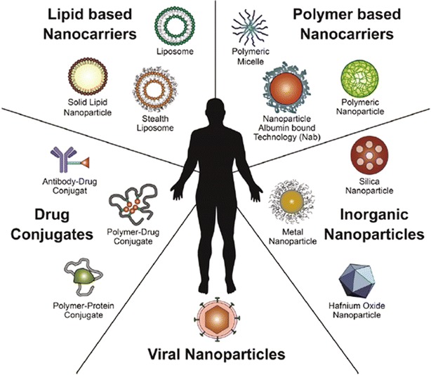
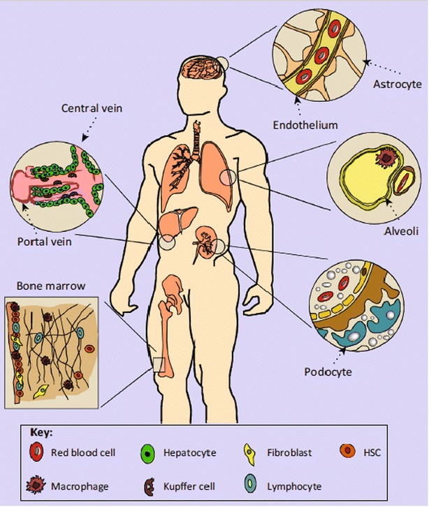
Cancer continues to be one of the most difficult global healthcare problems. Although there is a large library of drugs that can be used in cancer treatment, the problem is selectively killing all the cancer cells while reducing collateral toxicity to healthy cells. There are several biological barriers to effective drug delivery in cancer such as renal, hepatic, or immune clearance. Nanoparticles loaded with drugs can be designed to overcome these biological barriers to improve efficacy while reducing morbidity. Nanomedicine has ushered in a new era for drug delivery by improving the therapeutic indices of the active pharmaceutical ingredients engineered within nanoparticles. First generation nanomedicines have received widespread clinical approval over the past two decades, from Doxil ® (liposomal doxorubicin) in 1995 to Onivyde ® (liposomal irinotecan) in 2015. This review highlights the biological barriers to effective drug delivery in cancer, emphasizing the need for nanoparticles for improving therapeutic outcomes. A summary of different nanoparticles used for drug delivery applications in cancer are presented. The review summarizes recent successes in cancer nanomedicine in the clinic. The clinical trials of Onivyde leading to its approval in 2015 by the Food and Drug Adminstration are highlighted as a case study in the recent clinical success of nanomedicine against cancer. Next generation nanomedicines need to be better targeted to specifically destroy cancerous tissue, but face several obstacles in their clinical development, including identification of appropriate biomarkers to target, scale‐up of synthesis, and reproducible characterization. These hurdles need to be overcome through multidisciplinary collaborations across academia, pharmaceutical industry, and regulatory agencies in order to achieve the goal of eradicating cancer. This review discusses the current use of clinically approved nanomedicines, the investigation of nanomedicines in clinical trials, and the challenges that may hinder development of the nanomedicines for cancer treatment.
Overview
The claims and statistics of this paper haven’t been checked yet.
Run the check — free, no account needed. It reads the open-access text and checks the sources it cites.
Integrity signals
Registry
Cites
Statistics
Figures
Verification
Claims the paper makes
The claims and statistics of this paper haven’t been checked yet.
Run the check — free, no account needed. It reads the open-access text and checks the sources it cites.
Claims other papers make about this one
Each is checked against this paper’s own text: does it say what the citing paper says it does?
In the citing paper
“This, because of the vasculature of tumors, is often leaky, leading to accumulating nanoparticles in the bloodstream compared to normal tissue [45].”
From Targeted Nano-Drug Delivery of Colchicine against Colon Cancer Cells by Means of Mesoporous Silica Nanoparticles (2020) · cites this paper as [45]
Self-check
Percentages, sums and reported differences re-computed from the paper’s own numbers.
The self-check needs the paper’s full text.
Citations
Cites
Hallmarks of Cancer: The Next Generation
Hanahan, D., & Weinberg, R. A. (2011) · Cell · doi:10.1016/j.cell.2011.02.013 · Cited by 66,590
Principles of nanoparticle design for overcoming biological barriers to drug delivery
Blanco, E., Shen, H., & Ferrari, M. (2015) · Nature Biotechnology · doi:10.1038/nbt.3330 · Cited by 7,002 · cited 1×
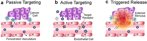
“This phenomenon, known as the enhanced permeability and retention (EPR) effect, has been utilized for passive targeting of nanoparticles to tumor tissue, but deeper penetration into the tumor is frequently restricted due to heterogeneity of the tumor microenvironment [46].”
BackgroundEngineered Nanoparticles for Drug Delivery in Cancer Therapy
Sun, T., Zhang, Y. S., Pang, B., Hyun, D. C., Yang, M., & Xia, Y. (2014) · Angewandte Chemie International Edition · doi:10.1002/anie.201403036 · Cited by 2,164
Wang-Gillam, A., Li, C.-P., Bodoky, G., Dean, A., Shan, Y.-S., Jameson, G., Macarulla, T., Lee, K.-H., Cunningham, D., Blanc, J. F., et al. (2016) · The Lancet · doi:10.1016/s0140-6736(15)00986-1 · Cited by 1,222 · cited 2×
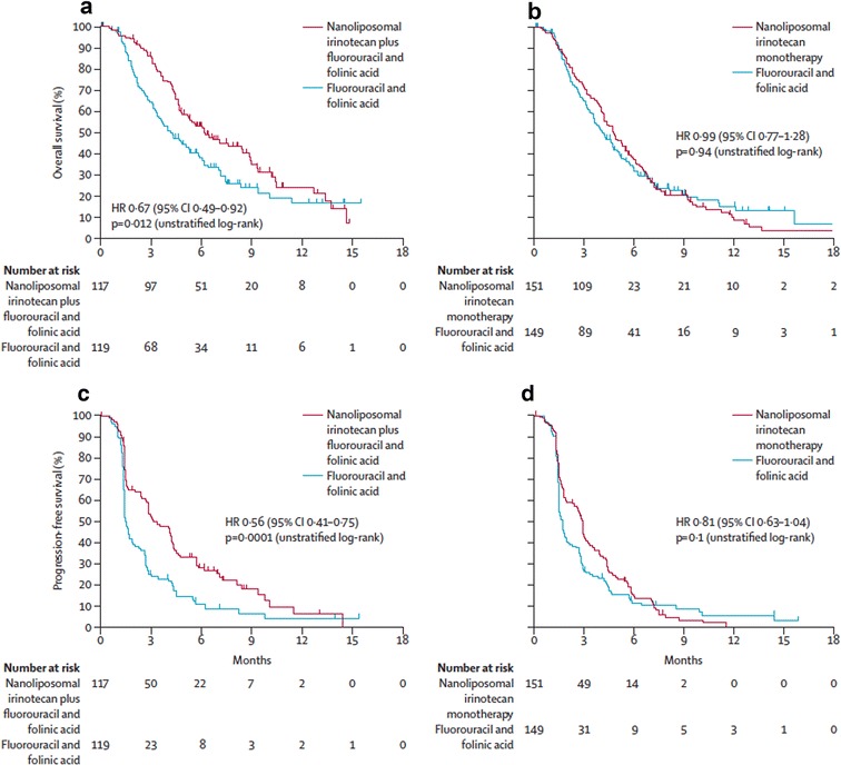
“survival with nanoliposomal irinotecan monotherapy versus fluorouracil and folinic acid (Reproduced with permission from [77])”
BackgroundMethodChauhan, V. P., Stylianopoulos, T., Martin, J. D., Popović, Z., Chen, O., Kamoun, W. S., Bawendi, M. G., Fukumura, D., & Jain, R. K. (2012) · Nature Nanotechnology · doi:10.1038/nnano.2012.45 · Cited by 1,101
Duan, X., & Li, Y. (2013) · Small · doi:10.1002/smll.201201390 · Cited by 926
Albumin-bound formulation of paclitaxel (Abraxane® ABI-007) in the treatment of breast cancer
Tomao, S. (2009) · International Journal of Nanomedicine · doi:10.2147/ijn.s3061 · Cited by 695 · cited 2×
“This effect may increase clearance from the bloodstream via recognition by the immune system or disrupt the ability of the nanoparticles to be internalized by the tissue of interest [26, 27].”
BackgroundA Review of Clinical Translation of Inorganic Nanoparticles
Anselmo, A. C., & Mitragotri, S. (2015) · The AAPS Journal · doi:10.1208/s12248-015-9780-2 · Cited by 509
Mechanism of polymeric nanoparticle-based drug transport across the blood-brain barrier (bbb).
Kreuter, J. (2013) · Journal of Microencapsulation · doi:10.3109/02652048.2012.692491 · Cited by 172
Phase I study of nanoliposomal irinotecan (PEP02) in advanced solid tumor patients
Chang, T. C., Shiah, H. S., Yang, C. H., Yeh, K. H., Cheng, A. L., Shen, B. N., Wang, Y. W., Yeh, C. G., Chiang, N. J., Chang, J. Y., et al. (2015) · Cancer Chemotherapy and Pharmacology · doi:10.1007/s00280-014-2671-x · Cited by 100
Research works printed but not linked: 80
- 10.3322/caac.21395 · DOI
- 10.1016/j.jconrel.2014.12.030 · DOI
- 10.1158/1535-7163.MCT-06-0141 · DOI
- 10.1146/annurev-bioeng-071811-150124 · DOI
- 10.1016/j.nano.2015.08.006 · DOI
- 10.1517/17425247.2014.950564 · DOI · Paper page
- 10.1016/j.bpj.2010.06.016 · DOI
- Locatelli E, 2012, Biodegradable PLGA‐b‐PEG polymeric nanoparticles: synthesis, properties, and nanomedical applications as drug delivery system, J Nanopart Res, 14, 1, 10.1007/s11051-012-1316-4 · DOI
- 10.1016/j.jconrel.2012.01.043 · DOI
- 10.1016/j.tibtech.2016.07.006 · DOI · Paper page
- 10.1158/1078-0432.CCR-07-1441 · DOI
- 10.1021/mp100253e · DOI
- 10.1039/C6TB00847J · DOI
- 10.1002/cbf.1149 · DOI
- Chen K‐J, 2012, A thermoresponsive bubble‐generating liposomal system for triggering localized extracellular drug delivery, ACS Nano, 7, 438, 10.1021/nn304474j · DOI
- 10.1016/j.jconrel.2014.05.002 · DOI
- 10.1093/jnci/djm043 · DOI
- 10.1038/srep02953 · DOI
- 10.1016/j.jconrel.2004.11.023 · DOI
- Wang L, 2017, Multifunctional telodendrimer nanocarriers restore synergy of bortezomib and doxorubicin in ovarian cancer treatment, Can Res, 77, 3293, 10.1158/0008-5472.CAN-16-3119 · DOI
- 10.1021/nn3044066 · DOI
- 10.1021/acs.molpharmaceut.6b00388 · DOI
- Ahmed N, 2012, Theranostic applications of nanoparticles in cancer, Drug Discov Today, 17, 928, 10.1016/j.drudis.2012.03.010 · DOI
- 10.1016/j.addr.2010.09.002 · DOI
- 10.1002/smll.201303540 · DOI
- 10.1504/IJNM.2009.028116 · DOI
- 10.1126/science.1229568 · DOI
- 10.1088/0957-4484/22/11/115101 · DOI
- 10.2217/nnm.13.191 · DOI
- 10.1152/ajprenal.00290.2001 · DOI
- Liu J, 2013, Renal clearable inorganic nanoparticles: a new frontier of bionanotechnology, Mater Today, 16, 477, 10.1016/j.mattod.2013.11.003 · DOI
- 10.1073/pnas.0913667107 · DOI
- 10.1016/B978-0-12-391860-4.00006-9 · DOI
- 10.1602/neurorx.2.1.3 · DOI · Paper page
- 10.1016/j.ejpb.2008.08.021 · DOI · Paper page
- 10.1124/jpet.105.097139 · DOI · Paper page
- 10.1016/j.jconrel.2008.10.016 · DOI
- 10.1007/s00018-007-6390-x · DOI
- 10.1186/s12951-015-0075-7 · DOI
- 10.1080/10611860400015936 · DOI
- ShankerSharma H, 2012, Neurotoxicity of engineered nanoparticles from metals, CNS Neurol Disord Drug Targets, 11, 65, 10.2174/187152712799960817 · DOI
- 10.1016/j.toxlet.2012.08.009 · DOI
- 10.1021/nn400548g · DOI
- Alley SC, 2010, Antibody–drug conjugates: targeted drug delivery for cancer, Curr Opin Chem Biol, 14, 529, 10.1016/j.cbpa.2010.06.170 · DOI
- 10.1016/j.cbpa.2009.03.023 · DOI
- 10.1615/CritRevTherDrugCarrierSyst.v26.i6.10 · DOI
- 10.1126/science.364652 · DOI
- 10.1016/j.addr.2008.03.007 · DOI
- 10.1021/mp400190z · DOI
- 10.1016/j.polymer.2008.01.027 · DOI
- 10.1021/ja053962k · DOI
- 10.2147/IJN.S112660 · DOI
- 10.1126/science.275.5297.187 · DOI
- Almajhdi F, 2014, In‐vitro anticancer and antimicrobial activities of PLGA/silver nanofiber composites prepared by electrospinning, J Mater Sci, 25, 1045
- Elbaz NM, 2016, Core‐shell silver/polymeric nanoparticles‐based combinatorial therapy against breast cancer in‐vitro, Sci Rep, 6, 30729, 10.1038/srep30729 · DOI
- 10.1016/S0021-9258(17)38654-4 · DOI · Paper page
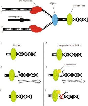
- Hsiang Y‐H, 1989, Arrest of replication forks by drugstabilized topoisomerase I‐DNA cleavable complexes as a mechanism of cell killing by camptothecin, Can Res, 49, 5077
- Zamboni WC, 2005, Relationship between plasma exposure of 9‐nitrocamptothecin and its 9‐aminocamptothecin metabolite and antitumor response in mice bearing human colon carcinoma xenografts, Clin Cancer Res, 11, 4867, 10.1158/1078-0432.CCR-05-0144 · DOI
- 10.2217/fon.15.333 · DOI
- 10.1158/0008-5472.CAN-05-4007 · DOI
- Chiang N‐J, 2016, A phase I dose‐escalation study of PEP02 (irinotecan liposome injection) in combination with 5‐fluorouracil and leucovorin in advanced solid tumors, BMC Cancer, 16, 907, 10.1186/s12885-016-2933-6 · DOI
- 10.1093/annonc/mdt002 · DOI
- Pan X, 2017, Construction of anti‐EGFR immunoliposomes via folate‐folate binding protein affinity, Int J Pharm, 336, 276, 10.1016/j.ijpharm.2006.12.007 · DOI
- 10.1038/bjc.2013.408 · DOI
- Stubbs M, 1994, Metabolic consequences of a reversed pH gradient in rat tumors, Cancer Res, 54, 4011
- 10.1038/nature07943 · DOI
- TinkleS McNeilSE MϋhlebachS BawaR BorchardG BarenholzYC TamarkinL DesaiN(2014)Nanomedicines: addressing the scientific and regulatory gap. Ann NY Acad Sci 1313:35–56 · DOI
- Ma MK, 2000, Pharmacokinetics of irinotecan and its metabolites SN‐38 and APC in children with recurrent solid tumors after protracted low‐dose irinotecan, Clin Cancer Res, 6, 813
- Cainelli F, 2009, Safety and efficacy of pegylated liposomal doxorubicin in HIV‐associated Kaposi’s sarcoma, Biologics, 3, 385
- Chen L, 2010, Phase I study of liposome irinotecan (PEP02) in combination with weekly infusion of 5‐FU/LV in advanced solid tumors, J Clin Oncol, 28, e13024, 10.1200/jco.2010.28.15_suppl.e13024 · DOI
- 10.1158/1078-0432.CCR-03-0655 · DOI · Paper page
- Ledet G, 2012, Nanomedicine: emerging therapeutics for the 21st century, US Pharm, 37, 7
- 10.1007/s00280-012-2042-4 · DOI
- Young C, 2011, CRLX101 (formerly IT‐101)—a novel nanopharmaceutical of camptothecin in clinical development, Current Bioactive Compd, 7, 8, 10.2174/157340711795163866 · DOI
- 10.1016/j.ijpharm.2006.12.007 · DOI
- Li C, 2014, Pegylated liposomal mitoxantrone is more therapeutically active than mitoxantrone in L1210 ascitic tumor and exhibits dose‐dependent activity saturation effect, Int J Pharm, 460, 165, 10.1016/j.ijpharm.2013.10.023 · DOI
- 10.1016/j.ejpb.2008.05.019 · DOI
- 10.1002/cncr.28974 · DOI · Paper page
- 10.1038/mt.2013.32 · DOI
- Young C, 2011, CRLX101 (formerly IT‐101) a novel nanopharmaceutical of camptothecin in clinical development, Curr Bioact Compd, 7, 8, 10.2174/157340711795163866 · DOI
Cited by
Sorted by citations
Nanoparticles for Cancer Therapy: Current Progress and Challenges
Gavas, S., Quazi, S., & Karpiński, T. M. (2021) · Nanoscale Research Letters · doi:10.1186/s11671-021-03628-6 · Cited by 1,069
Lombardo, D., Kiselev, M. A., & Caccamo, M. T. (2019) · Journal of Nanomaterials · doi:10.1155/2019/3702518 · Cited by 864
Fang, J., Islam, W., & Maeda, H. (2020) · Advanced Drug Delivery Reviews · doi:10.1016/j.addr.2020.06.005 · Cited by 757
Lipid-based nanoparticle formulations for small molecules and RNA drugs
Ickenstein, L. M., & Garidel, P. (2019) · Expert Opinion on Drug Delivery · doi:10.1080/17425247.2019.1669558 · Cited by 193
Cytokine and Chemokine Signals of T‐Cell Exclusion in Tumors
Zhang, Y., Guan, X.-Y., & Jiang, P. (2020) · Frontiers in Immunology · doi:10.3389/fimmu.2020.594609 · Cited by 133
Metadata
- Authors
- Tran, S., DeGiovanni, P., Piel, B., & Rai, P.
- Affiliations
- Department of Biological Sciences University of Massachusetts 01854 Lowell MA USA
- Department of Chemical Engineering University of Massachusetts 1 University ave 01854 Lowell MA USA
- ORCID
- Rai, Prakash
- Venue
- Clinical and Translational Medicine
- Publisher
- Wiley
- Published
- 2017-12
- Volume
- 6
- Issue
- 1
- ISSN
- 2001-1326
- PMC
- PMC5725398
- Cited by
- 879
- References
- 10
- Language
- en
- Licence
- Funders
- National Cancer Institute (R00CA153948, K99CA153948)
- National Institutes of Health (K99CA153948, R00CA153948)
About these checks
Registry, retraction, Cites and statistics results are computed, not judged, with one exception: a printed reference that no index lists is read once by a model before it counts as a citation error. Claims are extracted by a model and kept only when their quote appears verbatim in the text; each cited source is then checked, with a model’s help, for whether it says what the claim relies on. Those verdicts can be wrong — read the claim’s quote and the cited source before relying on one.
Statistics and arithmetic are recomputed, not judged.
Confidence bands are shown only for fields calibrated on our annotated test set. None is calibrated yet, so no band is shown.
What counts as a claim ›How we measure confidence ›How we check this ›
Are you an author, or do you see a problem with this page? Request a review
