

The DOI matches its Crossref record; 434 of the 440 references it cites were found in a registry or an index.
The Functional and Clinical Significance of the 24-Hour Rhythm of Circulating Glucocorticoids
Open accessAbstract
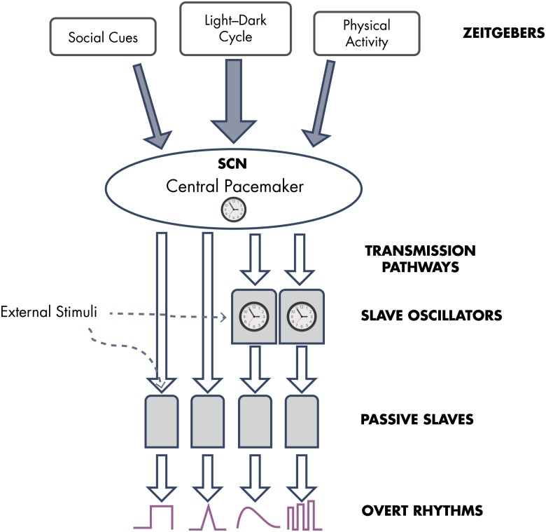
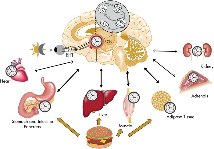
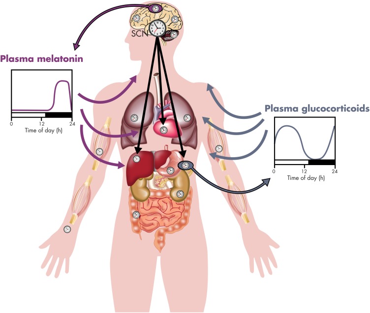
Adrenal glucocorticoids are major modulators of multiple functions, including energy metabolism, stress responses, immunity, and cognition. The endogenous secretion of glucocorticoids is normally characterized by a prominent and robust circadian (around 24 hours) oscillation, with a daily peak around the time of the habitual sleep-wake transition and minimal levels in the evening and early part of the night. It has long been recognized that this 24-hour rhythm partly reflects the activity of a master circadian pacemaker located in the suprachiasmatic nucleus of the hypothalamus. In the past decade, secondary circadian clocks based on the same molecular machinery as the central master pacemaker were found in other brain areas as well as in most peripheral tissues, including the adrenal glands. Evidence is rapidly accumulating to indicate that misalignment between central and peripheral clocks has a host of adverse effects. The robust rhythm in circulating glucocorticoid levels has been recognized as a major internal synchronizer of the circadian system. The present review examines the scientific foundation of these novel advances and their implications for health and disease prevention and treatment.
Overview
The claims and statistics of this paper haven’t been checked yet.
Run the check — free, no account needed. It reads the open-access text and checks the sources it cites.
Integrity signals
Registry
Cites
Statistics
Figures
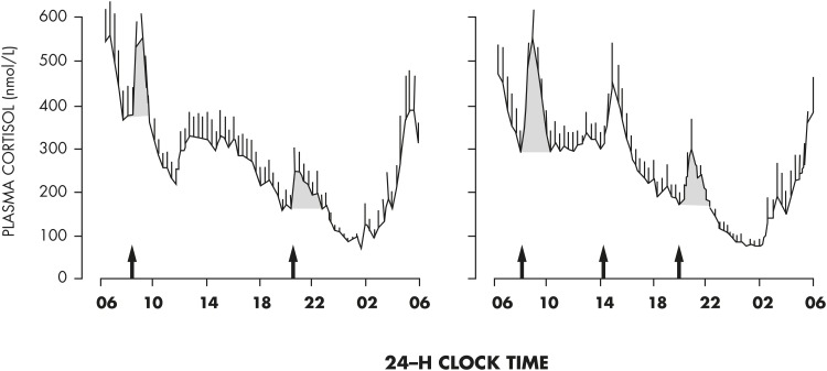
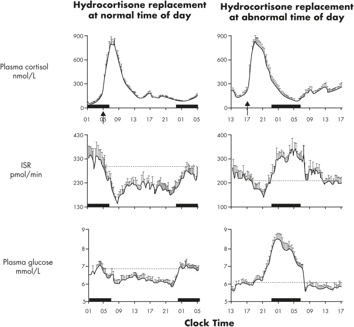
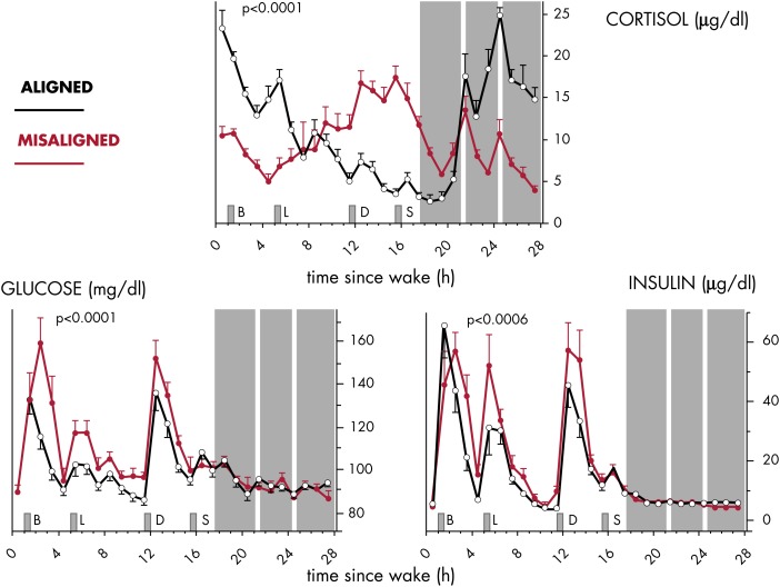
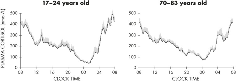
Verification
Claims the paper makes
The claims and statistics of this paper haven’t been checked yet.
Run the check — free, no account needed. It reads the open-access text and checks the sources it cites.
Claims other papers make about this one
Each is checked against this paper’s own text: does it say what the citing paper says it does?
The source backs part of the claim, not all of it — or backs it for a different population, condition or comparison.
In the citing paper
“This rhythm is important for its action because cortisol, not melatonin, is recognized to be the key metabolic central synchronizing signal [60–62].”
From Sleep, testosterone and cortisol balance, and ageing men (2022) · cites this paper as [60]
Experimental — accuracy not yet measuredThe source backs part of the claim, not all of it — or backs it for a different population, condition or comparison.
In the citing paper
“Experimental administration of glucocorticoid synchronizes the peripheral clocks located in liver, muscle, and adipose tissue which are the major storage sites for glycogen, protein, and fat, respectively, as well as other metabolically relevant organs such as pancreas and gut [60–63].”
From Sleep, testosterone and cortisol balance, and ageing men (2022) · cites this paper as [60]
Experimental — accuracy not yet measuredThe source backs part of the claim, not all of it — or backs it for a different population, condition or comparison.
In the citing paper
“These findings are highly relevant because hypercortisolemia during the late-afternoon and early-evening is harmful, and is believed to cause the insulin resistance observed with advanced age [60, 69, 70], impair physical performance [71] and trigger neurocognitive deficits [72].”
From Sleep, testosterone and cortisol balance, and ageing men (2022) · cites this paper as [60]
Experimental — accuracy not yet measuredThe source backs part of the claim, not all of it — or backs it for a different population, condition or comparison.
In the citing paper
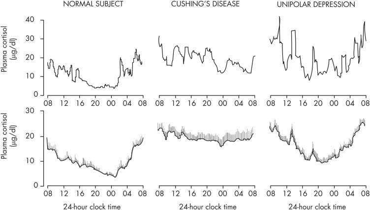
“Disruption of these hormone rhythms often leads to disease ( 2 ),”
From Dynamic modulation of glucose utilisation by glucocorticoid rhythms in health and disease (2020) · cites this paper as [2]
Experimental — accuracy not yet measuredThe source backs part of the claim, not all of it — or backs it for a different population, condition or comparison.
In the citing paper
“in hypercortisolism, it is not only that CORT levels are higher than normal, but in some cases, they also display abnormal rhythmicity. This varies depending on whether the underlying condition is associated with Cushing's syndrome, depression, PTSD, acute inflammation following major surgery, or chronic inflammatory diseases (e.g., rheumatoid arthritis) ( 2, 5-13, 57-60 ).”
From Dynamic modulation of glucose utilisation by glucocorticoid rhythms in health and disease (2020) · cites this paper as [2]
Experimental — accuracy not yet measured
The source backs part of the claim, not all of it — or backs it for a different population, condition or comparison.
In the citing paper
“many of these conditions are accompanied by a disruption of normal glucose metabolism while preserving some degree of CORT rhythmicity.”
From Dynamic modulation of glucose utilisation by glucocorticoid rhythms in health and disease (2020) · cites this paper as [2]
Experimental — accuracy not yet measuredThe source backs part of the claim, not all of it — or backs it for a different population, condition or comparison.
In the citing paper
“Similarly, mounting evidence suggests that circadian clock genes within the brain, skeletal muscle, adipose tissue, gut, liver and pancreas (to name a few) may play a major role in regulating insulin sensitivity, possibly cooperating with glucocorticoids in controlling circadian variability ( 2, 73, 74 ).”
From Dynamic modulation of glucose utilisation by glucocorticoid rhythms in health and disease (2020) · cites this paper as [2]
Experimental — accuracy not yet measured
Self-check
Percentages, sums and reported differences re-computed from the paper’s own numbers.
The self-check needs the paper’s full text.
Citations
Cites
Sapolsky, R. M., Romero, L. M., & Munck, A. U. (2000) · Endocrine Reviews · doi:10.1210/edrv.21.1.0389 · Cited by 7,541
Effects of stress throughout the lifespan on the brain, behaviour and cognition
Lupien, S. J., McEwen, B. S., Gunnar, M. R., & Heim, C. (2009) · Nature Reviews Neuroscience · doi:10.1038/nrn2639 · Cited by 6,256
Stress and the brain: from adaptation to disease
de Kloet, E. R., Joëls, M., & Holsboer, F. (2005) · Nature Reviews Neuroscience · doi:10.1038/nrn1683 · Cited by 4,621
Impact of sleep debt on metabolic and endocrine function
Spiegel, K., Leproult, R., & Van Cauter, E. (1999) · The Lancet · doi:10.1016/s0140-6736(99)01376-8 · Cited by 3,668
The diagnosis of Cushing's syndrome: an Endocrine Society Clinical Practice Guideline.
Nieman, L. K., Biller, B. M. K., Findling, J. W., Newell-Price, J., Savage, M. O., Stewart, P. M., & Montori, V. M. (2008) · The Journal of Clinical Endocrinology & Metabolism · doi:10.1210/jc.2008-0125 · Cited by 2,718 · cited 1×
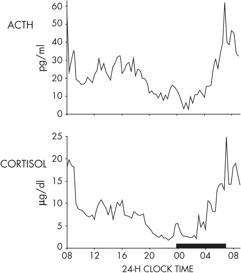
“The reduced amplitude of the circadian cortisol rhythm associated with higher nadir levels is, in clinical practice, an early marker of hypercortisolism in humans, and the evaluation of cortisol levels during the normal nadir of the rhythm is a common screening test to diagnose Cushing’s syndrome (357).”
The Concepts of Stress and Stress System Disorders: Overview of Physical and Behavioral Homeostasis
Chrousos, G. P. (1992) · JAMA · doi:10.1001/jama.1992.03480090092034 · Cited by 2,689
Brain corticosteroid receptor balance in health and disease
de Kloet, E. R., Vreugdenhil, E., Oitzl, M. S., & Joëls, M. (1998) · Endocrine Reviews · doi:10.1210/edrv.19.3.0331 · Cited by 2,614
Two receptor systems for corticosterone in rat brain: Microdistribution and differential occupation
REUL, J. M. H. M., & KLOET, E. R. D. (1985) · Endocrinology · doi:10.1210/endo-117-6-2505 · Cited by 2,580
Damiola, F., Le Minh, N., Preitner, N., Kornmann, B., Fleury-Olela, F., & Schibler, U. (2000) · Genes & Development · doi:10.1101/gad.183500 · Cited by 2,394
Adverse metabolic and cardiovascular consequences of circadian misalignment
Scheer, F. A. J. L., Hilton, M. F., Mantzoros, C. S., & Shea, S. A. (2009) · Proceedings of the National Academy of Sciences · doi:10.1073/pnas.0808180106 · Cited by 2,351
Research works printed but not linked: 7
- McEwen, 2016, Central role of the brain in stress and adaptation: allostasis, biological embedding, and cumulative change, 39
- de Kloet, 2016, Corticosteroid receptor balance hypothesis: implications for stress adaptation and health, 21
- Krieger, 1979, Endocrine Rhythms., 123
- Knapen, 1993, Addison Patients in the Netherlands: Social Report of the Survey, Summary and Recommendations
- Barthel, 2015, Endocrinology: Adult and Pediatric., 1763
- Linkowski, 1987, 24-Hour profiles of adrenocorticotropin, cortisol, and growth hormone in major depressive illness: effect of antidepressant treatment, J Clin Endocrinol Metab., 65, 141, 10.1210/jcem-65-1-141 · DOI
- Van Cauter, 1989, Physiology and pathology of circadian rhythms, 109
Cited by
Sorted by citations
Russell, G., & Lightman, S. (2019) · Nature Reviews Endocrinology · doi:10.1038/s41574-019-0228-0 · Cited by 1,032
Self-citationInfluence of Maternal Obesity on the Long-Term Health of Offspring
Godfrey, K. M., Reynolds, R. M., Prescott, S. L., Nyirenda, M., Jaddoe, V. W. V., Eriksson, J. G., & Broekman, B. F. P. (2017) · The Lancet Diabetes & Endocrinology · doi:10.1016/s2213-8587(16)30107-3 · Cited by 1,059
Diurnal transcriptome atlas of a primate across major neural and peripheral tissues
Mure, L. S., Le, H. D., Benegiamo, G., Chang, M. W., Ríos, L., Jillani, N., Ngotho, M., Kariuki, T., Dkhissi‐Benyahya, O., Cooper, H. M., et al. (2018) · Science · doi:10.1126/science.aao0318 · Cited by 791
Investigation of Cortisol Dynamics in Human Sweat Using a Graphene-Based Wireless mHealth System
Torrente-Rodríguez, R. M., Tu, J., Yang, Y., Min, J., Wang, M., Song, Y., Yu, Y., Xu, C., Ye, C., IsHak, W. W., et al. (2020) · Matter · doi:10.1016/j.matt.2020.01.021 · Cited by 479
Role of glucocorticoid negative feedback in the regulation of HPA axis pulsatility
Gjerstad, J. K., Lightman, S. L., & Spiga, F. (2018) · Stress · doi:10.1080/10253890.2018.1470238 · Cited by 437
Self-citationThe circadian regulation of food intake
Challet, E. (2019) · Nature Reviews Endocrinology · doi:10.1038/s41574-019-0210-x · Cited by 428
Signalling cognition: The gut microbiota and hypothalamic-pituitary-adrenal axis
Rusch, J. A., Layden, B. T., & Dugas, L. R. (2023) · Frontiers in Endocrinology · doi:10.3389/fendo.2023.1130689 · Cited by 415
Tsourdi, E. A., Zillikens, M. C., Meier, C., Body, J., Rodríguez, E. G., Anastasilakis, A. D., Abrahamsen, B., McCloskey, E. V., Hofbauer, L. C., Guañabens, N., et al. (2020) · The Journal of Clinical Endocrinology & Metabolism · doi:10.1210/clinem/dgaa756 · Cited by 391
Ballesta, A., Innominato, P. F., Dallmann, R., Rand, D. A. J., & Lévi, F. (2017) · Pharmacological Reviews · doi:10.1124/pr.116.013441 · Cited by 348
The multiple roles of life stress in metabolic disorders
Mika Kivimaki, B., Bartolomucci, A., & Kawachi, I. I. (2022) · Nature Reviews Endocrinology · doi:10.1038/s41574-022-00746-8 · Cited by 357
Light and Cognition: Roles for Circadian Rhythms, Sleep, and Arousal
Fisk, A. S., Tam, S. K. E., Brown, L. A., Vyazovskiy, V. V., Bannerman, D. M., & Peirson, S. N. (2018) · Frontiers in Neurology · doi:10.3389/fneur.2018.00056 · Cited by 295
de Kloet, E. R., Meijer, O. C., Nicola, A. F. D., Rijk, R. H. D., & Joëls, M. (2018) · Frontiers in Neuroendocrinology · doi:10.1016/j.yfrne.2018.02.003 · Cited by 287
Trade-Offs (and Constraints) in Organismal Biology
Garland, T., Downs, C. J., & Ives, A. R. (2021) · Physiological and Biochemical Zoology · doi:10.1086/717897 · Cited by 270
Dimitriadis, G. D., Maratou, E., Kountouri, A., Board, M., & Lambadiari, V. (2021) · Nutrients · doi:10.3390/nu13010159 · Cited by 241
Circadian regulation of depression: A role for serotonin
Daut, R. A., & Fonken, L. K. (2019) · Frontiers in Neuroendocrinology · doi:10.1016/j.yfrne.2019.04.003 · Cited by 237
Hahner, S., Ross, R. J., Arlt, W., Bancos, I., Burger-Stritt, S., Torpy, D. J., Husebye, E. S., & Quinkler, M. (2021) · Nature Reviews Disease Primers · doi:10.1038/s41572-021-00252-7 · Cited by 223
Isidori, A. M., Venneri, M. A., Graziadio, C., Simeoli, C., Fiore, D., Hasenmajer, V., Sbardella, E., Gianfrilli, D., Pozza, C., Pasqualetti, P., et al. (2018) · The Lancet Diabetes & Endocrinology · doi:10.1016/s2213-8587(17)30398-4 · Cited by 211
Treating the Side Effects of Exogenous Glucocorticoids; Can We Separate the Good From the Bad ?
Pofi, R., Caratti, G., Ray, D. W., & Tomlinson, J. (2023) · Endocrine Reviews · doi:10.1210/endrev/bnad016 · Cited by 237
The Mammalian Circadian Timing System and the Suprachiasmatic Nucleus as Its Pacemaker
Hastings, M. H., Maywood, E. S., & Brancaccio, M. (2019) · Biology · doi:10.3390/biology8010013 · Cited by 207
The Good and the Bad: Monocytes’ and Macrophages’ Diverse Functions in Inflammation
Austermann, J., Roth, J., & Barczyk‐Kahlert, K. (2022) · Cells · doi:10.3390/cells11121979 · Cited by 210
Mechanisms of Communication in the Mammalian Circadian Timing System
Astiz, M., Heyde, I., & Oster, H. (2019) · International Journal of Molecular Sciences · doi:10.3390/ijms20020343 · Cited by 180
Self-citationNew insights into the cell- and tissue-specificity of glucocorticoid actions
Quatrini, L., & Ugolini, S. (2020) · Cellular and Molecular Immunology · doi:10.1038/s41423-020-00526-2 · Cited by 174
Blood transcriptome based biomarkers for human circadian phase
Laing, E., Möller‐Levet, C. S., Poh, N., Santhi, N., Archer, S. N., & Dijk, D. (2017) · eLife · doi:10.7554/elife.20214 · Cited by 157
Self-citationCircadian clocks in the digestive system
Segers, A., & Depoortere, I. (2021) · Nature Reviews Gastroenterology & Hepatology · doi:10.1038/s41575-020-00401-5 · Cited by 165
Gulyaeva, N. V. (2018) · Neurochemical Research · doi:10.1007/s11064-018-2662-0 · Cited by 156
The role of the circadian clock system in physiology
Pilorz, V., Helfrich‐Förster, C., & Oster, H. (2018) · Pflügers Archiv - European Journal of Physiology · doi:10.1007/s00424-017-2103-y · Cited by 164
Self-citationNicolaides, N. C., Charmandari, E., Kino, T., & Chrousos, G. P. (2017) · Frontiers in Endocrinology · doi:10.3389/fendo.2017.00070 · Cited by 154
Chronopharmacology of glucocorticoids
Scherholz, M. L., Schlesinger, N., & Androulakis, I. P. (2019) · Advanced Drug Delivery Reviews · doi:10.1016/j.addr.2019.02.004 · Cited by 157
Sleep and circadian regulation of cortisol: A short review
O'Byrne, N. A., Yuen, F., Butt, W. Z., & Liu, P. Y. (2021) · Current Opinion in Endocrine and Metabolic Research · doi:10.1016/j.coemr.2021.03.011 · Cited by 145
Lananna, B. V., & Musiek, E. S. (2020) · Neurobiology of Disease · doi:10.1016/j.nbd.2020.104832 · Cited by 147
Jones, J. R., Chaturvedi, S. M., Granados‐Fuentes, D., & Herzog, E. D. (2021) · Nature Communications · doi:10.1038/s41467-021-25959-9 · Cited by 149
Novel Approaches for Assessing Circadian Rhythmicity in Humans: A Review
Dijk, D., & Duffy, J. F. (2020) · Journal of Biological Rhythms · doi:10.1177/0748730420940483 · Cited by 138
Self-citationSong, Q., Fan, C., Wang, P., Li, Y., Yang, M., & Yu, S. Y. (2018) · Journal of Neuroinflammation · doi:10.1186/s12974-018-1377-0 · Cited by 126
Inadequate sleep as a contributor to type 2 diabetes in children and adolescents
Dutil, C., & Chaput, J. (2017) · Nutrition and Diabetes · doi:10.1038/nutd.2017.19 · Cited by 120
Stress, the cortisol awakening response and cognitive function
Law, R. J., & Clow, A. (2020) · International review of neurobiology · doi:10.1016/bs.irn.2020.01.001 · Cited by 125
Spencer, R. L., Chun, L. E., Hartsock, M. J., & Woodruff, E. R. (2017) · Frontiers in Neuroendocrinology · doi:10.1016/j.yfrne.2017.12.005 · Cited by 125
The Cortisol Awakening Response: Regulation and Functional Significance
Stalder, T., Oster, H., Abelson, J. L., Huthsteiner, K., Klucken, T., & Clow, A. (2024) · Endocrine Reviews · doi:10.1210/endrev/bnae024 · Cited by 124
Self-citationGlucocorticoid impairs mitochondrial quality control in neurons
Choi, G. E., & Han, H. J. (2021) · Neurobiology of Disease · doi:10.1016/j.nbd.2021.105301 · Cited by 95
High-resolution daily profiles of tissue adrenal steroids by portable automated collection
Upton, T. J., Zavala, E., Methlie, P., Kämpe, O., Tsagarakis, S., Øksnes, M., Bensing, S., Vassiliadi, D. A., Grytaas, M. A., Botusan, I. R., et al. (2023) · Science Translational Medicine · doi:10.1126/scitranslmed.adg8464 · Cited by 94
Self-citationCircadian and Metabolic Effects of Light: Implications in Weight Homeostasis and Health
Plano, S. A., Casiraghi, L. P., Moro, P. G., Paladino, N., Golombek, D. A., & Chiesa, J. J. (2017) · Frontiers in Neurology · doi:10.3389/fneur.2017.00558 · Cited by 99
The Circadian Clock and Viral Infections
Borrmann, H., McKeating, J. A., & Zhuang, X. (2020) · Journal of Biological Rhythms · doi:10.1177/0748730420967768 · Cited by 93
Current methods for stress marker detection in saliva
Giacomello, G., Scholten, A., & Parr, M. K. (2020) · Journal of Pharmaceutical and Biomedical Analysis · doi:10.1016/j.jpba.2020.113604 · Cited by 99
Defining circadian disruption in neurodegenerative disorders
Colwell, C. S. (2021) · Journal of Clinical Investigation · doi:10.1172/jci148288 · Cited by 90
Multilevel Interactions of Stress and Circadian System: Implications for Traumatic Stress
Agorastos, A., Nicolaides, N. C., Bozikas, V. P., Chrousos, G. P., & Pervanidou, P. (2020) · Frontiers in Psychiatry · doi:10.3389/fpsyt.2019.01003 · Cited by 89
Sleep, testosterone and cortisol balance, and ageing men
Liu, P. Y., & Reddy, R. T. (2022) · Reviews in Endocrine and Metabolic Disorders · doi:10.1007/s11154-022-09755-4 · Cited by 80
“These findings are highly relevant because hypercortisolemia during the late-afternoon and early-evening is harmful, and is believed to cause the insulin resistance observed with advanced age [60, 69, 70], impair physical performance [71] and trigger neurocognitive deficits [72].”
BackgroundAggression, glucocorticoids, and the chronic costs of status competition for wild male chimpanzees
Muller, M. N., Enigk, D. K., Fox, S. A., Lucore, J. M., Machanda, Z. P., Wrangham, R. W., & Thompson, M. E. (2021) · Hormones and Behavior · doi:10.1016/j.yhbeh.2021.104965 · Cited by 82
Circadian clock, diurnal glucose metabolic rhythm, and dawn phenomenon
Peng, F., Li, X., Xiao, F., Zhao, R., & Sun, Z. (2022) · Trends in Neurosciences · doi:10.1016/j.tins.2022.03.010 · Cited by 88
Therapy options for adrenal insufficiency and recommendations for the management of adrenal crisis
(MTG1), O. B. O. E. E., Nowotny, H. F., Ahmed, S. F., Bensing, S., Beun, J. G., Brösamle, M., Chifu, I. O., der Grinten, H. L. C., Clemente, M., Falhammar, H., et al. (2021) · Endocrine · doi:10.1007/s12020-021-02649-6 · Cited by 85
Modified Cortisol Circadian Rhythm: The Hidden Toll of Night-Shift Work
Andreadi, A., Andreadi, S., Todaro, F., Ippoliti, L., Bellia, A., Magrini, A., Chrousos, G. P., & Lauro, D. (2025) · International Journal of Molecular Sciences · doi:10.3390/ijms26052090 · Cited by 102
Schouten, M., Bielefeld, P., García‐Corzo, L., Passchier, E. M. J., Gradari, S., Jungenitz, T., Pons‐Espinal, M., Gebara, E., Martín‐Suárez, S., Lucassen, P. J., et al. (2019) · Molecular Psychiatry · doi:10.1038/s41380-019-0440-2 · Cited by 77
The Effects of Light and the Circadian System on Rhythmic Brain Function
von Gall, C. (2022) · International Journal of Molecular Sciences · doi:10.3390/ijms23052778 · Cited by 82
Lee, E.-H., Park, J.-Y., Kwon, H.-J., & Han, P.-L. (2021) · Nature Communications · doi:10.1038/s41467-021-26968-4 · Cited by 82
From Obesity to Hippocampal Neurodegeneration: Pathogenesis and Non-Pharmacological Interventions
Lee, T. H.-Y., & Yau, S. (2020) · International Journal of Molecular Sciences · doi:10.3390/ijms22010201 · Cited by 78
Circadian Rhythm Disruption and Sepsis in Severe Trauma Patients
Coiffard, B., Diallo, A. B., Culver, A., Mezouar, S., Hammad, E., Vigne, C., Nicolino-Brunet, C., Dignat‐George, F., Baumstarck, K., Boucékine, M., et al. (2018) · Shock · doi:10.1097/shk.0000000000001241 · Cited by 76
Rao, R., & Androulakis, I. P. (2019) · Hormones and Behavior · doi:10.1016/j.yhbeh.2019.02.018 · Cited by 81
Mealtime: A circadian disruptor and determinant of energy balance?
Ruddick-Collins, L. C., Morgan, P. J., & Johnstone, A. M. (2020) · Journal of Neuroendocrinology · doi:10.1111/jne.12886 · Cited by 77
Sleep Disturbances Across a Woman's Lifespan: What Is the Role of Reproductive Hormones?
Haufe, A., & Leeners, B. (2023) · Journal of the Endocrine Society · doi:10.1210/jendso/bvad036 · Cited by 92
The Concept of Coupling in the Mammalian Circadian Clock Network
Pilorz, V., Astiz, M., Heinen, K. O., Rawashdeh, O., & Oster, H. (2020) · Journal of Molecular Biology · doi:10.1016/j.jmb.2019.12.037 · Cited by 71
Self-citationMolecular Mechanisms in Mood Regulation Involving the Circadian Clock
Albrecht, U. (2017) · Frontiers in Neurology · doi:10.3389/fneur.2017.00030 · Cited by 69
Circadian regulation of endocrine systems
Neumann, A., Schmidt, C. X., Brockmann, R. M., & Oster, H. (2018) · Autonomic Neuroscience · doi:10.1016/j.autneu.2018.10.001 · Cited by 69
Self-citationHypophosphatemia in critically ill adults and children – A systematic review
Blaser, A. R., Gunst, J., Ichaï, C., Casaer, M. P., Benstoem, C., Besch, G., Dauger, S., Fruhwald, S. M., Hiesmayr, M., Joannès-Boyau, O., et al. (2020) · Clinical Nutrition · doi:10.1016/j.clnu.2020.09.045 · Cited by 76
Adaptive immunity, chronic inflammation and the clock
Gray, K. J., & Gibbs, J. E. (2022) · Seminars in Immunopathology · doi:10.1007/s00281-022-00919-7 · Cited by 69
Cortisol and cardiometabolic disease: a target for advancing health equity
Ortiz, R., Kluwe, B., Lazarus, S. A., Teruel, M. N., & Joseph, J. J. (2022) · Trends in Endocrinology and Metabolism · doi:10.1016/j.tem.2022.08.002 · Cited by 73
Reciprocal Effects Between Loneliness and Sleep Disturbance in Older Americans
Griffin, S. C., Williams, A. B., Mladen, S. N., Perrin, P. B., Dzierzewski, J. M., & Rybarczyk, B. D. (2019) · Journal of Aging and Health · doi:10.1177/0898264319894486 · Cited by 74
Richter, J., Herzog, N., Janka, S., Baumann, T., Kistenmacher, A., & Oltmanns, K. M. (2020) · The Journal of Clinical Endocrinology & Metabolism · doi:10.1210/clinem/dgz311 · Cited by 71
Contributions of the lateral habenula to circadian timekeeping
Baño‐Otálora, B., & Piggins, H. D. (2017) · Pharmacology Biochemistry and Behavior · doi:10.1016/j.pbb.2017.06.007 · Cited by 66
Rhythmicity matters: Circadian and ultradian patterns of HPA axis activity
Focke, C. M. B., & Iremonger, K. J. (2019) · Molecular and Cellular Endocrinology · doi:10.1016/j.mce.2019.110652 · Cited by 64
Alexandraki, K., & Grossman, A. B. (2019) · Journal of Clinical Medicine · doi:10.3390/jcm8122153 · Cited by 66
Mood‐related central and peripheral clocks
Ketchesin, K. D., Becker‐Krail, D. D., & McClung, C. A. (2018) · European Journal of Neuroscience · doi:10.1111/ejn.14253 · Cited by 67
Quatrini, L., Wieduwild, É., Guia, S., Bernat, C., Glaichenhaus, N., Vivier, É., & Ugolini, S. (2017) · The Journal of Experimental Medicine · doi:10.1084/jem.20171048 · Cited by 62
Neuroendocrine regulation of innate lymphoid cells
Quatrini, L., Vivier, É., & Ugolini, S. (2018) · Immunological Reviews · doi:10.1111/imr.12707 · Cited by 58
Distinct regulation of hippocampal neuroplasticity and ciliary genes by corticosteroid receptors
Mifsud, K. R., Kennedy, C. L. M., Salatino, S., Sharma, E., Price, E. M., Haque, S. N., Gialeli, A., Goss, H. M., Panchenko, P. E., Broxholme, J., et al. (2021) · Nature Communications · doi:10.1038/s41467-021-24967-z · Cited by 57
Selection of appropriate biomatrices for studies of chronic stress in animals: a review
Ataallahi, M., Nejad, J. G., & Park, K.-H. (2022) · Journal of Animal Science and Technology · doi:10.5187/jast.2022.e38 · Cited by 58
Wearable technology and systems modeling for personalized chronotherapy
Kim, D. W., Zavala, E., & Kim, J. K. (2020) · Current Opinion in Systems Biology · doi:10.1016/j.coisb.2020.07.007 · Cited by 58
Circadian rhythm disruption in critically ill patients
Felten, M., Dame, C., Lachmann, G., Spies, C., Rubarth, K., Balzer, F., Kramer, A., & Witzenrath, M. (2023) · Acta Physiologica · doi:10.1111/apha.13962 · Cited by 65
Liu, P. Y. (2019) · The Journal of Clinical Endocrinology & Metabolism · doi:10.1210/jc.2019-00683 · Cited by 55
Mechanisms of circadian clock interactions with aryl hydrocarbon receptor signalling
Tischkau, S. A. (2019) · European Journal of Neuroscience · doi:10.1111/ejn.14361 · Cited by 56
Merabet, N., Lucassen, P. J., Crielaard, L., Stronks, K., Quax, R., Sloot, P. M. A., la Fleur, S. E., & Nicolaou, M. (2021) · Frontiers in Neuroendocrinology · doi:10.1016/j.yfrne.2021.100972 · Cited by 77
Circadian clock communication during homeostasis and ageing
Mortimer, T., Smith, J. G., Muñoz‐Cánoves, P., & Benitah, S. A. (2025) · Nature Reviews Molecular Cell Biology · doi:10.1038/s41580-024-00802-3 · Cited by 60
Hollanders, J. J., Heijboer, A. C., van der Voorn, B., Rotteveel, J., & Finken, M. J. J. (2017) · Best Practice & Research Clinical Endocrinology & Metabolism · doi:10.1016/j.beem.2017.10.001 · Cited by 52
Correa-Iriarte, S., Hidalgo-Fuentes, S., & Vilar, M. M. (2023) · Behavioral Sciences · doi:10.3390/bs13100839 · Cited by 66
Circadian rhythms and the HPA axis: A systems view
Androulakis, I. P. (2021) · WIREs Mechanisms of Disease · doi:10.1002/wsbm.1518 · Cited by 55
The Mammalian Circadian Time-Keeping System
Patton, A. P., & Hastings, M. H. (2023) · Journal of Huntington s Disease · doi:10.3233/jhd-230571 · Cited by 50
Oyama, Y., Bartman, C. M., Bonney, S. K., Lee, J. S., Walker, L. A., Han, J., Borchers, C. H., Buttrick, P. M., Aherne, C. M., Clendenen, N., et al. (2019) · Cell Reports · doi:10.1016/j.celrep.2019.07.020 · Cited by 49
The interplay between stress, circadian clocks, and energy metabolism
Oster, H. (2020) · Journal of Endocrinology · doi:10.1530/joe-20-0124 · Cited by 52
Self-citationThe Circadian Clock Drives Mast Cell Functions in Allergic Reactions
Christ, P., Sowa, A. S., Froy, O., & Lorentz, A. (2018) · Frontiers in Immunology · doi:10.3389/fimmu.2018.01526 · Cited by 53
Rao, R., & Androulakis, I. P. (2017) · Endocrinology · doi:10.1210/en.2017-00544 · Cited by 49
Multimodal Regulation of Circadian Glucocorticoid Rhythm by Central and Adrenal Clocks
Son, G. H., Cha, H. K., Chung, S., & Kim, K. (2018) · Journal of the Endocrine Society · doi:10.1210/js.2018-00021 · Cited by 49
Effects of surgery and general anaesthesia on sleep–wake timing: CLOCKS observational study
van Zuylen, M. L., Meewisse, A. J. G., ten Hoope, W., Eshuis, W. J., Hollmann, M. W., Preckel, B., Siegelaar, S. E., Stenvers, D. J., & Hermanides, J. (2021) · Anaesthesia · doi:10.1111/anae.15564 · Cited by 48
The circadian phase of antenatal glucocorticoid treatment affects the risk of behavioral disorders
Astiz, M., Heyde, I., Fortmann, M. I., Boßung, V., Roll, C., Stein, A., Grüttner, B., Göpel, W., Härtel, C., Obleser, J., et al. (2020) · Nature Communications · doi:10.1038/s41467-020-17429-5 · Cited by 48
Self-citationCircadian Regulation of Gene Expression and Metabolism in the Liver
Guan, D., & Lazar, M. A. (2022) · Seminars in Liver Disease · doi:10.1055/a-1792-4240 · Cited by 48
Relationship between obsessive compulsive disorder and cortisol: Systematic review and meta-analysis
Sousa-Lima, J., Moreira, P. S., Raposo-Lima, C., Sousa, N., & Morgado, P. F. (2019) · European Neuropsychopharmacology · doi:10.1016/j.euroneuro.2019.09.001 · Cited by 49
Daskalakis, N. P., Xu, C., Bader, H. N., Chatzinakos, C., Weber, P. C., Makotkine, I., Lehrner, A. L., Bierer, L. M., Binder, E. B., & Yehuda, R. (2020) · Neuropsychopharmacology · doi:10.1038/s41386-020-00900-8 · Cited by 48
Circadian Disruption in Critical Illness
Jobanputra, A. M., Scharf, M. T., Androulakis, I. P., & Sunderram, J. (2020) · Frontiers in Neurology · doi:10.3389/fneur.2020.00820 · Cited by 45
Pundir, M., Papagerakis, S. M., Rosa, M. C. D., Chronis, N., Kurabayashi, K., Abdulmawjood, S., Prince, M. E. P., Lobanova, L., Chen, D., & Papagerakis, P. (2022) · Biotechnology Advances · doi:10.1016/j.biotechadv.2022.107961 · Cited by 45
Murayama, Y., Yahagi, N., Takeuchi, Y., Aita, Y., Saber, Z. M., Wada, N., Li, E., Piao, X., Sawada, Y., Shikama, A., et al. (2019) · FEBS Letters · doi:10.1002/1873-3468.13328 · Cited by 43
On behalf of the Italian Society of Endocrinology, Isidori, A. M., Arnaldi, G., Boscaro, M., Falorni, A., Giordano, C. S. D. S., Giordano, R., Pivonello, R., Pozza, C., Sbardella, E., et al. (2019) · Journal of Endocrinological Investigation · doi:10.1007/s40618-019-01146-y · Cited by 44
Regulatory T cells confer a circadian signature on inflammatory arthritis
Hand, L. E., Gray, K. J., Dickson, S. H., Simpkins, D., Ray, D. W., Konkel, J. E., Hepworth, M. R., & Gibbs, J. E. (2020) · Nature Communications · doi:10.1038/s41467-020-15525-0 · Cited by 44
Mindfulness-Based Interventions and the Hypothalamic–Pituitary–Adrenal Axis: A Systematic Review
Vargas‐Uricoechea, H., Castellanos-Pinedo, A., Urrego-Noguera, K., Vargas-Sierra, H. D., Fernández, M. V. P., Barceló, E., & Giraldo, A. F. R. (2024) · Neurology International · doi:10.3390/neurolint16060115 · Cited by 52
Sleep duration and risk of diabetes: Observational and Mendelian randomization studies
Wang, J., Kwok, M. K., Yeung, S. L. A., Li, A. M., Lam, H. S., Leung, J. Y. Y., Hui, L. L., Leung, G. M., & Schooling, C. M. (2018) · Preventive Medicine · doi:10.1016/j.ypmed.2018.11.019 · Cited by 43
Metadata
- Authors
- Oster, H., Challet, E., Ott, V., Arvat, E., de Kloet, E. R., Dijk, D.-J., Lightman, S., Vgontzas, A., & Van Cauter, E.
- Affiliations
- Centre National de la Recherche Scientifique (FR)
- Medical Department I (H.O., V.O.), University of Lübeck, 23562 Lübeck, Germany; Institute for Cellular and Integrative Neuroscience (E.C.), Centre National de la Recherche Scientifique (CNRS) UPR 3212, University of Strasbourg, 67084 Strasbourg, France; Division of Endocrinology, Diabetology and Metabolism (E.A.), Department of Internal Medicine, University of Turin, 10043 Turin, Italy; Departmen
- ORCID
- Venue
- Endocrine Reviews
- Publisher
- The Endocrine Society
- Published
- 2016-10-17
- Volume
- 38
- Issue
- 1
- Pages
- 3-45
- ISSN
- 0163-769X (ISSN-L)
- 1945-7189
- PMC
- PMC5563520
- OpenAlex
- W2535896286
- Cited by
- 548
- References
- 440
- Citations by year
- 2026: 64 · 2025: 77 · 2024: 47 · 2023: 61 · 2022: 57 · 2021: 72 · 2020: 77 · 2019: 35 · 2018: 31 · 2017: 24 · 2016: 1
- FWCI
- 3.21
- Type
- journal article
- Language
- en
- Open access
- hybrid
- doi.org
- Locations
- Endocrine Reviews (publishedVersion, cc-by)
- PubMed Central (publishedVersion, cc-by)
- PubMed (publishedVersion)
- Explore Bristol Research (publishedVersion, cc-by)
- Data Archiving and Networked Services (DANS) (submittedVersion)
- HAL (submittedVersion, other-oa)
- Leiden Repository (Leiden University) (submittedVersion)
- Institutional Research Information System University of Turin (University of Turin) (submittedVersion)
- Bristol Research (University of Bristol) (submittedVersion, cc-by)
- Funders
- Medical Research Council (MR/J008893/1, MR/J012548/1)
- Biotechnology and Biological Sciences Research Council (BB/L007622/1)
- Topics
- Life Sciences › Neuroscience › Endocrine and Autonomic Systems › Circadian rhythm and melatonin
- Health Sciences › Medicine › Endocrinology, Diabetes and Metabolism › Adrenal Hormones and Disorders
- Life Sciences › Neuroscience › Behavioral Neuroscience › Stress Responses and Cortisol
- Keywords
- glucocorticoids, circadian rhythm, circadian clock, suprachiasmatic nucleus, adrenal gland, peripheral circadian clock, sleep-wake transition, circadian misalignment, adrenal steroid secretion
- MeSH terms
- Animals, Circadian Rhythm, Glucocorticoids, Humans, Circadian Clocks
About these checks
Registry, retraction, Cites and statistics results are computed, not judged, with one exception: a printed reference that no index lists is read once by a model before it counts as a citation error. Claims are extracted by a model and kept only when their quote appears verbatim in the text; each cited source is then checked, with a model’s help, for whether it says what the claim relies on. Those verdicts can be wrong — read the claim’s quote and the cited source before relying on one.
Statistics and arithmetic are recomputed, not judged.
Confidence bands are shown only for fields calibrated on our annotated test set. None is calibrated yet, so no band is shown.
What counts as a claim ›How we measure confidence ›How we check this ›
Are you an author, or do you see a problem with this page? Request a review
