

The DOI matches its Crossref record; 59 of the 59 references it cites were found in a registry or an index.
Parkinson's Disease–Associated Kinase PINK1 Regulates Miro Protein Level and Axonal Transport of Mitochondria
Open accessAbstract
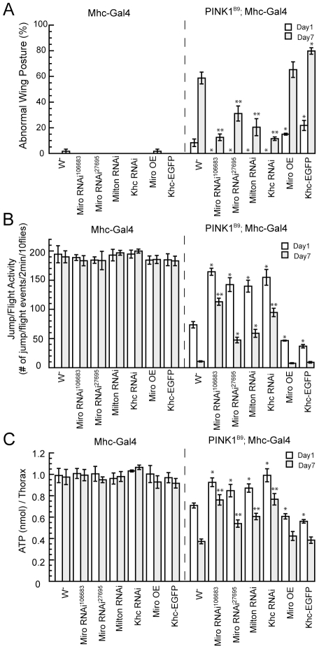
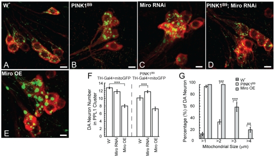
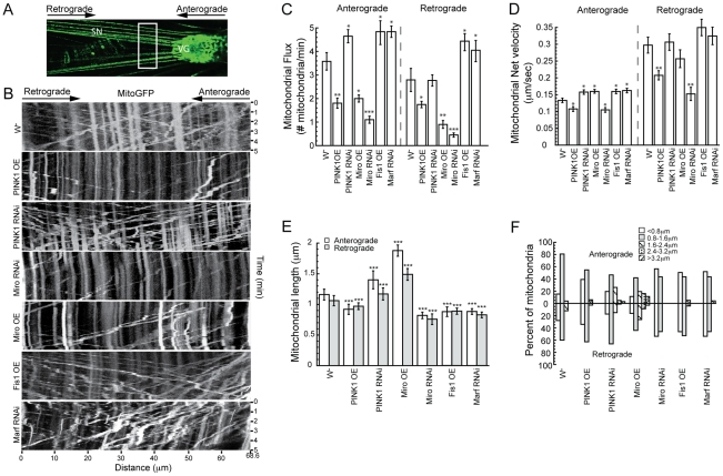
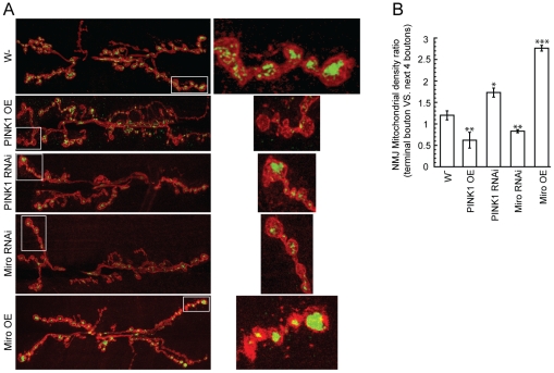
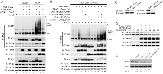
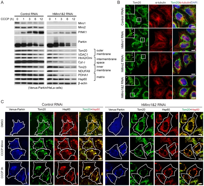
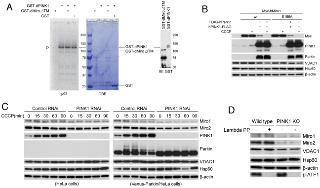
Mutations in Pten-induced kinase 1 (PINK1) are linked to early-onset familial Parkinson's disease (FPD). PINK1 has previously been implicated in mitochondrial fission/fusion dynamics, quality control, and electron transport chain function. However, it is not clear how these processes are interconnected and whether they are sufficient to explain all aspects of PINK1 pathogenesis. Here we show that PINK1 also controls mitochondrial motility. In Drosophila, downregulation of dMiro or other components of the mitochondrial transport machinery rescued dPINK1 mutant phenotypes in the muscle and dopaminergic (DA) neurons, whereas dMiro overexpression alone caused DA neuron loss. dMiro protein level was increased in dPINK1 mutant but decreased in dPINK1 or dParkin overexpression conditions. In Drosophila larval motor neurons, overexpression of dPINK1 inhibited axonal mitochondria transport in both anterograde and retrograde directions, whereas dPINK1 knockdown promoted anterograde transport. In HeLa cells, overexpressed hPINK1 worked together with hParkin, another FPD gene, to regulate the ubiquitination and degradation of hMiro1 and hMiro2, apparently in a Ser-156 phosphorylation-independent manner. Also in HeLa cells, loss of hMiro promoted the perinuclear clustering of mitochondria and facilitated autophagy of damaged mitochondria, effects previously associated with activation of the PINK1/Parkin pathway. These newly identified functions of PINK1/Parkin and Miro in mitochondrial transport and mitophagy contribute to our understanding of the complex interplays in mitochondrial quality control that are critically involved in PD pathogenesis, and they may explain the peripheral neuropathy symptoms seen in some PD patients carrying particular PINK1 or Parkin mutations. Moreover, the different effects of loss of PINK1 function on Miro protein level in Drosophila and mouse cells may offer one explanation of the distinct phenotypic manifestations of PINK1 mutants in these two species.
Overview
The claims and statistics of this paper haven’t been checked yet.
Run the check — free, no account needed. It reads the open-access text and checks the sources it cites.
Integrity signals
Registry
Cites
Statistics
Figures
Verification
Claims the paper makes
The claims and statistics of this paper haven’t been checked yet.
Run the check — free, no account needed. It reads the open-access text and checks the sources it cites.
Claims other papers make about this one
Each is checked against this paper’s own text: does it say what the citing paper says it does?
In the citing paper
“Once activated, Parkin has been reported to facilitate ubiquitination of numerous OMM proteins, including Mfn1 [126,127,128], Mfn2 [129], VDAC [130], Tom20 (translocase of the outer membrane 20) [121], and Miro [131].”
From Mitochondrial Dynamics and Mitophagy in Skeletal Muscle Health and Aging (2021) · cites this paper as [131]
Self-check
Percentages, sums and reported differences re-computed from the paper’s own numbers.
The self-check needs the paper’s full text.
Citations
Cites
Mutations in the parkin gene cause autosomal recessive juvenile parkinsonism
Kitada, T., Asakawa, S., Hattori, N., Matsumine, H., Yamamura, Y., Minoshima, S., Yokochi, M., Mizuno, Y., & Shimizu, N. (1998) · Nature · doi:10.1038/33416 · Cited by 5,227
Self-citationParkin is recruited selectively to impaired mitochondria and promotes their autophagy
Narendra, D., Tanaka, A., Suen, D.-F., & Youle, R. J. (2008) · The Journal of Cell Biology · doi:10.1083/jcb.200809125 · Cited by 4,026
Hereditary Early-Onset Parkinson's Disease Caused by Mutations in PINK1
Valente, E. M., Abou-Sleiman, P. M., Caputo, V., Muqit, M. M. K., Harvey, K., Gispert, S., Ali, Z., Del Turco, D., Bentivoglio, A. R., Healy, D. G., et al. (2004) · Science · doi:10.1126/science.1096284 · Cited by 3,481
Youle, R. J., & Narendra, D. P. (2011) · Nature Reviews Molecular Cell Biology · doi:10.1038/nrm3028 · Cited by 3,273
PINK1 Is Selectively Stabilized on Impaired Mitochondria to Activate Parkin
Narendra, D. P., Jin, S. M., Tanaka, A., Suen, D.-F., Gautier, C. A., Shen, J., Cookson, M. R., & Youle, R. J. (2010) · PLoS Biology · doi:10.1371/journal.pbio.1000298 · Cited by 2,944
PINK1/Parkin-mediated mitophagy is dependent on VDAC1 and p62/SQSTM1
Geisler, S., Holmström, K. M., Skujat, D., Fiesel, F. C., Rothfuss, O. C., Kahle, P. J., & Springer, W. (2010) · Nature Cell Biology · doi:10.1038/ncb2012 · Cited by 2,835
Matsuda, N., Sato, S., Shiba, K., Okatsu, K., Saisho, K., Gautier, C. A., Sou, Y.-S., Saiki, S., Kawajiri, S., Sato, F., et al. (2010) · Journal of Cell Biology · doi:10.1083/jcb.200910140 · Cited by 1,969
Self-citationDrosophila pink1 is required for mitochondrial function and interacts genetically with parkin
Clark, I. E., Dodson, M. W., Jiang, C., Cao, J. H., Huh, J. R., Seol, J. H., Yoo, S. J., Hay, B. A., & Guo, M. (2006) · Nature · doi:10.1038/nature04779 · Cited by 1,788
Mitochondrial dysfunction in Drosophila PINK1 mutants is complemented by parkin
Park, J., Lee, S. B., Lee, S., Kim, Y., Song, S., Kim, S., Bae, E., Kim, J., Shong, M., Kim, J.-M., et al. (2006) · Nature · doi:10.1038/nature04788 · Cited by 1,705
Self-citationKinesin and Dynein Superfamily Proteins and the Mechanism of Organelle Transport
Hirokawa, N. (1998) · Science · doi:10.1126/science.279.5350.519 · Cited by 1,689
Cited by
Sorted by citations
The Roles of PINK1, Parkin, and Mitochondrial Fidelity in Parkinson’s Disease
Pickrell, A. M., & Youle, R. J. (2015) · Neuron · doi:10.1016/j.neuron.2014.12.007 · Cited by 2,310
Synaptic Energy Use and Supply
Harris, J. J., Jolivet, R., & Attwell, D. (2012) · Neuron · doi:10.1016/j.neuron.2012.08.019 · Cited by 1,747
Landscape of the PARKIN-dependent ubiquitylome in response to mitochondrial depolarization
Sarraf, S. A., Raman, M., Guarani-Pereira, V., Sowa, M. E., Huttlin, E. L., Gygi, S. P., & Harper, J. W. (2013) · Nature · doi:10.1038/nature12043 · Cited by 1,010
Mitochondrial dynamics and mitochondrial quality control
Ni, H.-M., Williams, J. A., & Ding, W.-X. (2015) · Redox Biology · doi:10.1016/j.redox.2014.11.006 · Cited by 931
Kondapalli, C., Kazlauskaite, A., Zhang, N., Woodroof, H. I., Campbell, D. G., Gourlay, R., Burchell, L., Walden, H., Macartney, T. J., Deak, M., et al. (2012) · Open Biology · doi:10.1098/rsob.120080 · Cited by 940
Disturbed mitochondrial dynamics and neurodegenerative disorders
Burté, F., Carelli, V., Chinnery, P. F., & Yu‐Wai‐Man, P. (2014) · Nature Reviews Neurology · doi:10.1038/nrneurol.2014.228 · Cited by 686
Exner, N., Lutz, A. K., Haass, C., & Winklhofer, K. F. (2012) · The EMBO Journal · doi:10.1038/emboj.2012.170 · Cited by 677
Okatsu, K., Oka, T., Iguchi, M., Imamura, K., Kosako, H., Tani, N., Kimura, M., Go, E., Koyano, F., Funayama, M., et al. (2012) · Nature Communications · doi:10.1038/ncomms2016 · Cited by 629
Self-citationParkinson's disease: Mechanisms, translational models and management strategies
Raza, C., Anjum, R., & Shakeel, N. U. A. (2019) · Life Sciences · doi:10.1016/j.lfs.2019.03.057 · Cited by 628
Shiba-Fukushima, K., Imai, Y., Yoshida, S., Ishihama, Y., Kanao, T., Sato, S., & Hattori, N. (2012) · Scientific Reports · doi:10.1038/srep01002 · Cited by 577
Self-citationMitostasis in Neurons: Maintaining Mitochondria in an Extended Cellular Architecture
Misgeld, T., & Schwarz, T. L. (2017) · Neuron · doi:10.1016/j.neuron.2017.09.055 · Cited by 562
Mitochondrial dysfunction and mitophagy in Parkinson's: from familial to sporadic disease
Ryan, B. J., Hoek, S., Fon, E. A., & Wade-Martins, R. (2015) · Trends in Biochemical Sciences · doi:10.1016/j.tibs.2015.02.003 · Cited by 524
Mitochondrial fusion, fission, and mitochondrial toxicity
Meyer, J. N., Leuthner, T. C., & Luz, A. L. (2017) · Toxicology · doi:10.1016/j.tox.2017.07.019 · Cited by 535
Mitochondrial Trafficking in Neurons
Schwarz, T. L. (2013) · Cold Spring Harbor Perspectives in Biology · doi:10.1101/cshperspect.a011304 · Cited by 525
Hsieh, C.-H., Shaltouki, A., Gonzalez, A. E., da Cruz, A. B., Burbulla, L. F., Lawrence, E. S., Schüle, B., Krainc, D., Palmer, T. D., & Wang, X. (2016) · Cell stem cell · doi:10.1016/j.stem.2016.08.002 · Cited by 475
TRAK/Milton motor-adaptor proteins steer mitochondrial trafficking to axons and dendrites
van Spronsen, M., Mikhaylova, M., Lipka, J., Schlager, M. A., van den Heuvel, D. J., Kuijpers, M., Wulf, P. S., Keijzer, N., Demmers, J., Kapitein, L. C., et al. (2013) · Neuron · doi:10.1016/j.neuron.2012.11.027 · Cited by 450
Mitochondrial Diseases of the Brain
Chaturvedi, R. K., & Beal, M. F. (2013) · Free Radical Biology and Medicine · doi:10.1016/j.freeradbiomed.2013.03.018 · Cited by 426
Mitochondrial trafficking and anchoring in neurons: New insight and implications
Sheng, Z. (2014) · The Journal of Cell Biology · doi:10.1083/jcb.201312123 · Cited by 429
Parkin and PINK1: much more than mitophagy
Scarffe, L. A., Stevens, D. A., Dawson, V. L., & Dawson, T. M. (2014) · Trends in Neurosciences · doi:10.1016/j.tins.2014.03.004 · Cited by 392
New insights into the complex role of mitochondria in Parkinson’s disease
Grünewald, A., Kumar, K. R., & Sue, C. M. (2018) · Progress in Neurobiology · doi:10.1016/j.pneurobio.2018.09.003 · Cited by 375
Autophagy in Parkinson's Disease
Hou, X., Watzlawik, J. O., Fiesel, F. C., & Springer, W. (2020) · Journal of Molecular Biology · doi:10.1016/j.jmb.2020.01.037 · Cited by 361
Mitochondrial Quality Control Mediated by PINK1 and Parkin: Links to ParkinsonismPreprint
Narendra, D. P., Walker, J. E., & Youle, R. J. (2012) · Cold Spring Harbor Perspectives in Biology · doi:10.1101/cshperspect.a011338 · Cited by 360
Mechanisms of mitophagy: PINK1, Parkin, USP30 and beyond
Bingol, B., & Sheng, M. (2016) · Free Radical Biology and Medicine · doi:10.1016/j.freeradbiomed.2016.04.015 · Cited by 340
Determinants and Functions of Mitochondrial Behavior
Labbè, K., Murley, A., & Nunnari, J. M. (2014) · Annual Review of Cell and Developmental Biology · doi:10.1146/annurev-cellbio-101011-155756 · Cited by 324
RBR E3 ubiquitin ligases: new structures, new insights, new questions
Spratt, D. E., Walden, H., & Shaw, G. S. (2014) · Biochemical Journal · doi:10.1042/bj20140006 · Cited by 335
Godena, V. K., Brookes-Hocking, N., Möller, A., Shaw, G., Oswald, M. C. W., Sancho, R. M., Miller, C. C. J., Whitworth, A. J., & Vos, K. J. D. (2014) · Nature Communications · doi:10.1038/ncomms6245 · Cited by 279
BNIP3 Interacting with LC3 Triggers Excessive Mitophagy in Delayed Neuronal Death in Stroke
Shi, R., Zhu, S., Li, V., Gibson, S. B., Xu, X., & Kong, J. (2014) · CNS Neuroscience & Therapeutics · doi:10.1111/cns.12325 · Cited by 282
Parkin-mediated mitophagy in mutant hAPP neurons and Alzheimer's disease patient brains
Ye, X., Sun, X., Starovoytov, V., & Cai, Q. (2015) · Human Molecular Genetics · doi:10.1093/hmg/ddv056 · Cited by 274
Mitochondrial Aspects of Synaptic Dysfunction in Alzheimer’s Disease
Cai, Q., & Tammineni, P. (2016) · Journal of Alzheimer s Disease · doi:10.3233/jad-160726 · Cited by 281
Mitochondrial quality control in health and in Parkinson’s disease
Eldeeb, M. A., Thomas, R. A., Ragheb, M. A., Fallahi, A., & Fon, E. A. (2022) · Physiological Reviews · doi:10.1152/physrev.00041.2021 · Cited by 281
Mitochondrial Dynamics and Mitophagy in Skeletal Muscle Health and Aging
Leduc-Gaudet, J.-P., Hussain, S. N. A., Barreiro, E., & Gouspillou, G. (2021) · International Journal of Molecular Sciences · doi:10.3390/ijms22158179 · Cited by 268
“Once activated, Parkin has been reported to facilitate ubiquitination of numerous OMM proteins, including Mfn1 [126–128], Mfn2 [129], VDAC [130], Tom20 (translocase of the outer membrane 20) [121], and Miro [131].”
BackgroundMitophagy in Alzheimer’s Disease and Other Age-Related Neurodegenerative Diseases
Cai, Q., & Jeong, Y. Y. (2020) · Cells · doi:10.3390/cells9010150 · Cited by 270
Phosphorylated ubiquitin chain is the genuine Parkin receptor
Okatsu, K., Koyano, F., Kimura, M., Kosako, H., Saeki, Y., Tanaka, K., & Matsuda, N. (2015) · The Journal of Cell Biology · doi:10.1083/jcb.201410050 · Cited by 272
Mitochondrial Dysfunction in Parkinson’s Disease—Cause or Consequence?
Chen, C., Turnbull, D. M., & Reeve, A. K. (2019) · Biology · doi:10.3390/biology8020038 · Cited by 260
Wei, H., Liu, L., & Chen, Q. (2015) · Biochimica et Biophysica Acta (BBA) - Molecular Cell Research · doi:10.1016/j.bbamcr.2015.03.013 · Cited by 266
Programming axonal mitochondrial maintenance and bioenergetics in neurodegeneration and regeneration
Cheng, X., Huang, N., & Sheng, Z. (2022) · Neuron · doi:10.1016/j.neuron.2022.03.015 · Cited by 257
Sun, Y., Vashisht, A. A., Tchieu, J. H., Wohlschlegel, J. A., & Dreier, L. (2012) · Journal of Biological Chemistry · doi:10.1074/jbc.m112.419721 · Cited by 255
Babenko, V., Silachev, D., Popkov, V., Zorova, L., Pevzner, I., Plotnikov, E., Sukhikh, G., & Zorov, D. (2018) · Molecules · doi:10.3390/molecules23030687 · Cited by 242
Cutaneous neuropathy in Parkinson’s disease: a window into brain pathology
Doppler, K., Ebert, S., Üçeyler, N., Trenkwalder, C., Ebentheuer, J., Volkmann, J., & Sommer, C. (2014) · Acta Neuropathologica · doi:10.1007/s00401-014-1284-0 · Cited by 243
The ubiquitin signal and autophagy: an orchestrated dance leading to mitochondrial degradation
Yamano, K., Matsuda, N., & Tanaka, K. (2016) · EMBO Reports · doi:10.15252/embr.201541486 · Cited by 253
Lee, K., Huh, S., Lee, S., Wu, Z., Kim, A., Kang, H., & Lu, B. (2018) · Proceedings of the National Academy of Sciences · doi:10.1073/pnas.1721136115 · Cited by 237
Self-citationRegulation of mitochondrial transport in neurons
Lin, M.-Y., & Sheng, Z. (2015) · Experimental Cell Research · doi:10.1016/j.yexcr.2015.01.004 · Cited by 232
Alterations in Mitochondrial Quality Control in Alzheimer’s Disease
Cai, Q., & Tammineni, P. (2016) · Frontiers in Cellular Neuroscience · doi:10.3389/fncel.2016.00024 · Cited by 226
Gehrke, S., Wu, Z., Klinkenberg, M., Sun, Y., Auburger, G. W. J., Guo, S., & Lu, B. (2015) · Cell Metabolism · doi:10.1016/j.cmet.2014.12.007 · Cited by 223
Self-citationNeuronal Mitophagy in Neurodegenerative Diseases
Martínez‐Vicente, M. (2017) · Frontiers in Molecular Neuroscience · doi:10.3389/fnmol.2017.00064 · Cited by 222
Parkin-catalyzed Ubiquitin-Ester Transfer Is Triggered by PINK1-dependent Phosphorylation
Iguchi, M., Kujuro, Y., Okatsu, K., Koyano, F., Kosako, H., Kimura, M., Suzuki, N., Uchiyama, S., Tanaka, K., & Matsuda, N. (2013) · Journal of Biological Chemistry · doi:10.1074/jbc.m113.467530 · Cited by 209
PINK1 Primes Parkin-Mediated Ubiquitination of PARIS in Dopaminergic Neuronal Survival
Lee, Y., Stevens, D. A., Kang, S.-U., Jiang, H., Lee, Y.-I., Ko, H. S., Scarffe, L. A., Umanah, G. K. E., Kang, H., Ham, S., et al. (2017) · Cell Reports · doi:10.1016/j.celrep.2016.12.090 · Cited by 214
Mitochondria-associated membranes as hubs for neurodegeneration
Krols, M., Isterdael, G. V., Asselbergh, B., Kremer, A., Lippens, S., Timmerman, V., & Janssens, S. (2016) · Acta Neuropathologica · doi:10.1007/s00401-015-1528-7 · Cited by 210
Birsa, N., Norkett, R., Wauer, T., Mevissen, T. E. T., Wu, H.-C., Foltynie, T., Bhatia, K. P., Hirst, W. D., Komander, D., Plun‐Favreau, H., et al. (2014) · Journal of Biological Chemistry · doi:10.1074/jbc.m114.563031 · Cited by 203
Franco-Iborra, S., Vila, M., & Perier, C. (2018) · Frontiers in Neuroscience · doi:10.3389/fnins.2018.00342 · Cited by 197
Mitochondrial Dynamics in Mitochondrial Diseases
Suarez-Rivero, J. M., Paz, M. V., de la Cruz‐Ojeda, P., de la Mata, M., Cotán, D., Oropesa-Ávila, M., de Lavera, I., Álvarez-Córdoba, M., Luzón–Hidalgo, R., & Sánchez-Alcázar, J. A. (2016) · Diseases · doi:10.3390/diseases5010001 · Cited by 196
The genetic architecture of mitochondrial dysfunction in Parkinson’s disease
Larsen, S. B., Hanss, Z., & Krüger, R. (2018) · Cell and Tissue Research · doi:10.1007/s00441-017-2768-8 · Cited by 184
Phosphoproteomic screening identifies Rab GTPases as novel downstream targets of PINK1
Lai, Y., Kondapalli, C., Lehneck, R., Procter, J. B., Dill, B. D., Woodroof, H. I., Gourlay, R., Peggie, M. W., Macartney, T., Corti, O., et al. (2015) · The EMBO Journal · doi:10.15252/embj.201591593 · Cited by 182
Targeting of host organelles by pathogenic bacteria: a sophisticated subversion strategy
Escoll, P., Mondino, S., Rolando, M., & Buchrieser, C. (2015) · Nature Reviews Microbiology · doi:10.1038/nrmicro.2015.1 · Cited by 168
Anandhan, A., Jacome, M. S., Lei, S., Hernández-Franco, P., Pappa, A., Panayiotidis, M. I., Powers, R., & Franco, R. (2017) · Brain Research Bulletin · doi:10.1016/j.brainresbull.2017.03.009 · Cited by 165
Redox homeostasis, oxidative stress and mitophagy
Garza-Lombó, C., Pappa, A., Panayiotidis, M. I., & Franco, R. (2020) · Mitochondrion · doi:10.1016/j.mito.2020.01.002 · Cited by 168
Alpha-synuclein delays mitophagy and targeting Miro rescues neuron loss in Parkinson’s models
Shaltouki, A., Hsieh, C.-H., Kim, M. J., & Wang, X. (2018) · Acta Neuropathologica · doi:10.1007/s00401-018-1873-4 · Cited by 170
Atlastins remodel the endoplasmic reticulum for selective autophagy
Liang, J. R., Lingeman, E., Ahmed, S., & Corn, J. E. (2018) · Journal of Cell Biology · doi:10.1083/jcb.201804185 · Cited by 158
The role of mitophagy in the regulation of mitochondrial energetic status in neurons
Han, S., Zhang, M., Jeong, Y. Y., Margolis, D. J., & Cai, Q. (2021) · Autophagy · doi:10.1080/15548627.2021.1907167 · Cited by 151
Degradation of engulfed mitochondria is rate-limiting in Optineurin-mediated mitophagy in neurons
Evans, C. S., & Holzbaur, E. L. (2020) · eLife · doi:10.7554/elife.50260 · Cited by 137
“, 2014) and studies indicating that axonal mitochondria motility is arrested following mitochondrial damage (Hsieh et al., 2016; Liu et al., 2012; Wang et al., 2011), necessitating a local clearance mechanism.”
BackgroundParkin and mitochondrial quality control: toward assembling the puzzle
Winklhofer, K. F. (2014) · Trends in Cell Biology · doi:10.1016/j.tcb.2014.01.001 · Cited by 153
Miro phosphorylation sites regulate Parkin recruitment and mitochondrial motility
Shlevkov, E., Kramer, T., Schapansky, J., LaVoie, M. J., & Schwarz, T. L. (2016) · Proceedings of the National Academy of Sciences · doi:10.1073/pnas.1612283113 · Cited by 150
Regulation of mitochondrial dynamics in astrocytes: Mechanisms, consequences, and unknowns
Jackson, J. G., & Robinson, M. B. (2017) · Glia · doi:10.1002/glia.23252 · Cited by 153
A Mitocentric View of Parkinson's Disease
Haelterman, N. A. T., Yoon, W. H., Sandoval, H., Jaiswal, M., Shulman, J. M., & Bellen, H. J. (2014) · Annual Review of Neuroscience · doi:10.1146/annurev-neuro-071013-014317 · Cited by 141
Iijima-Ando, K., Sekiya, M., Maruko-Otake, A., Ohtake, Y., Suzuki, E., Lu, B., & Iijima, K. M. (2012) · PLoS Genetics · doi:10.1371/journal.pgen.1002918 · Cited by 142
Self-citationGautier, C., Erpapazoglou, Z., Mouton‐Liger, F., Mp, M., Cormier‐Dequaire, F., Bigou, S., Duffaure, S., Girard, M., Forêt, B., Iannielli, A., et al. (2016) · Human Molecular Genetics · doi:10.1093/hmg/ddw148 · Cited by 145
Kazlauskaite, A., Kelly, V. C., Johnson, C., Baillie, C., Hastie, C. J., Peggie, M. W., Macartney, T., Woodroof, H. I., Alessi, D. R., Pedrioli, P. G. A., et al. (2014) · Open Biology · doi:10.1098/rsob.130213 · Cited by 139
Back to the tubule: microtubule dynamics in Parkinson’s disease
Pellegrini, L., Wetzel, A., Grannò, S., Heaton, G., & Harvey, K. (2016) · Cellular and Molecular Life Sciences · doi:10.1007/s00018-016-2351-6 · Cited by 147
Mitochondria targeted therapeutic approaches in Parkinson's and Huntington's diseases
Chaturvedi, R. K., & Beal, M. F. (2012) · Molecular and Cellular Neuroscience · doi:10.1016/j.mcn.2012.11.011 · Cited by 139
Moghadam, A. K., Charsouei, S., Bell, B., & Jabalameli, M. R. (2018) · Cellular and Molecular Neurobiology · doi:10.1007/s10571-018-0587-4 · Cited by 143
Lionaki, E., Markaki, M., Palikaras, K., & Tavernarakis, N. (2015) · Biochimica et Biophysica Acta (BBA) - Bioenergetics · doi:10.1016/j.bbabio.2015.04.010 · Cited by 137
Stephen, T., Higgs, N., Sheehan, D. F., Awabdh, S. A., López‐Doménech, G., Arancibia‐Cárcamo, I. L., & Kittler, J. T. (2015) · Journal of Neuroscience · doi:10.1523/jneurosci.2068-15.2015 · Cited by 140
Targeting Mitochondrial Impairment in Parkinson's Disease: Challenges and Opportunities
Prasuhn, J., Davis, R. L., & Kumar, K. R. (2021) · Frontiers in Cell and Developmental Biology · doi:10.3389/fcell.2020.615461 · Cited by 135
Laar, V. S. V., & Berman, S. (2012) · Neurobiology of Disease · doi:10.1016/j.nbd.2012.05.015 · Cited by 137
Making connections: interorganelle contacts orchestrate mitochondrial behavior
Klecker, T., Böckler, S., & Westermann, B. (2014) · Trends in Cell Biology · doi:10.1016/j.tcb.2014.04.004 · Cited by 137
Miro proteins prime mitochondria for Parkin translocation and mitophagy
Safiulina, D., Kuum, M., Choubey, V., Gogichaishvili, N., Liiv, J., Hickey, M. A., Cagalinec, M., Mandel, M., Zeb, A., Liiv, M., et al. (2018) · The EMBO Journal · doi:10.15252/embj.201899384 · Cited by 133
Laar, V. S. V., Roy, N., Liu, A., Rajprohat, S., Arnold, B. A., Dukes, A. A., Holbein, C. D., & Berman, S. B. (2014) · Neurobiology of Disease · doi:10.1016/j.nbd.2014.11.015 · Cited by 121
Beyond the mitochondrion: cytosolic PINK 1 remodels dendrites through Protein Kinase A
Dagda, R. K., Pien, I., Wang, R., Zhu, J., Wang, K. Z. Q., Callio, J., Banerjee, T. D., Dagda, R. Y., & Chu, C. T. (2013) · Journal of Neurochemistry · doi:10.1111/jnc.12494 · Cited by 122
Motor proteins at the mitochondria–cytoskeleton interface
Kruppa, A. J., & Buß, F. (2021) · Journal of Cell Science · doi:10.1242/jcs.226084 · Cited by 127
Aparicio, I. M., Espino, J., Bejarano, I., Gallardo, A., Campo, M. L., Salido, G. M., Pariente, J. A., Peña, F. J., & Tapia, J. A. (2016) · Scientific Reports · doi:10.1038/srep33647 · Cited by 121
Regulation of long-distance transport of mitochondria along microtubules
Melkov, A., & Abdu, U. (2017) · Cellular and Molecular Life Sciences · doi:10.1007/s00018-017-2590-1 · Cited by 129
Axon and dendritic trafficking
Maeder, C. I., Shen, K., & Hoogenraad, C. C. (2014) · Current Opinion in Neurobiology · doi:10.1016/j.conb.2014.03.015 · Cited by 116
Mitochondrial trafficking in neurons and the role of the Miro family of GTPase proteins
Birsa, N., Norkett, R., Higgs, N., López‐Doménech, G., & Kittler, J. T. (2013) · Biochemical Society Transactions · doi:10.1042/bst20130234 · Cited by 116
Mitotic catastrophe and cancer drug resistance: A link that must to be broken
Denisenko, T. V., Sorokina, I. V., Gogvadze, V., & Zhivotovsky, B. (2016) · Drug Resistance Updates · doi:10.1016/j.drup.2015.11.002 · Cited by 108
Identification of Miro1 and Miro2 as mitochondrial receptors for myosin XIX
Oeding, S. J., Majstrowicz, K., Hu, X.-P., Schwarz, V., Freitag, A., Honnert, U., Nikolaus, P., & Bähler, M. (2018) · Journal of Cell Science · doi:10.1242/jcs.219469 · Cited by 118
Yang, D., Ying, J., Wang, X., Zhao, T., Yoon, S., Yang, F., Zheng, Q., Liu, X., Yu, W., & Hua, F. (2021) · Frontiers in Neuroscience · doi:10.3389/fnins.2021.654785 · Cited by 121
Self-citationPINK1: The guard of mitochondria
Wang, N., Zhu, P., Huang, R., Wang, C., Sun, L., Lan, B., He, Y., Zhao, H., & Gao, Y. (2020) · Life Sciences · doi:10.1016/j.lfs.2020.118247 · Cited by 107
Lee, S., Lee, K., Huh, S., Liu, S., Lee, D.-Y., Hong, S. H., Yu, K., & Lu, B. (2016) · Developmental Cell · doi:10.1016/j.devcel.2016.03.023 · Cited by 107
Self-citationvan der Merwe, C., Dashti, Z. J. S., Christoffels, A. G., Loos, B., & Bardien, S. (2015) · European Journal of Neuroscience · doi:10.1111/ejn.12872 · Cited by 110
Suzuki, M., Danilchanka, O., & Mekalanos, J. J. (2014) · Cell Host & Microbe · doi:10.1016/j.chom.2014.09.015 · Cited by 106
Drosophila as a Model to Study Mitochondrial Dysfunction in Parkinson's DiseasePreprint
Guo, M. (2012) · Cold Spring Harbor Perspectives in Medicine · doi:10.1101/cshperspect.a009944 · Cited by 111
A PGAM5–KEAP1–Nrf2 complex is required for stress-induced mitochondrial retrograde trafficking
O'Mealey, G. B., Plafker, K. S., Berry, W. L., Janknecht, R., Chan, J. Y., & Plafker, S. M. (2017) · Journal of Cell Science · doi:10.1242/jcs.203216 · Cited by 99
Unravelling mitochondrial pathways to P arkinson's disease
Celardo, I., Martins, L. M., & Gandhi, S. (2013) · British Journal of Pharmacology · doi:10.1111/bph.12433 · Cited by 105
Quality Control in Neurons: Mitophagy and Other Selective Autophagy Mechanisms
Evans, C. S., & Holzbaur, E. L. F. (2019) · Journal of Molecular Biology · doi:10.1016/j.jmb.2019.06.031 · Cited by 109
Regulation of mitochondrial functions by protein phosphorylation and dephosphorylation
Lim, S., Smith, K. R., Lim, S., Tian, R., Lu, J., & Tan, M. (2016) · Cell & Bioscience · doi:10.1186/s13578-016-0089-3 · Cited by 106
Mitophagy in neurodegenerative diseases
Rodolfo, C., Campello, S., & Cecconi, F. (2017) · Neurochemistry International · doi:10.1016/j.neuint.2017.08.004 · Cited by 104
WldS and PGC-1α Regulate Mitochondrial Transport and Oxidation State after Axonal Injury
O’Donnell, K. C., Vargas, M. E., & Sagasti, A. (2013) · Journal of Neuroscience · doi:10.1523/jneurosci.1331-13.2013 · Cited by 105
Mitochondria as central hubs in synaptic modulation
Duarte, F. V., Ciampi, D., & Duarte, C. B. (2023) · Cellular and Molecular Life Sciences · doi:10.1007/s00018-023-04814-8 · Cited by 109
Mitophagy regulates integrity of mitochondria at synapses and is critical for synaptic maintenance
Han, S., Jeong, Y. Y., Sheshadri, P., Su, X. Y., & Cai, Q. (2020) · EMBO Reports · doi:10.15252/embr.201949801 · Cited by 96
Mechanisms of Parkinson's Disease
Hewitt, V. L., & Whitworth, A. J. (2016) · Current topics in developmental biology/Current Topics in Developmental Biology · doi:10.1016/bs.ctdb.2016.07.005 · Cited by 97
Metadata
- Authors
- Liu, S., Sawada, T., Lee, S., Yu, W., Silverio, G., Alapatt, P., Millan, I., Shen, A., Saxton, W., Kanao, T., Takahashi, R., Hattori, N., Imai, Y., & Lu, B.
- Affiliations
- Stanford Medicine (US)
- Kyoto University (JP)
- Washington University in St. Louis (US)
- University of California, Santa Cruz (US)
- Juntendo University (JP)
- Corresponding author
- Imai, Yuzuru
- Lu, Bingwei
- Venue
- PLoS Genetics
- Publisher
- Public Library of Science (PLoS)
- Published
- 2012-03-01
- Volume
- 8
- Issue
- 3
- Pages
- e1002537
- ISSN
- 1553-7390 (ISSN-L)
- 1553-7404
- PMC
- PMC3291531
- OpenAlex
- W2148819754
- Cited by
- 371
- References
- 59
- Citations by year
- 2026: 11 · 2025: 10 · 2024: 10 · 2023: 11 · 2022: 18 · 2021: 35 · 2020: 29 · 2019: 22 · 2018: 28 · 2017: 30 · 2016: 36 · 2015: 32 · 2014: 42 · 2013: 33 · 2012: 23
- FWCI
- 27.18
- Type
- journal article
- Language
- en
- Open access
- gold
- doi.org
- Locations
- PLoS Genetics (publishedVersion, cc-by)
- Europe PMC (PubMed Central) (publishedVersion, cc-by)
- PubMed Central (publishedVersion, cc-by)
- PubMed (publishedVersion)
- DOAJ (DOAJ: Directory of Open Access Journals) (submittedVersion, cc-by-sa)
- Figshare (submittedVersion, cc-by)
- Funders
- Ministry of Education, Culture, Sports, Science and Technology (CREST)
- Dainippon Sumitomo Pharma
- Ministry of Health, Labour and Welfare
- National Institutes of Health
- Japan Society for the Promotion of Science (23390232, 23111002, 23890206, 23790973)
- National Institute of Mental Health (R01MH080378)
- National Institute of General Medical Sciences (R01GM084947, GM084947)
- National Institute of Arthritis and Musculoskeletal and Skin Diseases (R01AR054926)
- Core Research for Evolutional Science and Technology
- Topics
- Health Sciences › Medicine › Clinical Neurology › Parkinson's Disease Mechanisms and Treatments
- Life Sciences › Biochemistry, Genetics and Molecular Biology › Cell Biology › Autophagy in Disease and Therapy
- Life Sciences › Biochemistry, Genetics and Molecular Biology › Molecular Biology › Mitochondrial Function and Pathology
- Keywords
- PINK1, Miro1, Miro2, Parkinson's disease, mitochondrial motility, Parkin, mitophagy, mitochondrial quality control, dopaminergic neurons, motor neurons, ubiquitin-mediated proteolysis, peripheral neuropathy, Drosophila
- MeSH terms
- PTEN-Induced Putative Kinase, Animals, Autophagy, Axonal Transport, Carbonyl Cyanide m-Chlorophenyl Hydrazone, Disease Models, Animal, Drosophila, Gene Expression Regulation, HeLa Cells, Humans, Mitochondria, Motor Neurons, Parkinson Disease, Protein Serine-Threonine Kinases, Mice, Knockout, rho GTP-Binding Proteins, Drosophila Proteins, Ubiquitin-Protein Ligases, Mutant Proteins, Mice, Dopaminergic Neurons, Proton Ionophores
About these checks
Registry, retraction, Cites and statistics results are computed, not judged, with one exception: a printed reference that no index lists is read once by a model before it counts as a citation error. Claims are extracted by a model and kept only when their quote appears verbatim in the text; each cited source is then checked, with a model’s help, for whether it says what the claim relies on. Those verdicts can be wrong — read the claim’s quote and the cited source before relying on one.
Statistics and arithmetic are recomputed, not judged.
Confidence bands are shown only for fields calibrated on our annotated test set. None is calibrated yet, so no band is shown.
What counts as a claim ›How we measure confidence ›How we check this ›
Are you an author, or do you see a problem with this page? Request a review
