

The DOI matches its Crossref record; 56 of the 59 references it cites were found in a registry or an index.
An Immune Basis for Lung Parenchymal Destruction in Chronic Obstructive Pulmonary Disease and Emphysema
Open accessAbstract
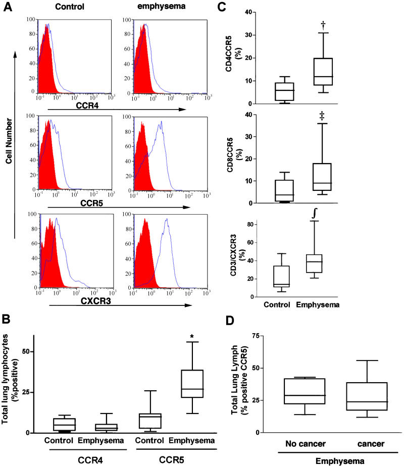
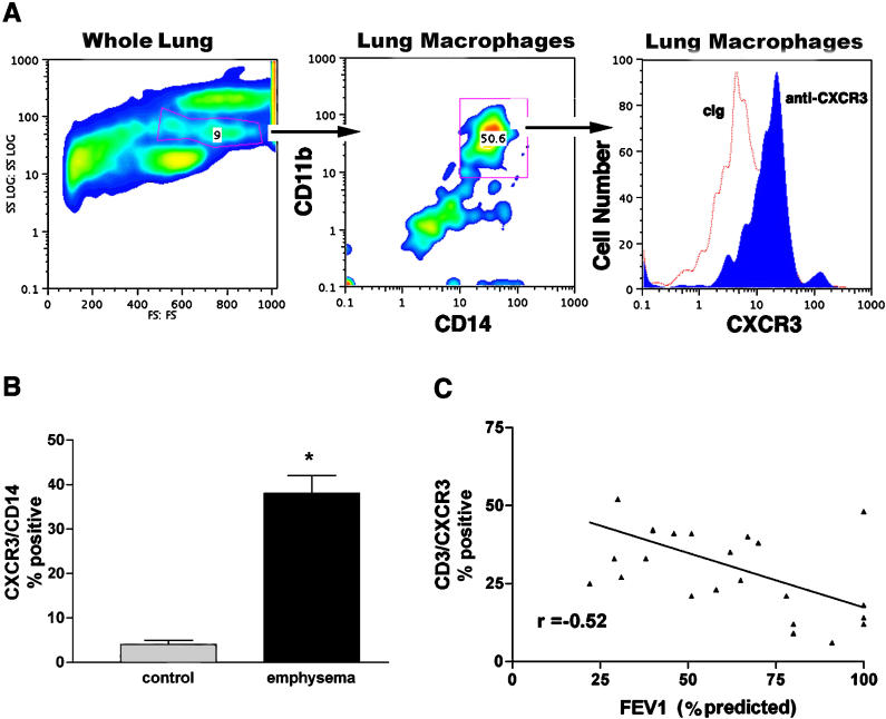
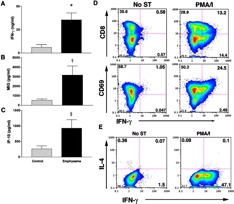
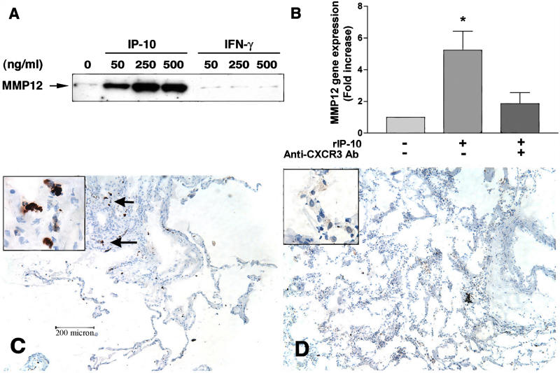
BACKGROUND: Chronic obstructive pulmonary disease and emphysema are a frequent result of long-term smoking, but the exact mechanisms, specifically which types of cells are associated with the lung destruction, are unclear. METHODS AND FINDINGS: We studied different subsets of lymphocytes taken from portions of human lungs removed surgically to find out which lymphocytes were the most frequent, which cell-surface markers these lymphocytes expressed, and whether the lymphocytes secreted any specific factors that could be associated with disease. We found that loss of lung function in patients with chronic obstructive pulmonary disease and emphysema was associated with a high percentage of CD4+ and CD8+ T lymphocytes that expressed chemokine receptors CCR5 and CXCR3 (both markers of T helper 1 cells), but not CCR3 or CCR4 (markers of T helper 2 cells). Lung lymphocytes in patients with chronic obstructive pulmonary disease and emphysema secrete more interferon gamma--often associated with T helper 1 cells--and interferon-inducible protein 10 and monokine induced by interferon, both of which bind to CXCR3 and are involved in attracting T helper 1 cells. In response to interferon-inducible protein 10 and monokine induced by interferon, but not interferon gamma, lung macrophages secreted macrophage metalloelastase (matrix metalloproteinase-12), a potent elastin-degrading enzyme that causes tissue destruction and which has been linked to emphysema. CONCLUSIONS: These data suggest that Th1 lymphoctytes in the lungs of people with smoking-related damage drive progression of emphysema through CXCR3 ligands, interferon-inducible protein 10, and monokine induced by interferon.
Overview
The claims and statistics of this paper haven’t been checked yet.
Run the check — free, no account needed. It reads the open-access text and checks the sources it cites.
Integrity signals
Registry
Cites
Statistics
Figures
Verification
Claims the paper makes
The claims and statistics of this paper haven’t been checked yet.
Run the check — free, no account needed. It reads the open-access text and checks the sources it cites.
Claims other papers make about this one
Each is checked against this paper’s own text: does it say what the citing paper says it does?
In the citing paper
“Specifically, macrophages, which can produce multiple proinflammatory cytokines and secrete MMPs, are the most abundant cell type in the BAL fluid of COPD patients, and increased numbers correlate with disease severity (85, 86, 104).”
From Aging and Lung Disease (2020) · cites this paper as [85]
In the citing paper
“Impaired phagocytosis and clearance of cellular debris by airway macrophages in COPD result in persistent inflammation (85, 104).”
From Aging and Lung Disease (2020) · cites this paper as [85]
Self-check
Percentages, sums and reported differences re-computed from the paper’s own numbers.
The self-check needs the paper’s full text.
Citations
Cites
Vestbo, J., Hurd, S. S., Agustí, A. G., Jones, P. W., Vogelmeier, C., Anzueto, A., Barnes, P. J., Fabbri, L. M., Martinez, F. J., Nishimura, M., et al. (2013) · American Journal of Respiratory and Critical Care Medicine · doi:10.1164/rccm.201204-0596pp · Cited by 15,983
PAUWELS, R. A., Buist, A. S., CALVERLEY, P. M. A., Jenkins, C. R., & Hurd, S. S. (2001) · American Journal of Respiratory and Critical Care Medicine · doi:10.1164/ajrccm.163.5.2101039 · Cited by 6,578 · cited 1×
“COPD was diagnosed according to the criteria recommended by the National Institutes of Health/World Health Organization workshop summary [31].”
MethodHow Matrix Metalloproteinases Regulate Cell Behavior
Sternlicht, M. D., & Werb, Z. (2001) · Annual Review of Cell and Developmental Biology · doi:10.1146/annurev.cellbio.17.1.463 · Cited by 3,959 · cited 1×
“Cytokines and chemokines are substrates for MMPs, but they also regulate expression of MMPs under pathological conditions [47,48].”
BackgroundZlotnik, A., & Yoshie, O. (2000) · Immunity · doi:10.1016/s1074-7613(00)80165-x · Cited by 3,923 · cited 1×
“Interferon-inducible protein 10 (IP10), monokine induced by interferon gamma (MIG), and interferon-inducible T cell alpha chemoattractant (I-TAC) are three known ligands for CXCR3 produced by normal and injured epithelial cells and T cells that are required for homing of Th1 cells [27,28,29].”
BackgroundChronic Obstructive Pulmonary Disease
Flavin, K., Morkane, C. M., & Marsh, S. (2018) · Cambridge University Press eBooks · doi:10.1017/9781108233712.025 · Cited by 2,687
Anthonisen, N. R. (1994) · JAMA · doi:10.1001/jama.272.19.1497 · Cited by 1,960
Requirement for IL-13 Independently of IL-4 in Experimental Asthma
Grünig, G., Warnock, M. L., Wakil, A. E., Venkayya, R., Brombacher, F., Rennick, D. M., Sheppard, D., Mohrs, M., Donaldson, D. D., Locksley, R. M., et al. (1998) · Science · doi:10.1126/science.282.5397.2261 · Cited by 1,845
Self-citationRequirement for Macrophage Elastase for Cigarette Smoke-Induced Emphysema in Mice
Hautamaki, R. D., Kobayashi, D. K., Senior, R. M., & Shapiro, S. D. (1997) · Science · doi:10.1126/science.277.5334.2002 · Cited by 1,470 · cited 1×
“MMP12 deficiency has been shown to protect mice against emphysema after chronic exposure to cigarette smoke, implying that MMP12 may be the key proteinase in the development of emphysema in this species [49,50].”
BackgroundChronic Obstructive Pulmonary Disease
Barnes, P. J. (2000) · New England Journal of Medicine · doi:10.1056/nejm200007273430407 · Cited by 1,078
Cole, K. E., Strick, C. A., Paradis, T. J., Ogborne, K. T., Loetscher, M., Gladue, R. P., Lin, W., Boyd, J. G., Moser, B., Wood, D. E., et al. (1998) · The Journal of Experimental Medicine · doi:10.1084/jem.187.12.2009 · Cited by 870 · cited 1×
“]. In addition to T cells, a wide variety of other inflammatory cells have been shown to express distinct chemokine receptors that are critical for their homing, suggesting a universal mechanism for regulating immune responses. Interferon-inducible protein 10 (IP-10), monokine induced by interferon gamma (MIG), and in…”
Background
Research works printed but not linked: 3
- Kay, 1997, Ciba Found Symp, 206, 56
- Anthonisen, 1994, JAMA, 272, 1497, 10.1001/jama.1994.03520190043033 · DOI
- Fiocchi, 1985, Acta Chir Scand Suppl, 525, 11
Cited by
Sorted by citations
Items 101–200 of 484
Elias, J. A., Kang, M. J., Crothers, K. A., Homer, R., & Lee, C. G. (2006) · Proceedings of the American Thoracic Society · doi:10.1513/pats.200603-068ms · Cited by 86
McComb, J. G., Ranganathan, M., Liu, X. H., Pilewski, J. M., Ray, P., Watkins, S. C., Choi, A. M. K., & Lee, J. S. (2008) · American Journal Of Pathology · doi:10.2353/ajpath.2008.071034 · Cited by 86
“5,6,16 In our study, we show increased CX3CL1 gene expression in murine lungs after prolonged cigarette smoke exposure.”
Duncan, S. R. (2008) · International Journal of COPD · doi:10.2147/copd.s1759 · Cited by 86
“Pulmonary lymphocytes isolated from emphysematous lung tissue are frequently activated (Sullivan et al 2005) and capable of secreting mediators that have been implicated in the pathogenesis of COPD (Grumelli et al 2004).”
BackgroundResultInfluentialDysregulated Functions of Lung Macrophage Populations in COPD
Kapellos, T. S., Bassler, K. E., Aschenbrenner, A. C., Fujii, W., & Schultze, J. L. (2018) · Journal of Immunology Research · doi:10.1155/2018/2349045 · Cited by 87
“Macrophages are the main source of MMP-12 in the lungs of emphysematous mice [113, 182], and this MMP was shown to be important for connective tissue breakdown and neutrophil recruitment [99].”
BackgroundHarrell, C. R., Miloradovic, D., Sadikot, R. T., Fellabaum, C., Markovic, B. S., Miloradović, D., Acović, A., Djonov, V., Arsenijević, N., & Volarević, V. (2020) · Analytical Cellular Pathology · doi:10.1155/2020/3153891 · Cited by 84
“Lunginfiltrated CD8+CTLs induce the cell death of alveolar epithelial cells either directly, through the secretion of cytotoxic molecules (TNF-α, perforins, and granzymes), or indirectly, through the production of IFN-γ or IL-17 which promotes tissue destruction by promoting the secretion of matrix metalloproteinases…”
BackgroundChronic Bronchitis and Emphysema
Shapiro, S. D., Reilly, J. J., & Rennard, S. I. (2010) · Elsevier eBooks · doi:10.1016/b978-1-4160-4710-0.00039-0 · Cited by 80
The immune microenvironment in non‐small cell lung cancer is predictive of prognosis after surgery
Öjlert, Å. K., Halvorsen, A. R., Nebdal, D. J. H., Lund‐Iversen, M., Solberg, S. K., Brustugun, O. T., Lingjærde, O. C., & Helland, Å. (2019) · Molecular Oncology · doi:10.1002/1878-0261.12475 · Cited by 82
“However, the immune response seen in smokers compared to in nonsmokers is different from what we expect to find in a tumor (Grumelli et al., 2004; Shaykhiev et al., 2009), and when samples from normal lung were clustered together with tumor samples based on immune cell estimates, these did not cluster with their paire…”
BackgroundEltom, S. M., Belvisi, M. G., Stevenson, C. S., Maher, S. A., Dubuis, E. D., Fitzgerald, K. A., & Birrell, M. A. (2014) · PLoS ONE · doi:10.1371/journal.pone.0112829 · Cited by 85
“Currently significant research focus and pharmaceutical company direction is geared towards the discovery of effective anti-inflammatory therapeutic targets given the minimal efficacy of existing treatments [4].”
BackgroundMatrix Metalloproteinase-2 Protein in Lung Periphery Is Related to COPD Progression
Baraldo, S., Bazzan, E., Zanin, M. E., Turato, G., Garbisa, S., Maestrelli, P., Papi, A. A., Miniati, M., Fabbri, L. M., Zuin, R., et al. (2007) · CHEST Journal · doi:10.1378/chest.06-2819 · Cited by 81
Aspects on pathophysiological mechanisms in COPD
Larsson, K. (2007) · Journal of Internal Medicine · doi:10.1111/j.1365-2796.2007.01837.x · Cited by 83
Bracke, K. R., D'hulst, A. I., Maes, T., Demedts, I., Moerloose, K., Kuziel, W. A., Joos, G., & Brusselle, G. (2007) · Clinical & Experimental Allergy · doi:10.1111/j.1365-2222.2007.02808.x · Cited by 80
Lung CD8+ T cells in COPD have increased expression of bacterial TLRs
Freeman, C. M., Martínez, F. J., Han, M. K., Washko, G. R., McCubbrey, A. L., Chensue, S. W., Arenberg, D. A., Meldrum, C. A., McCloskey, L., & Curtis, J. L. (2013) · Respiratory Research · doi:10.1186/1465-9921-14-13 · Cited by 85
“Collectively, our findings support previous studies [33-37] on the importance of adaptive immune responses in emphysematous lung destruction.”
ResultAgonistic induction of PPARγ reverses cigarette smoke–induced emphysema
Shan, M., You, R., Yuan, X., Frazier, M. V., Porter, P., Seryshev, A. B., Hong, J.-S., Song, L., Zhang, Y., Hilsenbeck, S. G., et al. (2014) · Journal of Clinical Investigation · doi:10.1172/jci70587 · Cited by 79
“Macrophages and mDCs comprise some of the most abundant inflammatory cells in the lungs of smokers and in response to IL-17A can express proteolytic enzymes (e.g., MMP12 and MMP9) that are strongly associated with emphysema (3, 11, 30).”
Self-citationAksoy, M. O., Yang, Y., Ji, R., Reddy, P. J., Shahabuddin, S., Litvin, J., Rogers, T. J., & Kelsen, S. G. (2005) · American Journal of Physiology-Lung Cellular and Molecular Physiology · doi:10.1152/ajplung.00430.2005 · Cited by 78
“In particular, the CXC chemokine receptor CXCR3 (21, 22) is highly expressed by T lymphocytes of the Th1/Tc1 phenotype, natural killer cells (NK cells), dendritic cells, mast cells, alveolar macrophages, and eosinophils (7, 10, 13, 16, 21, 26).”
BackgroundEnhanced monocyte migration to CXCR3 and CCR5 chemokines in COPD
da Costa, C. H., Traves, S. L., Tudhope, S. J., Fenwick, P. S., Belchamber, K. B. R., Russell, R. E. K., Barnes, P. J., & Donnelly, L. (2016) · European Respiratory Journal · doi:10.1183/13993003.01642-2015 · Cited by 78
COPD as a Disease of Immunosenescence
Cho, W., Lee, C. G., & Kim, L. K. (2019) · Yonsei Medical Journal · doi:10.3349/ymj.2019.60.5.407 · Cited by 79
“Th1 cells secrete interferon gamma and Th17 releases IL-17, thus leading to increased inflammation at the site of injury in response to cigarette exposure.(23,24) In addition, smokers with COPD have significantly fewer T-regulatory cells in the lungs; this may contribute to the perpetuation of inflammation and the ind…”
BackgroundNontypeable Haemophilus influenzae in chronic obstructive pulmonary disease and lung cancer
Moghaddam, S. J. (2011) · International Journal of COPD · doi:10.2147/copd.s15417 · Cited by 75
“CCR5 and CXCR3 (markers of Th1 cells), but not CCR3 or CCR4 (markers of Th2 cells).(33,34) CD8+ cells are typically”
Motz, G. T., Eppert, B. L., Wesselkamper, S. C., Flury, J. L., & Borchers, M. T. (2010) · American Journal of Respiratory and Critical Care Medicine · doi:10.1164/rccm.200910-1485oc · Cited by 77
“Indirect mechanisms, whereby lymphocyte activation contributes to COPD, are based on data that indicate production of Th1 cytokines, such as IFN-g, is increased in T cells of patients with COPD (34, 47).”
BackgroundBorchers, M. T., Wesselkamper, S. C., Curull, V., Ramírez-Sarmiento, A., Sánchez‐Font, A., García‐Aymerich, J., Coronell, C., Lloreta, J., Agustí, A. G. N., Gea, J., et al. (2009) · Journal of Clinical Investigation · doi:10.1172/jci34462 · Cited by 74
Pneumocystis: A Novel Pathogen in Chronic Obstructive Pulmonary Disease?
Morris, A. M., Sciurba, F. C., & Norris, K. A. (2008) · COPD Journal of Chronic Obstructive Pulmonary Disease · doi:10.1080/15412550701817656 · Cited by 74
Singh, M., Lee, S., Porter, P., Xu, C., Ohno, A., Atmar, R. L., Greenberg, S. B., Bandi, V., Gern, J. E., Amineva, S. P., et al. (2010) · Journal of Allergy and Clinical Immunology · doi:10.1016/j.jaci.2010.02.035 · Cited by 73
“The latter finding was unexpected because we and others have previously shown that the lung immune phenotype of subjects with stable COPD and emphysema is highly TH1 biased and that T cells extracted from the lungs of patients with COPD and emphysema specifically do not secrete IL-4.”
ResultSelf-citationHoffmann, R. F., Jonker, M. R., Brandenburg, S. M., de Bruin, H. G., ten Hacken, N. H. T., van Oosterhout, A. J. M., & Heijink, I. H. (2019) · Scientific Reports · doi:10.1038/s41598-019-51517-x · Cited by 71
Neff‐LaFord, H. D., Teske, S., Bushnell, T. P., & Lawrence, B. P. (2007) · The Journal of Immunology · doi:10.4049/jimmunol.179.1.247 · Cited by 75
Southworth, T. W., Metryka, A., Lea, S., Farrow, S. N., Plumb, J., & Singh, D. (2012) · British Journal of Pharmacology · doi:10.1111/j.1476-5381.2012.01907.x · Cited by 74
“Furthermore, viruses are a major cause of COPD exacerbations, and the levels of IFN-g are increased in COPD patients during virus-triggered exacerbations (Grumelli et al., 2004; Makris et al., 2008; Singh et al., 2010).”
Role of Apoptosis in Amplifying Inflammatory Responses in Lung Diseases
Schmidt, E. P., & Tuder, R. M. (2010) · Journal of Cell Death · doi:10.4137/jcd.s5375 · Cited by 70
Pathogenesis of Inflammation and Repair in Advanced COPD
Cornwell, W. D., Kim, V., Song, C., & Rogers, T. J. (2010) · Seminars in Respiratory and Critical Care Medicine · doi:10.1055/s-0030-1254066 · Cited by 69
“These CD4þ cells are activated and appear to be of a Th1 subtype because they express IFN-gamma and CXCR3.(20,21) Like CD8þ cells, CD4þ cell numbers in the lung correlate directly with disease severity.”
Marjanović, N., Bosnar, M., Michielin, F., Willé, D. R., Anić-Milić, T., Čulić, O., Popović‐Grle, S., Bogdan, M., Parnham, M. J., & Haber, V. E. (2011) · Pharmacological Research · doi:10.1016/j.phrs.2011.02.001 · Cited by 71
Clauss, M. A., Voswinckel, R., Gangaraju, R., Sigua, N. L., Fehrenbach, H., Rush, N. I., Schweitzer, K. S., Yildirim, A. Ö., Kamocki, K., Fisher, A. J., et al. (2011) · Journal of Clinical Investigation · doi:10.1172/jci43881 · Cited by 68
MyD88 as a therapeutic target for inflammatory lung diseases
Padova, F. D., Quesniaux, V. F. J., & Ryffel, B. (2018) · Expert Opinion on Therapeutic Targets · doi:10.1080/14728222.2018.1464139 · Cited by 67
Smoke exposure as a determinant of autoantibody titre in α1-antitrypsin deficiency and COPD
Wood, A. M., de Pablo, P., Buckley, C. D., Ahmad, A., & Stockley, R. A. (2010) · European Respiratory Journal · doi:10.1183/09031936.00033710 · Cited by 67
Differences in systemic inflammation between cigarette and biomass smoke-induced COPD
Golpe, R., Martín-Robles, I., Sanjuán-López, P., Pérez-de-Llano, L., González‐Juanatey, C., López-Campos, J. L., & Arellano‐Orden, E. (2017) · International Journal of COPD · doi:10.2147/copd.s141068 · Cited by 67
“Th1 lymphocytes have been implicated in the pathogenesis of smoking-related emphysema.(21) On the other side, in asthma, there is a predominance of Th2 cells.”
Pappas, K., Papaioannou, A. Ι., Κostikas, Κ., & Tzanakis, Ν. (2013) · Cytokine · doi:10.1016/j.cyto.2013.09.010 · Cited by 71
“Pulmonary macrophages are able to induce airway smooth muscle contraction, which leads to airway hyperresponsiveness by inducing histamine release from basophils and mast cells [176], by impairing airway-adrenergic function through the production of oxygen radicals [53], and by increasing the function of”
BackgroundRitter, M., Göggel, R., Chaudhary, N. I., Wiedenmann, A., Jung, B., Weith, A., & Seither, P. (2005) · Biochemical and Biophysical Research Communications · doi:10.1016/j.bbrc.2005.06.084 · Cited by 66
Overexpression of Methyl-CpG Binding Protein 2 Impairs T H 1 Responses
Yang, T., Ramocki, M. B., Neul, J. L., Lu, W., Roberts, L., Knight, J., Ward, C. S., Zoghbi, H. Y., Kheradmand, F., & Corry, D. B. (2012) · Science Translational Medicine · doi:10.1126/scitranslmed.3004430 · Cited by 64
Self-citationProteases and Their Inhibitors in Chronic Obstructive Pulmonary Disease
Dey, T., Kalita, J., Weldon, S., & Taggart, C. C. (2018) · Journal of Clinical Medicine · doi:10.3390/jcm7090244 · Cited by 64
“Increased numbers of CD8+ T lymphocytes in peripheral airways was found to be associated with COPD [156] [157] [158] .”
BackgroundKelsen, S. G., Aksoy, M. O., Georgy, M., Hershman, R., Ji, R., Li, X., Hurford, M. T., Solomides, C., Chatila, W. M., & Kim, V. (2009) · American Journal of Respiratory and Critical Care Medicine · doi:10.1164/rccm.200807-1089oc · Cited by 62
“For example, Th1 cells generally coexpress CXCR3 and CCR5 (2, 20, 24, 38, 43, 46).”
BackgroundInfluentialThe cellular and molecular determinants of emphysematous destruction in COPD
Suzuki, M., Sze, M. A., Campbell, J. D., Brothers, J. F., Lenburg, M. E., McDonough, J. E., Elliott, W. M., Cooper, J. D., Spira, A. E., & Hogg, J. C. (2017) · Scientific Reports · doi:10.1038/s41598-017-10126-2 · Cited by 64
Freeman, C. M., Stolberg, V. R., Crudgington, S., Martínez, F. J., Han, M. K., Chensue, S. W., Arenberg, D. A., Meldrum, C. A., McCloskey, L., & Curtis, J. L. (2014) · PLoS ONE · doi:10.1371/journal.pone.0103840 · Cited by 65
“Production by lung CD8+ T cells of IFN-c and chemokines such as CXCL10 can induce production of elastinolytic matrix metalloproteinases by lung macrophages [55–57].”
BackgroundFitzpatrick, M. E., Singh, V. K., Bertolet, M. H., Lucht, L. A., Kessinger, C. J., Michel, J. J., Logar, A., Weinman, R., McMahon, D. K., Norris, K. A., et al. (2014) · AIDS · doi:10.1097/qad.0000000000000471 · Cited by 62
“Indeed, lung CD8þ T cells have been associated with COPD development in animal and human studies [75,76] and are thought to contribute to lung destruction via elaboration of inflammatory mediators [76–78].”
BackgroundA Role for Matrix Metalloproteinase 9 in IFNγ-Mediated Injury in Developing Lungs
Harijith, A. K., Choo-Wing, R., Çataltepe, S., Yasumatsu, R., Aghai, Z. H., Janér, J., ANDERSSON, S. M., Homer, R., & Bhandari, V. (2011) · American Journal of Respiratory Cell and Molecular Biology · doi:10.1165/rcmb.2010-0058oc · Cited by 60
Role of the Lung Microbiome in the Pathogenesis of Chronic Obstructive Pulmonary Disease
Wang, L., Hao, K., Yang, T., & Wang, C. (2017) · Chinese Medical Journal · doi:10.4103/0366-6999.211452 · Cited by 61
“Immune response is also related with COPD progression.[4,5] In addition, protease‐antiprotease imbalance in the lungs of COPD patients can break down connective tissue component, Role of the Lung Microbiome in the Pathogenesis of Chronic”
Lee, J. S., Rosengart, M. R., Kondragunta, V., Zhang, Y., McMurray, J. M., Branch, R. A., Choi, A. M. K., & Sciurba, F. C. (2007) · Respiratory Research · doi:10.1186/1465-9921-8-64 · Cited by 59
“Plasma concentrations of soluble Fas were associated with the % DLCO, whereas CXCL9/monokine induced by interferon-γ (CXCL9/Mig), granulocyte- colony stimulating factor (G-CSF) and IL13 showed inverse relationships with the % DLCO.”
InfluentialXu, Y., Li, J., Lin, Z., Liang, W., Qin, L., Ding, J., Chen, S., & Zhou, L. (2022) · Frontiers in Pharmacology · doi:10.3389/fphar.2022.860362 · Cited by 56
“However, T lymphocytes also mediate cell death directly via the production of cytotoxic mediators and secretion of Fas (Grumelli et al., 2004; Gadgil and Duncan, 2008).”
BackgroundMorris, A., Kinnear, G., Wan, W.-Y. H., Wyss, D., Bahra, P., & Stevenson, C. S. (2008) · Journal of Pharmacology and Experimental Therapeutics · doi:10.1124/jpet.108.140848 · Cited by 57
“MMP-9 and -12, in particular, have been implicated in COPD because the expression of these proteinases is elevated in the lungs of COPD patients and appear to localize to areas of the lung that are diseased (Segura-Valdez et al., 2000; Grumelli et al., 2004).”
Backgroundp38 MAPK signaling in chronic obstructive pulmonary disease pathogenesis and inhibitor therapeutics
Ahmadi, A., Ahrari, S., Salimian, J., Salehi, Z., Karimi, M., Emamvirdizadeh, A., Jamalkandi, S. A., & Ghanei, M. (2023) · Cell Communication and Signaling · doi:10.1186/s12964-023-01337-4 · Cited by 59
“These studies have found that the increased numbers of these cells in COPD patients are the only significant difference in inflammatory cell infiltration between asymptomatic smokers and those with COPD [108, 109].”
BackgroundDust events, pulmonary diseases and immune system.
Esmaeil, N., Gharagozloo, M., Rezaei, A., & Grünig, G. (2014) · Europe PMC (PubMed Central) · Cited by 55
Regulation of IL-17 in chronic inflammation in the human lung
Pridgeon, C., Bugeon, L., Donnelly, L., Straschil, U., Tudhope, S. J., Fenwick, P. S., Lamb, J. R., Barnes, P. J., & Dallman, M. J. (2011) · Clinical Science · doi:10.1042/cs20100417 · Cited by 55
Secretory Leucocyte Protease Inhibitor Inhibits Interferon-γ-induced Cathepsin S Expression
Geraghty, P. M., Greene, C. M., O’Mahony, M. J., O'Neill, S. J., Taggart, C. C., & McElvaney, N. G. (2007) · Journal of Biological Chemistry · doi:10.1074/jbc.m706884200 · Cited by 55
Garbacki, N., Valentin, E. D., Piette, J. G., Cataldo, D. D., Crahay, C., & Colige, A. (2009) · Pulmonary Pharmacology & Therapeutics · doi:10.1016/j.pupt.2009.03.001 · Cited by 57
“On the other hand, CXCL9 and CXCL10, by binding to CXCR3 chemokine receptor present on macrophage surface, are able to induce MMP-12 secretion [58,75].”
BackgroundGoldklang, M. P., Golovatch, P., Zelonina, T., Trischler, J., Rabinowitz, D., Lemaître, V., & D'Armiento, J. M. (2012) · American Journal of Physiology-Lung Cellular and Molecular Physiology · doi:10.1152/ajplung.00454.2010 · Cited by 54
A hypothesis for the initiation of COPD
Tzortzaki, E. G., & Siafakas, N. M. (2009) · European Respiratory Journal · doi:10.1183/09031936.00067008 · Cited by 54
Martínez, F. J., Han, M. K., Flaherty, K. R., & Curtis, J. L. (2006) · Expert Review of Anti-infective Therapy · doi:10.1586/14787210.4.1.101 · Cited by 54
Solleti, S. K., Simon, D. M., Srisuma, S., Arikan, M. C., Bhattacharya, S., Rangasamy, T., Bijli, K. M., Rahman, A., Crossno, J. T., Shapiro, S. D., et al. (2015) · American Journal of Physiology-Lung Cellular and Molecular Physiology · doi:10.1152/ajplung.00287.2014 · Cited by 59
ROLE FOR CATHEPSIN K IN EMPHYSEMA IN SMOKE-EXPOSED GUINEA PIGS
Golovatch, P., Mercer, B. A., Lemaître, V., Wallace, A. M. R., Foronjy, R., & D'Armiento, J. M. (2009) · Experimental Lung Research · doi:10.3109/01902140902822304 · Cited by 52
“Multiple studies indicate that lung inflammation is a hallmark of smoke-induced emphysema [16,17,18].”
Matrix remodeling in chronic lung diseases
Gu, B., Madison, M. C., Corry, D. B., & Kheradmand, F. (2018) · Matrix Biology · doi:10.1016/j.matbio.2018.03.012 · Cited by 52
“Here, we describe the roles of collagen and its associated fragments in the pathogenesis of idiopathic pulmonary fibrosis, COPD, and asthma.”
BackgroundInfluentialSelf-citationRojas, J., Ochsner, S. A., New, F., Divakar, P., Yang, C. X., Wu, T. D., Robinson, J. W., Chandrashekar, D. S., Banovich, N. E., Rosas, I. O., et al. (2023) · American Journal of Respiratory and Critical Care Medicine · doi:10.1164/rccm.202303-0507le · Cited by 50
“The emphysematous lung is characterized by off-targeted immune responses contributing to parenchymal damage (3, 4).”
BackgroundSelf-citationSolleiro-Villavicencio, H., Quintana-Carrillo, R., Falfán‐Valencia, R., & Vargas-Rojas, M. I. (2015) · Clinical Immunology · doi:10.1016/j.clim.2015.07.009 · Cited by 50
Kwon, B., Hong, S., Shin, K., Choi, E., Hwang, J.-J., & Lee, S. (2013) · American Journal of Respiratory and Critical Care Medicine · doi:10.1164/rccm.201302-0295oc · Cited by 49
Bhavani, S. V., Tsai, C., Perusich, S., Hesselbacher, S. E., Coxson, H. O., Pandit, L. M., Corry, D. B., & Kheradmand, F. (2015) · American Journal of Respiratory and Critical Care Medicine · doi:10.1164/rccm.201504-0736oc · Cited by 49
“by type 1 and 17 cytokines in emphysema as we previously demonstrated, specifically, their role in up-regulating elastolytic matrix metalloproteinase activity (23, 25) Conversely, in former smokers, we found a negative association between IFN-g production and emphysema progression, whereas IL-6 did not reach statistic…”
BackgroundSelf-citationInnate immunity proteins in chronic obstructive pulmonary disease and idiopathic pulmonary fibrosis
Tsoumakidou, M., Bouloukaki, I., Thimaki, K., Tzanakis, N., & Siafakas, N. M. (2010) · Experimental Lung Research · doi:10.3109/01902141003690389 · Cited by 48
“Both Th1 immunity and DC numbers have been shown to be increased in COPD [17, 18].”
BackgroundLimjunyawong, N., Craig, J. M., Lagassé, H. A. D., Scott, A. L., & Mitzner, W. (2015) · American Journal of Physiology-Lung Cellular and Molecular Physiology · doi:10.1152/ajplung.00214.2015 · Cited by 48
Freeman, C. M., Crudgington, S., Stolberg, V. R., Brown, J. P., Sonstein, J., Alexis, N. E., Doerschuk, C. M., Basta, P. V., Carretta, E. E., Couper, D. J., et al. (2015) · Journal of Translational Medicine · doi:10.1186/s12967-014-0374-z · Cited by 48
“especially of emphysema, in multiple studies [14-19].”
Liu, Y., Kheradmand, F., Davis, C., Scheurer, M. E., Wheeler, D. A., Tsavachidis, S., Armstrong, G., Simpson, C. L., Mandal, D. M., Kupert, E. Y., et al. (2016) · Journal of Thoracic Oncology · doi:10.1016/j.jtho.2015.09.015 · Cited by 48
Self-citationExperimental Therapeutics of Nrf2 as a Target for Prevention of Bacterial Exacerbations in COPD
Biswal, S. S., Thimmulappa, R. K., & Harvey, C. J. (2012) · Proceedings of the American Thoracic Society · doi:10.1513/pats.201201-009ms · Cited by 47
“CD4(1) Th1 cells secrete IFN-g and augment innate immune responses (7).”
BackgroundO’Connell, D., Bouazza, B., Kokalari, B., Amrani, Y., Khatib, A., Ganther, J. D., & Tliba, O. (2015) · American Journal of Physiology-Lung Cellular and Molecular Physiology · doi:10.1152/ajplung.00099.2015 · Cited by 46
“Similarly, an increase in IFNγ levels 89 has been observed in patients with chronic obstructive pulmonary disease (COPD) and 90 emphysema, two chronic inflammatory airway diseases associated with a high rate of GC 91 insensitivity (14, 25).”
BackgroundEppert, B. L., Wortham, B. W., Flury, J. L., & Borchers, M. T. (2012) · The Journal of Immunology · doi:10.4049/jimmunol.1202442 · Cited by 46
“CD4 T cells in the lungs of COPD patients are skewed toward a Th1 phenotype, and T cell–derived cytokines were linked to the expression of proteases by macrophages (13).”
BackgroundPerforin, granzyme B, and FasL expression by peripheral blood T lymphocytes in emphysema
Morissette, M. C., Parent, J., & Milot, J. (2007) · Respiratory Research · doi:10.1186/1465-9921-8-62 · Cited by 45
“[32] also showed that lymphocytes isolated from the lung of emphysematous subjects express higher levels of CCR5, another Th1 receptor, and of CXCR3 than those of normal subjects.”
BackgroundAttenuation of acute lung inflammation induced by cigarette smoke in CXCR3 knockout mice
Nie, L., Xiang, R., Zhou, W., Lu, B., Cheng, D., & Gao, J. (2008) · Respiratory Research · doi:10.1186/1465-9921-9-82 · Cited by 45
“CXCL10, a CXCR3 ligand, was abundantly expressed in bronchiolar epithelial cells and airway smooth muscle cells [9,10].”
BackgroundChronic Obstructive Pulmonary Disease: Evidence for an Autoimmune Component
Stefanska, A. M., & Walsh, P. T. (2009) · Cellular and Molecular Immunology · doi:10.1038/cmi.2009.11 · Cited by 45
“Most studies in both human and animal models indicate that a Th1 type response predominates in COPD, although the presence of lung infiltrating Th2 cells has also been demonstrated indicating a degree of mechanistic heterogeneity (21, 37, 38).”
BackgroundNicola, T., Wenger, N. M., Xu, X., Evans, M. T., Qiao, L., Rezonzew, G., Yang, Y., Jilling, T., Margaroli, C., Genschmer, K. R., et al. (2024) · Nature Communications · doi:10.1038/s41467-024-51169-0 · Cited by 44
“CXCL10 isa CXCR3 ligand involved in immunomodulation; it activates T cells and is highly expressed in the small airway tissue of COPD patients 25,26 .”
Borchers, M. T., Wesselkamper, S. C., Harris, N. L., Deshmukh, H. S., Beckman, E., Vitucci, M., Tichelaar, J. W., & Leikauf, G. D. (2007) · Experimental and Molecular Pathology · doi:10.1016/j.yexmp.2007.08.020 · Cited by 44
“Additionally, T cells from patients with emphysema secrete more IFNγ and IP-10 compared to control patients (7).”
BackgroundSmall Airway Mucous Metaplasia and Inflammation in Chronic Obstructive Pulmonary Disease
Kim, V., Kelemen, S. E., Abuel‐Haija, M., Gaughan, J., Sharafkaneh, A., Evans, C. M., Dickey, B. F., Solomides, C., Rogers, T. J., & Criner, G. J. (2008) · COPD Journal of Chronic Obstructive Pulmonary Disease · doi:10.1080/15412550802522445 · Cited by 44
“Some studies on COPD have reported a Th2 phenotype of lymphocytic cytokine production (20, 21), but the majority have shown a Th1 phenotype, which has not been implicated in mucous metaplasia in any study to date (22, 23).”
BackgroundRegulatory T Cells Prevent Inducible BALT Formation by Dampening Neutrophilic Inflammation
Foo, S. Y., Zhang, V., Lalwani, A., Lynch, J. P., Zhuang, A., Lam, C. E., Foster, P. S., King, C., Steptoe, R. J., Mazzone, S. B., et al. (2015) · The Journal of Immunology · doi:10.4049/jimmunol.1400909 · Cited by 43
“In addition to neutrophils, CXCR3 expression (the cognate receptor for CXCL9) is elevated on CD4 Th1 cells, on CD8 T cells, and on the B lymphocytes at the center of the follicles within iBALT (31, 41), further supporting an important role for this receptor– ligand interaction in iBALT formation and maintenance.”
BackgroundPodolin, P. L., Foley, J. P., Carpenter, D. C., Bolognese, B. J., Logan, G. A., Long, E., Harrison, O. J., & Walsh, P. T. (2013) · American Journal of Physiology-Lung Cellular and Molecular Physiology · doi:10.1152/ajplung.00152.2012 · Cited by 43
“Whereas a number of groups have previously demonstrated a positive correlation between the numbers of infiltrating CD8 cytotoxic T cells in the lungs and the degree of airflow limitation and disease progression in COPD patients (32, 37), there is increasing evidence that CD4 T helper cells may also play an important r…”
BackgroundImmune response in chronic obstructive pulmonary disease
Faner, R., Cruz, T., & Agustí, A. G. N. (2013) · Expert Review of Clinical Immunology · doi:10.1586/1744666x.2013.828875 · Cited by 43
Wohnhaas, C. T., Bassler, K. E., Watson, C. K., Shen, Y., Leparc, G., Tilp, C., Heinemann, F., Kind, D., Stierstorfer, B., Delić, D., et al. (2024) · Frontiers in Immunology · doi:10.3389/fimmu.2024.1325090 · Cited by 45
“Indeed, functional relevance of the N5 subset for smoke-induced lung pathology is supported by its strong expression of Cxcl10 , which is increased in sputum of COPD patients and smokers and known to induce emphysema associated MMP-12 (81, 82).”
BackgroundQin, S., Alcorn, J. F., Craigo, J. K., Tjoeng, C., Tarwater, P. M., Kolls, J. K., & Reinhart, T. A. (2011) · The Journal of Immunology · doi:10.4049/jimmunol.1002876 · Cited by 42
Protective role of γδ T cells in cigarette smoke and influenza infection
Hong, M. J., Gu, B., Madison, M. C., Landers, C. T., Tung, H. Y., Kim, M.-J., Yuan, X., You, R., Machado, A. A., Gilbert, B. E., et al. (2017) · Mucosal Immunology · doi:10.1038/mi.2017.93 · Cited by 42
“…lung surface area (e.g. from normal pink to maroon color), were scored using the following ranges: 0 (pink; no evidence of discoloration), 25, 50, 75, 100% (complete maroon discoloration) 37,38 Mouse immune cell isolation from lung, draining lymph nodes, and spleen Mouse lung, draining…”
Self-citationKachuri, L., Johansson, M., Rashkin, S. R., Graff, R. E., Bossé, Y., Manem, V. S., Caporaso, N. E., Landi, M. T., Christiani, D. C., Vineis, P., et al. (2020) · Nature Communications · doi:10.1038/s41467-019-13855-2 · Cited by 44
Chemotactic Mediators of Th1 T-cell Trafficking in Smokers and COPD Patients
Brozyna, S., Ahern, J., Hodge, G. L., Nairn, J., Holmes, M., Reynolds, P. N., & Hodge, S. J. (2009) · COPD Journal of Chronic Obstructive Pulmonary Disease · doi:10.1080/15412550902724164 · Cited by 41
“Other studies that have investigated IP-10 in COPD have found increased expression compared to non-COPD patients, however these studies investigated lung sections and bronchial biopsies (13,14).”
BackgroundMethodResultInfluentialTudhope, S. J., Catley, M. C., Fenwick, P. S., Russell, R. E. K., Rumsey, W. L., Newton, R. C., Barnes, P. J., & Donnelly, L. (2007) · The Journal of Immunology · doi:10.4049/jimmunol.179.9.6237 · Cited by 43
“flammation and tissue destruction in COPD (9).”
Bai, J., Qiu, S., Zhong, X.-N., Huang, Q., He, Z., Zhang, J., Liu, G., Li, M., & Deng, J. (2012) · Mediators of Inflammation · doi:10.1155/2012/410232 · Cited by 40
“mechanical fragmentation, enzymatic digestion, and centrifugation, as described in previous studies [5, 15].”
MethodSimilarities and differences in inflammatory mechanisms of asthma and COPD
Barnes, P. J. (2011) · Breathe · doi:10.1183/20734735.026410 · Cited by 42
“and express CXCR3, suggesting that they are attracted to the lungs by CXCR3-binding chemokines [44, 61].”
BackgroundLymphangiogenesis in COPD: Another link in the pathogenesis of the disease
Hardavella, G. F., Tzortzaki, E. G., Siozopoulou, V., Galanis, P., Vlachaki, E. M., Avgousti, M., Stefanou, D., & Siafakas, N. M. (2011) · Respiratory Medicine · doi:10.1016/j.rmed.2011.11.011 · Cited by 39
“It has been proposed that COPD has an autoimmune component and that LF participate in this process.(4,28,29) Up to now, there is scant information regarding the presence of lymphatics and lymphangiogenesis in the pathogenesis of chronic lung diseases, such as COPD.”
BackgroundDifferent regulation of cigarette smoke induced inflammation in upper versus lower airways
Huvenne, W., Perez-Novo, C. A., Derycke, L., Ruyck, N. D., Krysko, O., Maes, T., Pauwels, N., Robays, L. J., Bracke, K. R., Joos, G., et al. (2010) · Respiratory Research · doi:10.1186/1465-9921-11-100 · Cited by 39
“COPD is frequently considered a Th1/Tc1 disease [11], although recent developments in cytokine biology imply that COPD might be better explained by the pro-inflammatory T helper 17 (Th17) phenotype [12], * Correspondence: Wouter.”
BackgroundPinilla-Vera, M., Xiong, Z., Zhao, Y., Zhao, J., Donahoe, M. P., Barge, S., Horne, W. T., Kolls, J. K., McVerry, B. J., Birukova, A., et al. (2016) · PLoS ONE · doi:10.1371/journal.pone.0159329 · Cited by 36
“However, a subset of these cells showed surface CXCR3 (Fig 1D and 1E) as previously reported [20].”
ResultFitzpatrick, M. E., Tedrow, J. R., Hillenbrand, M. E., Lucht, L. A., Richards, T. J., Norris, K. A., Zhang, Y., Sciurba, F. C., Kaminski, N., & Morris, A. (2014) · Microbiology and Immunology · doi:10.1111/1348-0421.12135 · Cited by 39
“Our data strongly suggest that colonization with Pneumocystis induces expression of CXCL3 ligands, which are known to attract effector Tcells to the lungs and also to induce alveolar macrophages to produce a metalloproteinase that is key in emphysema, MMP12 (27).”
BackgroundIL-23 Is Essential for the Development of Elastase-Induced Pulmonary Inflammation and Emphysema
Fujii, U., Miyahara, N., Taniguchi, A., Waseda, K., Morichika, D., Kurimoto, E., 古賀, 光., Kataoka, M., Gelfand, E. W., Cua, D. J., et al. (2016) · American Journal of Respiratory Cell and Molecular Biology · doi:10.1165/rcmb.2016-0015oc · Cited by 38
Hacker, S., Lambers, C., Pollreisz, A., Höetzenecker, K., Lichtenauer, M., Mangold, A., Niederpold, T., Hacker, A., Láng, G., Dworschak, M., et al. (2009) · Journal of Clinical Laboratory Analysis · doi:10.1002/jcla.20348 · Cited by 37
“Previous studies have demonstrated a predominance of TH1 cytokine milieu in COPD patients (35).”
BackgroundAirway inflammation in chronic obstructive pulmonary disease
Górska, K., Maskey‐Warzęchowska, M., & Krenke, R. (2010) · Current Opinion in Pulmonary Medicine · doi:10.1097/mcp.0b013e3283341ba0 · Cited by 37
Torvinen, M., Campwala, H., & Kilty, I. (2007) · Respiratory Research · doi:10.1186/1465-9921-8-80 · Cited by 38
“[16] Th1 cytokine phenotype has been demonstrated in peripheral blood [17] and in lung portions removed surgically from patients with COPD [18].”
BackgroundResultInfluentialPolverino, F., Mirra, D., Yang, C. X., Esposito, R., Spaziano, G., Rojas, J., Sgambato, M., Piegari, E., Cozzolino, A., Cione, E., et al. (2022) · Scientific Reports · doi:10.1038/s41598-022-26650-9 · Cited by 36
Jaques, J. A. D. S., Rezer, J. F. P., Ruchel, J. B., Gutierres, J. M., de Bairros, A. V., Farias, I. L. G., da Luz, S. C. A., Bertoncheli, C. D. M., Schetinger, M. R. C., Morsch, V. M., et al. (2010) · Analytical Biochemistry · doi:10.1016/j.ab.2010.10.039 · Cited by 38
“E-mail address: dbitencourtrosaleal@gmail.com (D 1 Abbreviations used: PMN, polymorphonuclear cell; eosinophil; COPD, chronic obstructive pulmonary dise side triphosphate diphosphohydrolase; WBC, white solution; EDTA, ethylenediaminetetraacetic acid; PBM clear cell; Pi, inorganic phosphate; TCA, trichloroacetic cell.…”
BackgroundPorter, J. C., Falzon, M., & Hall, A. (2008) · The Journal of Immunology · doi:10.4049/jimmunol.180.3.1866 · Cited by 35
“The bronchial T cells associated with COPD, tuberculosis, and viral disease are of the Th1/Tc1 phenotype (10–13) and express high levels of CXCR3 (10, 13, 14).”
BackgroundAutoreactive T Cells in Human Smokers is Predictive of Clinical Outcome
Xu, C., Hesselbacher, S. E., Tsai, C., Shan, M., Spitz, M. R., Scheurer, M. E., Roberts, L., Perusich, S., Zarinkamar, N., Coxson, H. O., et al. (2012) · Frontiers in Immunology · doi:10.3389/fimmu.2012.00267 · Cited by 35
“…neutrophils that can directly secrete elastin degrading enzymes, emphysematous lung harbors Th1 and Th17 cells that secrete cytokines and chemokines that further enhance the release of matrix metalloproteinases (Grumelli et al., 2004; Freeman et al., 2007; Kelsen et al., 2009; Shan et al., 2009).”
BackgroundInfluentialSelf-citationForsslund, H., Yang, M., Mikko, M., Karimi, R., Nyrén, S., Engvall, B., Grünewald, J., Merikallio, H., Kaarteenaho, R. L., Wahlström, J., et al. (2016) · International Journal of COPD · doi:10.2147/copd.s113625 · Cited by 35
Immune-mediated inflammation in the pathogenesis of emphysema: insights from mouse models
Craig, J. M., Scott, A. L., & Mitzner, W. (2017) · Cell and Tissue Research · doi:10.1007/s00441-016-2567-7 · Cited by 38
“In addition to these cell types, the inflammatory activity of CD4 Th1 and Th17 cells has also been associated with the progression of emphysema in human COPD (Di Stefano et al. 2009; Grumelli et al. 2004).”
Rare Variants in Known Susceptibility Loci and Their Contribution to Risk of Lung Cancer
Liu, Y., Lusk, C. M., Cho, M. H., Silverman, E. K., Qiao, D., Zhang, R., Scheurer, M. E., Kheradmand, F., Wheeler, D. A., Tsavachidis, S., et al. (2018) · Journal of Thoracic Oncology · doi:10.1016/j.jtho.2018.06.016 · Cited by 35
Self-citationDwivedi, V., Manickam, C., Binjawadagi, B., Dechamma, H. J., & Renukaradhya, G. J. (2012) · PLoS ONE · doi:10.1371/journal.pone.0051794 · Cited by 35
“Frozen lung sections were immunostained as described previously [29].”
MethodRole of 5-Lipoxygenase in IL-13-Induced Pulmonary Inflammation and Remodeling
Shim, Y. M., Zhu, Z., Zheng, T., Lee, C. G., Homer, R., Ma, B., & Elias, J. A. (2006) · The Journal of Immunology · doi:10.4049/jimmunol.177.3.1918 · Cited by 34
Metadata
- Authors
- Grumelli, S., Corry, D. B., Song, L.-Z., Song, L., Green, L., Huh, J., Hacken, J., Espada, R., Bag, R., Lewis, D. E., & Kheradmand, F.
- Affiliations
- Baylor College of Medicine (US)
- Michael E. DeBakey VA Medical Center (US)
- Corresponding author
- Kheradmand, Farrah
- Venue
- PLoS Medicine
- Publisher
- Public Library of Science (PLoS)
- Published
- 2004-10-19
- Volume
- 1
- Issue
- 1
- Pages
- e8
- ISSN
- 1549-1277 (ISSN-L)
- 1549-1676
- PMC
- PMC523885
- OpenAlex
- W2108679807
- Cited by
- 484
- References
- 59
- Citations by year
- 2026: 6 · 2025: 12 · 2024: 21 · 2023: 12 · 2022: 11 · 2021: 21 · 2020: 14 · 2019: 22 · 2018: 16 · 2017: 25 · 2016: 21 · 2015: 28 · 2014: 15 · 2013: 29 · 2012: 31
- FWCI
- 19.11
- Type
- journal article
- Language
- en
- Open access
- gold
- doi.org
- Locations
- PLoS Medicine (publishedVersion, cc-by)
- PubMed Central (publishedVersion, cc-by)
- PubMed (publishedVersion)
- DOAJ (DOAJ: Directory of Open Access Journals) (submittedVersion, cc-by-sa)
- Funders
- U.S. Public Health Service (69585, 64061, 72419)
- National Institute of Allergy and Infectious Diseases (P30AI036211, AI 36211)
- Topics
- Health Sciences › Medicine › Pulmonary and Respiratory Medicine › Chronic Obstructive Pulmonary Disease (COPD) Research
- Health Sciences › Medicine › Pulmonary and Respiratory Medicine › Asthma and respiratory diseases
- Health Sciences › Medicine › Surgery › Transplantation: Methods and Outcomes
- Keywords
- chronic obstructive pulmonary disease, emphysema, Th1 cells, CXCR3, CCR5, interferon-gamma, CXCL10, macrophage metalloelastase, matrix metalloproteinase-12, elastin degradation, smoking, lung lymphocytes, chemokine receptors
- MeSH terms
- Aged, Female, Humans, Male, Middle Aged, Pulmonary Emphysema, Smoking, Tomography, X-Ray Computed, Lymphocyte Subsets, Cytokines, Macrophages, Alveolar, Th1 Cells, Disease Progression, Receptors, Chemokine, Chemokines, CXC, Pulmonary Disease, Chronic Obstructive, Chemokine CXCL10, Receptors, CXCR3
About these checks
Registry, retraction, Cites and statistics results are computed, not judged, with one exception: a printed reference that no index lists is read once by a model before it counts as a citation error. Claims are extracted by a model and kept only when their quote appears verbatim in the text; each cited source is then checked, with a model’s help, for whether it says what the claim relies on. Those verdicts can be wrong — read the claim’s quote and the cited source before relying on one.
Statistics and arithmetic are recomputed, not judged.
Confidence bands are shown only for fields calibrated on our annotated test set. None is calibrated yet, so no band is shown.
What counts as a claim ›How we measure confidence ›How we check this ›
Are you an author, or do you see a problem with this page? Request a review
