

The DOI matches its Crossref record; 56 of the 59 references it cites were found in a registry or an index.
An Immune Basis for Lung Parenchymal Destruction in Chronic Obstructive Pulmonary Disease and Emphysema
Open accessAbstract
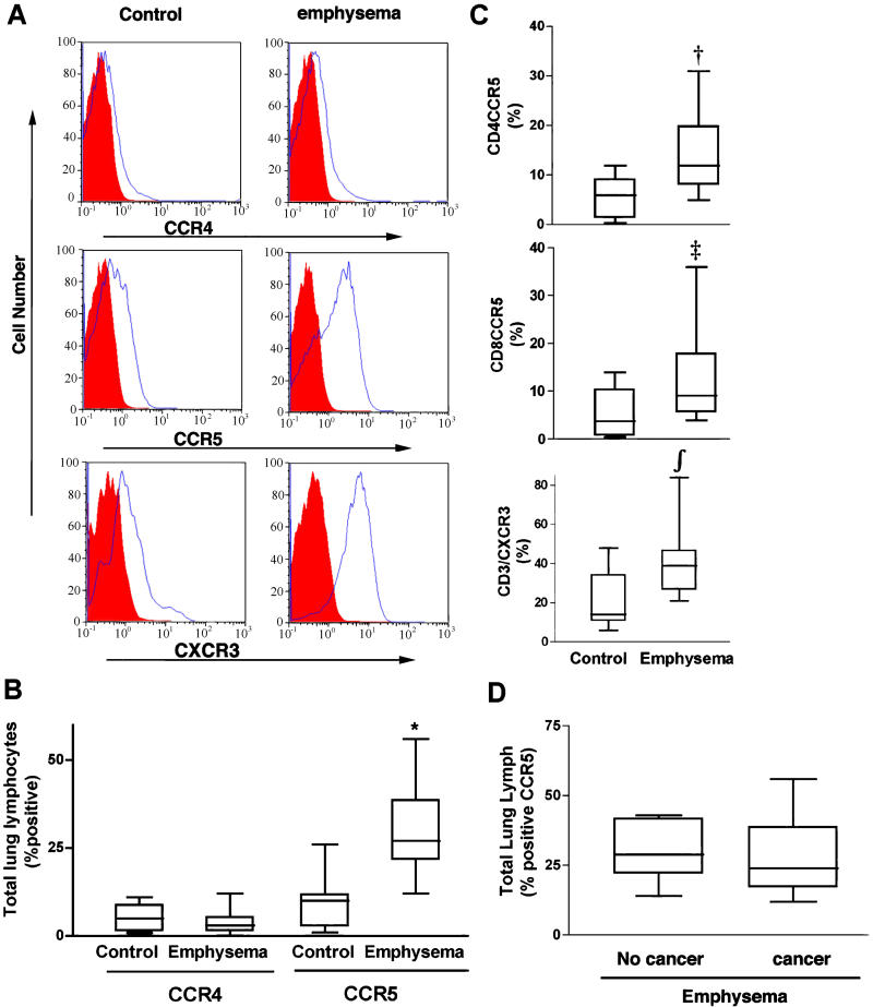
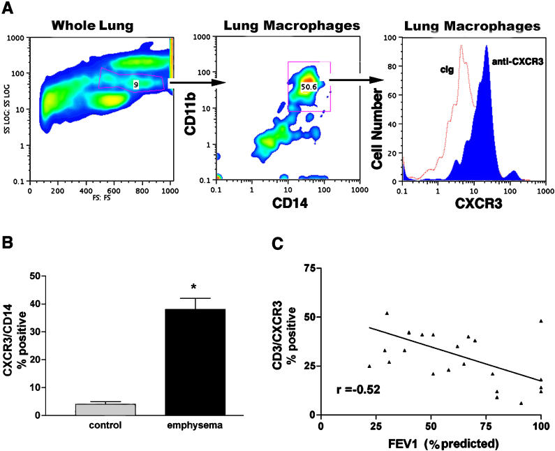
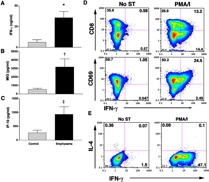
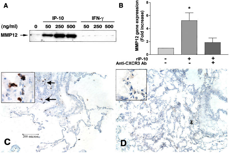
BACKGROUND: Chronic obstructive pulmonary disease and emphysema are a frequent result of long-term smoking, but the exact mechanisms, specifically which types of cells are associated with the lung destruction, are unclear. METHODS AND FINDINGS: We studied different subsets of lymphocytes taken from portions of human lungs removed surgically to find out which lymphocytes were the most frequent, which cell-surface markers these lymphocytes expressed, and whether the lymphocytes secreted any specific factors that could be associated with disease. We found that loss of lung function in patients with chronic obstructive pulmonary disease and emphysema was associated with a high percentage of CD4+ and CD8+ T lymphocytes that expressed chemokine receptors CCR5 and CXCR3 (both markers of T helper 1 cells), but not CCR3 or CCR4 (markers of T helper 2 cells). Lung lymphocytes in patients with chronic obstructive pulmonary disease and emphysema secrete more interferon gamma--often associated with T helper 1 cells--and interferon-inducible protein 10 and monokine induced by interferon, both of which bind to CXCR3 and are involved in attracting T helper 1 cells. In response to interferon-inducible protein 10 and monokine induced by interferon, but not interferon gamma, lung macrophages secreted macrophage metalloelastase (matrix metalloproteinase-12), a potent elastin-degrading enzyme that causes tissue destruction and which has been linked to emphysema. CONCLUSIONS: These data suggest that Th1 lymphoctytes in the lungs of people with smoking-related damage drive progression of emphysema through CXCR3 ligands, interferon-inducible protein 10, and monokine induced by interferon.
Overview
The claims and statistics of this paper haven’t been checked yet.
Run the check — free, no account needed. It reads the open-access text and checks the sources it cites.
Integrity signals
Registry
Cites
Statistics
Figures
Verification
Claims the paper makes
The claims and statistics of this paper haven’t been checked yet.
Run the check — free, no account needed. It reads the open-access text and checks the sources it cites.
Claims other papers make about this one
Each is checked against this paper’s own text: does it say what the citing paper says it does?
In the citing paper
“Specifically, macrophages, which can produce multiple proinflammatory cytokines and secrete MMPs, are the most abundant cell type in the BAL fluid of COPD patients, and increased numbers correlate with disease severity (85, 86, 104).”
From Aging and Lung Disease (2020) · cites this paper as [85]
In the citing paper
“Impaired phagocytosis and clearance of cellular debris by airway macrophages in COPD result in persistent inflammation (85, 104).”
From Aging and Lung Disease (2020) · cites this paper as [85]
Self-check
Percentages, sums and reported differences re-computed from the paper’s own numbers.
The self-check needs the paper’s full text.
Citations
Cites
Vestbo, J., Hurd, S. S., Agustí, A. G., Jones, P. W., Vogelmeier, C., Anzueto, A., Barnes, P. J., Fabbri, L. M., Martinez, F. J., Nishimura, M., et al. (2013) · American Journal of Respiratory and Critical Care Medicine · doi:10.1164/rccm.201204-0596pp · Cited by 15,983
PAUWELS, R. A., Buist, A. S., CALVERLEY, P. M. A., Jenkins, C. R., & Hurd, S. S. (2001) · American Journal of Respiratory and Critical Care Medicine · doi:10.1164/ajrccm.163.5.2101039 · Cited by 6,572
How Matrix Metalloproteinases Regulate Cell Behavior
Sternlicht, M. D., & Werb, Z. (2001) · Annual Review of Cell and Developmental Biology · doi:10.1146/annurev.cellbio.17.1.463 · Cited by 3,959
Zlotnik, A., & Yoshie, O. (2000) · Immunity · doi:10.1016/s1074-7613(00)80165-x · Cited by 3,923
Chronic Obstructive Pulmonary Disease
Flavin, K., Morkane, C. M., & Marsh, S. (2018) · Cambridge University Press eBooks · doi:10.1017/9781108233712.025 · Cited by 2,687
Anthonisen, N. R. (1994) · JAMA · doi:10.1001/jama.272.19.1497 · Cited by 1,960
Requirement for IL-13 Independently of IL-4 in Experimental Asthma
Grünig, G., Warnock, M. L., Wakil, A. E., Venkayya, R., Brombacher, F., Rennick, D. M., Sheppard, D., Mohrs, M., Donaldson, D. D., Locksley, R. M., et al. (1998) · Science · doi:10.1126/science.282.5397.2261 · Cited by 1,845
Self-citationRequirement for Macrophage Elastase for Cigarette Smoke-Induced Emphysema in Mice
Hautamaki, R. D., Kobayashi, D. K., Senior, R. M., & Shapiro, S. D. (1997) · Science · doi:10.1126/science.277.5334.2002 · Cited by 1,470
Chronic Obstructive Pulmonary Disease
Barnes, P. J. (2000) · New England Journal of Medicine · doi:10.1056/nejm200007273430407 · Cited by 1,078
Cole, K. E., Strick, C. A., Paradis, T. J., Ogborne, K. T., Loetscher, M., Gladue, R. P., Lin, W., Boyd, J. G., Moser, B., Wood, D. E., et al. (1998) · The Journal of Experimental Medicine · doi:10.1084/jem.187.12.2009 · Cited by 870
Research works printed but not linked: 3
- Kay, 1997, Ciba Found Symp, 206, 56
- Anthonisen, 1994, JAMA, 272, 1497, 10.1001/jama.1994.03520190043033 · DOI
- Fiocchi, 1985, Acta Chir Scand Suppl, 525, 11
Cited by
Sorted by citations
Items 301–400 of 484
Pathogenesis of chronic obstructive pulmonary disease (COPD)
Daheshia, M. (2005) · Clinical and Applied Immunology Reviews · doi:10.1016/j.cair.2005.09.003 · Cited by 13
Jeffery, P. K. (2005) · Expert Opinion on Investigational Drugs · doi:10.1517/13543784.14.5.619 · Cited by 13
Xie, W., Deng, L., Qian, R., Huang, X., Liu, W., & Tang, S. (2024) · Antioxidants · doi:10.3390/antiox13040420 · Cited by 13
Cells and mediators of chronic obstructive pulmonary disease
Barnes, P. J., & Cosio, M. G. (2006) · European Respiratory Society eBooks · doi:10.1183/1025448x.00038008 · Cited by 12
Pathological remodeling of distal lung matrix in end-stage cystic fibrosis patients
Pinezich, M. R., Tamargo, M. A., Fleischer, S., Reimer, J. A., Hudock, M. R., Hozain, A. E., Kaslow, S. R., Tipograf, Y., Soni, R. K., Gavaudan, O. P., et al. (2022) · Journal of Cystic Fibrosis · doi:10.1016/j.jcf.2022.04.016 · Cited by 12
Interferon gamma polymorphisms and their interaction with smoking are associated with lung function
He, J., Burkett, K. M., Connett, J. E., Anthonisen, N. R., Paré, P. D., & Sandford, A. J. (2006) · Human Genetics · doi:10.1007/s00439-006-0143-z · Cited by 12
Modulation of eotaxin-3 (CCL26) in alveolar type II epithelial cells
Abonyo, B. O., Lebby, K., Tonry, J. H., Ahmad, M., & Heiman, A. S. (2006) · Cytokine · doi:10.1016/j.cyto.2007.01.001 · Cited by 12
Shi, L., Hu, X., Wang, Z., Wu, M., & Zhang, J. (2015) · Inflammation Research · doi:10.1007/s00011-015-0816-y · Cited by 12
Hypoxia but not cigarette smoke modulates VEGF secretion from human T cells
Mikko, M., Wahlström, J., Grünewald, J., & Sköld, C. M. (2009) · Growth Factors · doi:10.3109/08977190903210863 · Cited by 12
SP-D Polymorphisms and the Risk of COPD
Shakoori, T. A., Sin, D. D., Bokhari, S. N. H., Ghafoor, F., & Shakoori, A. R. (2012) · Disease Markers · doi:10.1155/2012/802145 · Cited by 12
Valaciclovir for Epstein-Barr Virus Suppression in Moderate-to-Severe COPD
Linden, D. A., Parke, H. G., McKelvey, M. C., Einarsson, G. G., Lee, A. J., Fairley, D., Brown, V., Lundy, G., Campbell, C., Logan, D., et al. (2023) · CHEST Journal · doi:10.1016/j.chest.2023.03.040 · Cited by 11
Park, J., Kim, Y., Shin, I.-S., Kwon, O., Hong, J. M., Shin, N., Oh, S., Ha, U., Kim, J., & Ahn, K. (2016) · The Journal of Infectious Diseases · doi:10.1093/infdis/jiw278 · Cited by 11
Lipid let loose in pulmonary emphysema
Elias, J. A., & Lee, C. G. (2005) · Nature Medicine · doi:10.1038/nm0505-471 · Cited by 11
Wang, Z., Liang, W., Ma, C., Wang, J., Gao, X. M., & Wei, L. (2021) · Frontiers in Molecular Biosciences · doi:10.3389/fmolb.2021.663987 · Cited by 10
Pu, J., Xu, J., Chen, L., Zhou, H., Cao, W., Hao, B., Li, N., Wu, J., Zheng, J., Hong, W., et al. (2021) · Toxicology Letters · doi:10.1016/j.toxlet.2021.05.010 · Cited by 10
Ma, X., Yamaguchi, A., Maeshige, N., Uemura, M., Noguchi, H., Kondo, H., & Fujino, H. (2022) · Journal of Medical Ultrasonics · doi:10.1007/s10396-022-01189-4 · Cited by 10
Immunological pathways in virus‐induced COPD exacerbations: a role for IL‐15
Zdrenghea, M., Mallia, P., & Johnston, S. L. (2012) · European Journal of Clinical Investigation · doi:10.1111/j.1365-2362.2012.02672.x · Cited by 10
Autoantibody profiles and their association with blood eosinophils in asthma and COPD
Tamai, K., Yoshimatsu, H., Saito, T., Matsuoka, H., Okada, N., Koma, Y., Otsuka, A., Oda, N., Inoue, S., Kume, S., et al. (2016) · Allergology International · doi:10.1016/j.alit.2016.08.005 · Cited by 10
Nie, L., Liu, Z.-J., Zhou, W., Xiang, R., Xiao, Y. P., Lu, B., Pang, B., & Gao, J. (2010) · Acta Pharmacologica Sinica · doi:10.1038/aps.2009.192 · Cited by 10
Barnes, P. J., & Rennard, S. I. (2009) · Elsevier eBooks · doi:10.1016/b978-0-12-374001-4.00034-1 · Cited by 10
Ren, X., Wu, J., Levin, D. B., Santos, S. C., de Faria, R. L., Zhang, M., & Lin, F. (2019) · Annals of the New York Academy of Sciences · doi:10.1111/nyas.14029 · Cited by 10
Gutiérrez-Romero, K. J., Falfán‐Valencia, R., Ramírez‐Venegas, A., Hernández-Zenteno, R. D. J., Flores-Trujillo, F., Sansores-Martínez, R. H., Ramos-Martı́nez, E., & Pérez-Rubio, G. (2024) · Frontiers in Immunology · doi:10.3389/fimmu.2024.1411408 · Cited by 9
Understanding COPD Etiology, Pathophysiology, and Definition
Curtis, J. L. (2023) · Respiratory Care · doi:10.4187/respcare.10873 · Cited by 20
Stress Responses Affecting Homeostasis of the Alveolar Capillary Unit
Tuder, R. M., & Yoshida, T. (2011) · Proceedings of the American Thoracic Society · doi:10.1513/pats.201103-029mw · Cited by 9
Pathogenesis and pathophysiology of COPD
Yoo, C. G. (2009) · The Korean Journal of Internal Medicine · Cited by 8
Non-infectious pulmonary disorders in HIV
Thao, C., Shorr, A. F., & Woods, C. J. (2017) · Expert Review of Respiratory Medicine · doi:10.1080/17476348.2017.1288101 · Cited by 9
Triptolide protects against TGF‑β1‑induced pulmonary fibrosis by regulating FAK/calpain signaling
Zhang, P., Liu, J., & Zong, R. (2019) · Experimental and Therapeutic Medicine · doi:10.3892/etm.2019.8127 · Cited by 9
Pérez-Rial, S., del Puerto‐Nevado, L., González‐Mangado, N., & Peces‐Barba, G. (2011) · Molecular Imaging · doi:10.2310/7290.2011.00010 · Cited by 8
Grumelli, S. (2016) · Current Respiratory Medicine Reviews · doi:10.2174/1573398x12999160506104327 · Cited by 8
Self-citationUzeloto, J. S., de Toledo, A. C., Silva, B. S. D. A., Golim, M. D. A., Braz, A. M. M., de Lima, F. F., Grigoletto, I., & Ramos, E. M. C. (2020) · International Journal of COPD · doi:10.2147/copd.s268955 · Cited by 8
The Lung Elastin Matrix Undergoes Rapid Degradation Upon Adult Loss of Hox5 Function
Li, M., Marty‐Santos, L., van Ginkel, P. R., McDermott, A. E., Rasky, A. J., Lukacs, N. W., & Wellik, D. M. (2021) · Frontiers in Cell and Developmental Biology · doi:10.3389/fcell.2021.767454 · Cited by 8
Cosío, B. G., & Agustí, A. G. N. (2005) · Archivos de Bronconeumología · doi:10.1016/s0300-2896(05)70762-6 · Cited by 8
Wang, X., Rojas, J., Zhang, D., Nakajima, T., Walker, K. H., Peh, H. Y., Li, Y., Fucci, Q., Tesfaigzi, Y., & Owen, C. A. (2020) · Mucosal Immunology · doi:10.1038/s41385-020-0325-3 · Cited by 8
Chen, K.-Y., Sun, W.-L., Wu, S., Feng, P., Lin, C., Chen, T.-T., Lu, Y., Ho, S., Chen, Y.-H., & Lee, K. (2024) · Cells · doi:10.3390/cells13100878 · Cited by 7
Dost, A. F. M., Balážová, K., Casellas, C. P., Rooijen, L. V., Epskamp, W., van Son, G. J. F., van de Wetering, W. J., López‐Iglesias, C., Begthel, H., Peters, P. J., et al. (2026) · The EMBO Journal · doi:10.1038/s44318-026-00774-4 · Cited by 7
Efectores celulares de la respuesta inflamatoria en la enfermedad pulmonar obstructiva crónica
Rafael Silva, O., Montes, J. F., García‐Valero, J., & Olloquequi, J. (2015) · Revista médica de Chile · doi:10.4067/s0034-98872015000900009 · Cited by 7
Transcription Factor T-bet Attenuates the Development of Elastase-induced Emphysema in Mice
Hayashi, S., Matsuno, Y., Tsunoda, Y., Sakurai, H., Kiwamoto, T., Morishima, Y., Ishii, Y., Yoh, K., Takahashi, S., & Hizawa, N. (2019) · American Journal of Respiratory Cell and Molecular Biology · doi:10.1165/rcmb.2018-0109oc · Cited by 7
Chang, C.-C., Chen, C., Chiu, H., Dai, S.-X., Liu, M., & Yang, C. (2011) · Inhalation Toxicology · doi:10.3109/08958378.2011.598965 · Cited by 8
Regenerative therapeutics for chronic obstructive pulmonary disease
van der Koog, L., Showell, H., Nugraha, D. F., Lehmann, M., Conlon, T. M., Yildirim, A. Ö., Fuentes‐Mateos, R., Baarsma, H. A., Ng-Blichfeldt, J.-P., Melgert, B. N., et al. (2026) · Pharmacological Reviews · doi:10.1016/j.pharmr.2026.100124 · Cited by 8
Blocking IL ‐23 Signaling Mitigates Cigarette Smoke‐Induced Murine Emphysema
Tian, X., Wang, S., Zhang, C., Prakash, Y. S., & Vassallo, R. (2024) · Environmental Toxicology · doi:10.1002/tox.24405 · Cited by 6
Chen, H., Wang, D., & Wang, X. (2010) · Journal of Epithelial Biology & Pharmacology · doi:10.2174/1875044301003010026 · Cited by 6
Dhamodharan, P., & Arumugam, M. (2020) · COPD Journal of Chronic Obstructive Pulmonary Disease · doi:10.1080/15412555.2020.1793314 · Cited by 6
COPD: Immunopathogenesis and Immunological Markers
Sng, J. J., & Thomas, P. S. (2014) · Advances in Research · doi:10.9734/air/2015/12320 · Cited by 6
HIV Prevalence and Morbidity in Older Inpatients in a High HIV Prevalence Setting
Naidoo, V. A., Martinson, N. A., Moodley, P., Joyimbana, W., Mothlaoleng, K., Abraham, P., Otwombe, K. N., & Variava, E. (2019) · AIDS Research and Human Retroviruses · doi:10.1089/aid.2019.0137 · Cited by 6
Airway Inflammation Biomarker for Precise Management of Neutrophil-Predominant COPD
Liang, X., Liu, T., Zhang, Z., & Yu, Z. (2020) · Methods in molecular biology · doi:10.1007/978-1-0716-0904-0_16 · Cited by 6
Chronic Obstructive Pulmonary Disease
Bracke, K. R., & Brusselle, G. (2015) · Elsevier eBooks · doi:10.1016/b978-0-12-415847-4.00097-5 · Cited by 6
Bordag, N., Jandl, K., Syarif, A. H., Gindlhuber, J., Schnoegl, D., Mutgan, A. C., Foris, V., Höetzenecker, K., Boehm, P. M., Breyer‐Kohansal, R., et al. (2025) · iScience · doi:10.1016/j.isci.2025.112966 · Cited by 7
Clinical relevance of miR-423-5p levels in chronic obstructive pulmonary disease patients
Zhang, X., Shi, Q., Xiong, L., Shi, S., Li, Y., Wang, Y., & Zhang, M. (2022) · Clinics · doi:10.1016/j.clinsp.2022.100102 · Cited by 6
Sputum Biomarkers in Wood and Tobacco Smoke Etiotypes of Chronic Obstructive Pulmonary Disease
Montoya, Á. M. G., Torres-Duque, C. A., Giraldo‐Cadavid, L. F., Laucho-Contreras, M. E., González-Flórez, A., Santos, A. M., Tuta-Quintero, E., Celli, B. R., & González-García, M. (2023) · International Journal of COPD · doi:10.2147/copd.s439064 · Cited by 5
Erice, P. A., Huang, X., Seasock, M. J., Robertson, M. J., Tung, H.-Y., Perez-Negron, M. A., Lotlikar, S. L., Corry, D. B., Kheradmand, F., & Rodríguez, A. (2024) · eLife · doi:10.7554/elife.92879 · Cited by 5
Self-citationMacrophages in Lung Repair and Fibrosis
de Sá, Y. A. P. J., Creyns, B., Hogaboam, C. M., Parks, W. C., & Hohmann, M. S. (2024) · Results and problems in cell differentiation · doi:10.1007/978-3-031-65944-7_10 · Cited by 5
Xiang, Y., Kogel, U., Gebel, S., Peck, M. J., Peitsch, M. C., Akmaev, V. R., & Hoeng, J. (2014) · Gene Regulation and Systems Biology · doi:10.4137/grsb.s13140 · Cited by 5
Duan, M., Huang, Y., Zhong, X., & Tang, H. (2013) · Inflammation · doi:10.1007/s10753-013-9793-x · Cited by 5
Endogenous Modifiers of Cigarette Smoke Exposure within the Lung
Houghton, A. M. (2012) · Proceedings of the American Thoracic Society · doi:10.1513/pats.201108-046ms · Cited by 5
At the Checkpoint: Lung CD8+ T Cells, Respiratory Viruses, and Chronic Obstructive Pulmonary Disease
Curtis, J. L. (2016) · American Journal of Respiratory and Critical Care Medicine · doi:10.1164/rccm.201511-2122ed · Cited by 5
Кириллова, Н. А., Deyev, I. A., Kremer, Y. E., Ogorodova, L. M., & Chernogoryuk, G. E. (2011) · Bulletin of Siberian Medicine · doi:10.20538/1682-0363-2011-1-48-54 · Cited by 5
Li, L., Liu, Y., Chiu, C., Jin, Y., Zhou, W., Peng, M., Chen, L., Sun, Q., & Gao, J. (2021) · Inflammation · doi:10.1007/s10753-020-01393-9 · Cited by 5
Williams, A. E. (2021) · Elsevier eBooks · doi:10.1016/b978-0-08-102723-3.00252-3 · Cited by 5
Elwakil, M. M. A., Ni, J., Ramsay-Burrough, S., Hurst, P. J., McClellan, R. L., Khasnavis, S. R., Jia, Y., Masud, S. R., Blake, T. R., Sallets, A., et al. (2025) · Journal of the American Chemical Society · doi:10.1021/jacs.5c06654 · Cited by 5
Role of Airway Epithelium-Origin Chemokines and their Receptors in COPD
Chen, H., Wang, D., & Wang, X. (2010) · Journal of Epithelial Biology & Pharmacology · Cited by 4
Zhang, Y., Li, X., Zhou, Y., Lu, X., Xiao, M., Guo, J., Zhang, J., & Chen, G. (2020) · Medicine · doi:10.1097/md.0000000000023442 · Cited by 4
Li, D., Chen, L., Miao, S.-Y., Zhou, M., Wu, J., Sun, S., Liu, L., Qi, C., & Xiong, X. (2022) · International Journal of COPD · doi:10.2147/copd.s371801 · Cited by 4
Inflammation and Lung Cancer: The Relationship to Chronic Obstructive Pulmonary Disease
Houghton, A. M., & Shapiro, S. D. (2015) · Springer eBooks · doi:10.1007/978-1-4939-2724-1_1 · Cited by 4
Cigarette Smoking and Lower Respiratory Tract Infection
Fernando Saldías, P., & Orlando Díaz, P. (2011) · InTech eBooks · doi:10.5772/17652 · Cited by 4
Some Features of the Key Phenotypes of Allergic Rhinitis Among Children in a Metropolis
Tataurschikova, N. S., & Berezhanskiy, P. V. (2021) · Springer geography · doi:10.1007/978-3-030-75285-9_19 · Cited by 4
Mechanisms in COPD compared with asthma
Barnes, P. J. (2008) · Breathe · doi:10.1183/18106838/breathe.5.2.134 · Cited by 4
Yin, C., Jiacui, S., Hasegawa, T., Yao, L., Kondo, T., & Li, H. (2025) · Frontiers in Immunology · doi:10.3389/fimmu.2025.1576849 · Cited by 3
Lung NR3C1+ and CXCR6high T cells distinguish immunopathogenesis of human emphysema
Zhang, Y., Sauler, M., Corry, D. B., Ochsner, S. A., Perusich, S., Song, L., Malo, J., Estépar, R. S. J., Polverino, F., & Kheradmand, F. (2025) · Communications Biology · doi:10.1038/s42003-025-08698-1 · Cited by 4
Self-citationMechanisms and mediators of disease
Devulder, J. V., & Donnelly, L. (2024) · European Respiratory Society eBooks · doi:10.1183/2312508x.10006423 · Cited by 3
Inflammatory Mechanisms in Chronic Obstructive Pulmonary Disease
Barnes, P. J. (2017) · Wiley-VCH eBooks · doi:10.1002/9783527692156.ch45 · Cited by 3
Chronic obstructive pulmonary disease and diseases of the airways
Timens, W., Sietsma, H., & Wright, J. L. (2013) · Cambridge University Press eBooks · doi:10.1017/cbo9781139018760.020 · Cited by 3
Matrix Degrading Proteinases in COPD and Asthma
Shapiro, S. D. (2009) · Elsevier eBooks · doi:10.1016/b978-0-12-374001-4.00028-6 · Cited by 3
The Multiple Components of COPD
Fabbri, L. M., Luppi, F., Beghé, B., & Rabe, K. F. (2010) · Humana Press eBooks · doi:10.1007/978-1-59745-357-8_1 · Cited by 3
Braun, A. C., Ernst, H., Schaudien, D., Hoymann, H.-G., & Rittinghausen, S. (2012) · Elsevier eBooks · doi:10.1016/b978-0-12-382008-2.00012-x · Cited by 3
Some pathogenic aspects of pulmonary emphysema in COPD patients
Averyanov, A. V., Samsonova, M. V., Chernyaev, A. L., Чучалин, А. Г., Polivanova, A. E., Николаев, Г. В., Perepechin, V. I., Polivanov, G. E., & Dvorakovskaya, I. (2008) · PULMONOLOGIYA · doi:10.18093/0869-0189-2008-0-3-48-53 · Cited by 3
Emphysema and Diseases of Large Airways
Green, L. (2008) · Elsevier eBooks · doi:10.1016/b978-0-443-06741-9.50026-4 · Cited by 3
Self-citationEmphysema and Diseases of Large Airways
Litzky, L. A., & Green, L. (2018) · Elsevier eBooks · doi:10.1016/b978-0-323-39308-9.00020-0 · Cited by 3
Self-citationEpithelial Rho GTPases and the Transepithelial Migration of Lymphocytes
Porter, J. C. (2008) · Methods in enzymology on CD-ROM/Methods in enzymology · doi:10.1016/s0076-6879(07)00416-8 · Cited by 3
Deng, L., Sun, B., Xia, J., Zhuo, W., Jiang, M., Yin, S., & Yang, F. (2025) · Scientific Reports · doi:10.1038/s41598-025-21752-6 · Cited by 4
Li, J., Li, G., Liu, J., Li, L., Zhou, H., Fei, X., Wen, Y., & Zhao, D. (2025) · International Journal of COPD · doi:10.2147/copd.s548160 · Cited by 2
Divergent roles of serum CXCL9 as a biomarker in ILD and COPD: a comparative study
Yin, C., Kang, X., Zhang, Y., Song, J., Hasegawa, T., Yao, L., Hu, Y., & Li, H. (2026) · Frontiers in Pharmacology · doi:10.3389/fphar.2026.1730688 · Cited by 2
Effects of Root of Curcumin longa on LPS-induced Lung Injury
Ji-Seok, Yang, S.-Y., Kim, M., Namgung, U., & Park, Y. (2013) · Cited by 2
Aging in COPD and Idiopathic Pulmonary Fibrosis
Sánchez, C. G. (2015) · Springer eBooks · doi:10.1007/978-3-319-23246-1_15 · Cited by 2
MacNee, W., Vestbo, J., & Agustí, A. G. N. (2015) · Elsevier eBooks · doi:10.1016/b978-1-4557-3383-5.00043-9 · Cited by 2
Lee, J. (2017) · Springer eBooks · doi:10.1007/978-3-662-47178-4_4 · Cited by 2
Roll of Toll‐like Receptor 9 in Mouse Lung Inflammation in Response to Chicken Barn Air
Schneberger, D., Aulakh, G., Channabasappa, S., Caldwell, S., & Singh, B. (2010) · The FASEB Journal · doi:10.1096/fasebj.24.1_supplement.753.7 · Cited by 1
CHEMOKINES, CXC | CXCL10 (IP-10)
Kheradmand, F., & Corry, D. B. (2006) · Elsevier eBooks · doi:10.1016/b0-12-370879-6/00472-5 · Cited by 2
Self-citationMolecular mechanisms mediating development of pulmonary cachexia in COPD
Basic, V. (2014) · KTH Publication Database DiVA (KTH Royal Institute of Technology) · Cited by 2
Inflammatory and Immune Mechanisms in COPD
Clarke, S. E., & Barnes, P. J. (2021) · Elsevier eBooks · doi:10.1016/b978-0-08-102723-3.00221-3 · Cited by 2
Natural product-based compounds for chronic respiratory disorders
Bisht, D., Joshi, D. C., Bisht, M. S., Joshi, N., Azizov, S., Lalhlenmawia, H., Kumar, D., Dua, K., Shetty, S. R., & Suares, D. (2024) · Elsevier eBooks · doi:10.1016/b978-0-443-27345-2.00010-2 · Cited by 1
Nyaupane, U., Johannson, K. A., Kelly, M. M., & Kai, F. (2025) · Experimental Lung Research · doi:10.1080/01902148.2025.2582970 · Cited by 1
TIGIT/SHIP-1/RelB regulating Th1 inflammation in smoking induced COPD
Ke, J., Lei, S., He, Z., Li, Q., Huang, H., Huang, S., Lin, S., Li, Y., Lu, L., Wu, Y., et al. (2025) · Respiratory Research · doi:10.1186/s12931-025-03400-9 · Cited by 1
Kadushkin, A. G., Шман, Т. В., Novikov, V. P., Ибрагимова, Ж. А., & Taganovich, A. D. (2013) · PULMONOLOGIYA · doi:10.18093/0869-0189-2013-0-3-25-30 · Cited by 1
Diverse Activities for Proteinases in the Pathogenesis of Chronic Obstructive Pulmonary Disease
Kelly, E., & Caroline, A. (2012) · InTech eBooks · doi:10.5772/30305 · Cited by 1
SIRT1 and Inflammaging in Chronic Obstructive Pulmonary Disease
Hwang, J., Sundar, I. K., Yao, H., & Rahman, I. (2013) · Elsevier eBooks · doi:10.1016/b978-0-12-397803-5.00015-0 · Cited by 1
PPARγ in emphysema: blunts the damage and triggers repair?
Kelly, N. J., & Shapiro, S. D. (2014) · Journal of Clinical Investigation · doi:10.1172/jci74417 · Cited by 1
La bronchopneumopathie chronique obstructive : une maladie auto-immune ?
Guennec, L. L., Pestre, V., & Mouthon, L. (2012) · Revue des Maladies Respiratoires · doi:10.1016/j.rmr.2012.02.001 · Cited by 1
Popper, H. (2016) · Springer eBooks · doi:10.1007/978-3-662-50491-8_7 · Cited by 1
Togo, S., Namba, Y., & Takahashi, K. (2016) · Respiratory disease series · doi:10.1007/978-981-10-0839-9_18 · Cited by 1
Respiratory Disorders of the Immune System and Their Pharmacological Treatment
Mathias, C. B. (2019) · Springer eBooks · doi:10.1007/978-3-030-19922-7_4 · Cited by 1
Metadata
- Authors
- Grumelli, S., Corry, D. B., Song, L.-Z., Song, L., Green, L., Huh, J., Hacken, J., Espada, R., Bag, R., Lewis, D. E., & Kheradmand, F.
- Affiliations
- Baylor College of Medicine (US)
- Michael E. DeBakey VA Medical Center (US)
- Corresponding author
- Kheradmand, Farrah
- Venue
- PLoS Medicine
- Publisher
- Public Library of Science (PLoS)
- Published
- 2004-10-19
- Volume
- 1
- Issue
- 1
- Pages
- e8
- ISSN
- 1549-1277 (ISSN-L)
- 1549-1676
- PMC
- PMC523885
- OpenAlex
- W2108679807
- Cited by
- 484
- References
- 59
- Citations by year
- 2026: 6 · 2025: 12 · 2024: 21 · 2023: 12 · 2022: 11 · 2021: 21 · 2020: 14 · 2019: 22 · 2018: 16 · 2017: 25 · 2016: 21 · 2015: 28 · 2014: 15 · 2013: 29 · 2012: 31
- FWCI
- 19.11
- Type
- journal article
- Language
- en
- Open access
- gold
- doi.org
- Locations
- PLoS Medicine (publishedVersion, cc-by)
- PubMed Central (publishedVersion, cc-by)
- PubMed (publishedVersion)
- DOAJ (DOAJ: Directory of Open Access Journals) (submittedVersion, cc-by-sa)
- Funders
- U.S. Public Health Service (69585, 64061, 72419)
- National Institute of Allergy and Infectious Diseases (P30AI036211, AI 36211)
- Topics
- Health Sciences › Medicine › Pulmonary and Respiratory Medicine › Chronic Obstructive Pulmonary Disease (COPD) Research
- Health Sciences › Medicine › Pulmonary and Respiratory Medicine › Asthma and respiratory diseases
- Health Sciences › Medicine › Surgery › Transplantation: Methods and Outcomes
- Keywords
- chronic obstructive pulmonary disease, emphysema, Th1 cells, CXCR3, CCR5, interferon-gamma, CXCL10, macrophage metalloelastase, matrix metalloproteinase-12, elastin degradation, smoking, lung lymphocytes, chemokine receptors
- MeSH terms
- Aged, Female, Humans, Male, Middle Aged, Pulmonary Emphysema, Smoking, Tomography, X-Ray Computed, Lymphocyte Subsets, Cytokines, Macrophages, Alveolar, Th1 Cells, Disease Progression, Receptors, Chemokine, Chemokines, CXC, Pulmonary Disease, Chronic Obstructive, Chemokine CXCL10, Receptors, CXCR3
About these checks
Registry, retraction, Cites and statistics results are computed, not judged, with one exception: a printed reference that no index lists is read once by a model before it counts as a citation error. Claims are extracted by a model and kept only when their quote appears verbatim in the text; each cited source is then checked, with a model’s help, for whether it says what the claim relies on. Those verdicts can be wrong — read the claim’s quote and the cited source before relying on one.
Statistics and arithmetic are recomputed, not judged.
Confidence bands are shown only for fields calibrated on our annotated test set. None is calibrated yet, so no band is shown.
What counts as a claim ›How we measure confidence ›How we check this ›
Are you an author, or do you see a problem with this page? Request a review
