

The DOI matches its Crossref record; 83 of the 83 references it cites were found in a registry or an index.
Experimental ‘Jet Lag’ Inhibits Adult Neurogenesis and Produces Long-Term Cognitive Deficits in Female Hamsters
Open accessAbstract
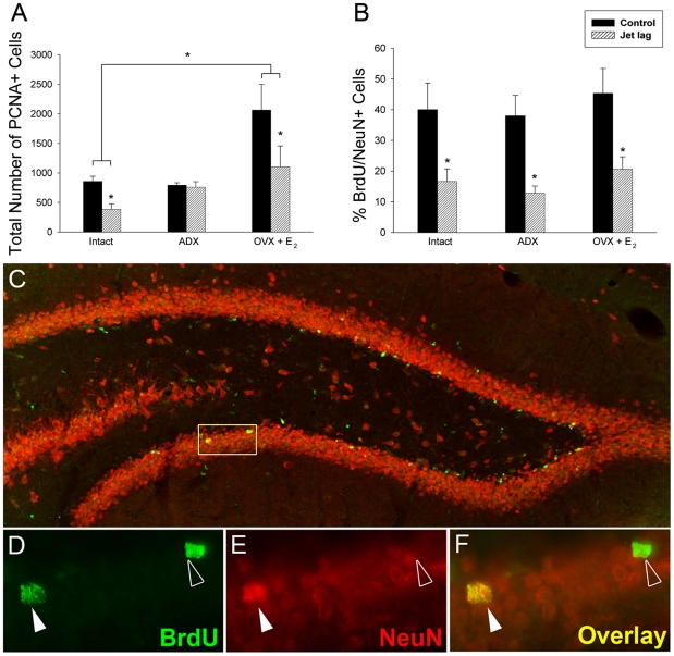
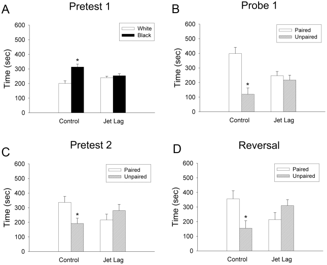
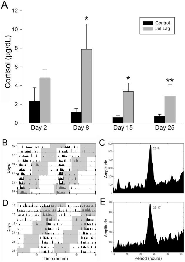
BACKGROUND: Circadian disruptions through frequent transmeridian travel, rotating shift work, and poor sleep hygiene are associated with an array of physical and mental health maladies, including marked deficits in human cognitive function. Despite anecdotal and correlational reports suggesting a negative impact of circadian disruptions on brain function, this possibility has not been experimentally examined. METHODOLOGY/PRINCIPAL FINDINGS: In the present study, we investigated whether experimental 'jet lag' (i.e., phase advances of the light:dark cycle) negatively impacts learning and memory and whether any deficits observed are associated with reductions in hippocampal cell proliferation and neurogenesis. Because insults to circadian timing alter circulating glucocorticoid and sex steroid concentrations, both of which influence neurogenesis and learning/memory, we assessed the contribution of these endocrine factors to any observed alterations. Circadian disruption resulted in pronounced deficits in learning and memory paralleled by marked reductions in hippocampal cell proliferation and neurogenesis. Significantly, deficits in hippocampal-dependent learning and memory were not only seen during the period of the circadian disruption, but also persisted well after the cessation of jet lag, suggesting long-lasting negative consequences on brain function. CONCLUSIONS/SIGNIFICANCE: Together, these findings support the view that circadian disruptions suppress hippocampal neurogenesis via a glucocorticoid-independent mechanism, imposing pronounced and persistent impairments on learning and memory.
Overview
The claims and statistics of this paper haven’t been checked yet.
Run the check — free, no account needed. It reads the open-access text and checks the sources it cites.
Integrity signals
Registry
Cites
Statistics
Figures
Verification
Claims the paper makes
The claims and statistics of this paper haven’t been checked yet.
Run the check — free, no account needed. It reads the open-access text and checks the sources it cites.
Claims other papers make about this one
Each is checked against this paper’s own text: does it say what the citing paper says it does?
In the citing paper
“When these disruptions are acute (e.g., in the case of a single ‘jet lag’), adults can achieve internal realignment with limited negative impact to health4.”
From Maternal and Early-Life Circadian Disruption Have Long-Lasting Negative Consequences on Offspring Development and Adult Behavior in Mice (2017) · cites this paper as [4]
Self-check
Percentages, sums and reported differences re-computed from the paper’s own numbers.
The self-check needs the paper’s full text.
Citations
Cites
Coordination of circadian timing in mammals
Reppert, S. M., & Weaver, D. R. (2002) · Nature · doi:10.1038/nature00965 · Cited by 4,379
The efficiency of systematic sampling in stereology and its prediction*
Gundersen, H. J. G., & Jensen, E. B. V. (1987) · Journal of Microscopy · doi:10.1111/j.1365-2818.1987.tb02837.x · Cited by 3,519
Social Jetlag: Misalignment of Biological and Social Time
Wittmann, M., Dinich, J., Merrow, M., & Roenneberg, T. (2006) · Chronobiology International · doi:10.1080/07420520500545979 · Cited by 2,625
Dibner, C., Schibler, U., & Albrecht, U. (2010) · Annual Review of Physiology · doi:10.1146/annurev-physiol-021909-135821 · Cited by 2,421
Learning enhances adult neurogenesis in the hippocampal formation
Gould, E., Beylin, A., Tanapat, P., Reeves, A., & Shors, T. J. (1999) · Nature Neuroscience · doi:10.1038/6365 · Cited by 2,157
Loss of a circadian adrenal corticosterone rhythm following suprachiasmatic lesions in the rat
Moore, R. Y., & Eichler, V. B. (1972) · Brain Research · doi:10.1016/0006-8993(72)90054-6 · Cited by 2,135
Neurogenesis in the adult is involved in the formation of trace memories
Shors, T. J., Miesegaes, G. R., Beylin, A. V., Zhao, M., Rydel, T. A., & Gould, E. (2001) · Nature · doi:10.1038/35066584 · Cited by 2,014
Resetting central and peripheral circadian oscillators in transgenic rats
Yamazaki, S., Numano, R., Abe, M., Hida, A., Takahashi, R.-I., Ueda, M., Block, G. D., Sakaki, Y., Menaker, M., & Tei, H. (2000) · Science · doi:10.1126/science.288.5466.682 · Cited by 1,907
A Functional Role for Adult Hippocampal Neurogenesis in Spatial Pattern Separation
Clelland, C. D., Choi, M., Romberg, C., Clemenson, G. D., Fragniere, A., Tyers, P., Jessberger, S., Saksida, L. M., Barker, R. A., Gage, F. H., et al. (2009) · Science · doi:10.1126/science.1173215 · Cited by 1,642
Walker, M. P., & Stickgold, R. (2006) · Annual Review of Psychology · doi:10.1146/annurev.psych.56.091103.070307 · Cited by 1,075 · cited 1×
“It is noteworthy that sleep deprivation has also been shown to disrupt neurogenesis in both a glucocorticoid-dependent and independent manner [69,70,81].”
Background
Research works printed but not linked: 1
- M Orsini, 1961, The external vaginal phenomena characterizing the stages of the estrous cycle, pregnancy, pseudopregnancy, lactation and anestrous hamster, Mesocricetus auratus Waterhouse., Proceedings of Animal Care Panel, 14, 193
Cited by
Sorted by citations
Light as a central modulator of circadian rhythms, sleep and affect
LeGates, T. A., Fernandez, D. C., & Hattar, S. (2014) · Nature Reviews Neuroscience · doi:10.1038/nrn3743 · Cited by 1,001
Mechanisms linking circadian clocks, sleep, and neurodegeneration
Musiek, E. S., & Holtzman, D. M. (2016) · Science · doi:10.1126/science.aah4968 · Cited by 878
Circadian clock control of endocrine factors
Gamble, K. L., Berry, R., Frank, S. J., & Young, M. E. (2014) · Nature Reviews Endocrinology · doi:10.1038/nrendo.2014.78 · Cited by 539
How might circadian rhythms control mood? Let me count the ways
McClung, C. A. (2013) · Biological Psychiatry · doi:10.1016/j.biopsych.2013.02.019 · Cited by 507
Circadian rest-activity pattern changes in aging and preclinical Alzheimer disease
Musiek, E. S., Bhimasani, M., Zangrilli, M. A., Morris, J. C., Holtzman, D. M., & Ju, Y.-E. S. (2018) · JAMA Neurology · doi:10.1001/jamaneurol.2017.4719 · Cited by 465
Metadata
- Authors
- Gibson, E. M., Wang, C., Tjho, S., Khattar, N., & Kriegsfeld, L. J.
- Affiliations
- University of California, Berkeley (US)
- Corresponding author
- Kriegsfeld, Lance J.
- Venue
- PLoS ONE
- Publisher
- Public Library of Science (PLoS)
- Published
- 2010-12-01
- Volume
- 5
- Issue
- 12
- Pages
- e15267
- ISSN
- 1932-6203 (ISSN-L)
- PMC
- PMC2995744
- OpenAlex
- W2116127629
- Cited by
- 196
- References
- 83
- Citations by year
- 2026: 2 · 2025: 6 · 2024: 8 · 2023: 17 · 2022: 12 · 2021: 12 · 2020: 17 · 2019: 10 · 2018: 18 · 2017: 16 · 2016: 10 · 2015: 17 · 2014: 20 · 2013: 15 · 2012: 10
- FWCI
- 3.14
- Type
- journal article
- Language
- en
- Open access
- gold
- doi.org
- Locations
- PLoS ONE (publishedVersion, cc-by)
- PubMed Central (publishedVersion, cc-by)
- PubMed (publishedVersion)
- DOAJ (DOAJ: Directory of Open Access Journals) (submittedVersion, cc-by-sa)
- eScholarship (California Digital Library) (submittedVersion)
- eScholarship (California Digital Library) (submittedVersion, cc-by)
- Figshare (submittedVersion, cc-by)
- Funders
- Northwestern University
- National Institutes of Health (050470, HD 050470)
- Eunice Kennedy Shriver National Institute of Child Health and Human Development (R01HD050470, HD 050470)
- Topics
- Life Sciences › Neuroscience › Developmental Neuroscience › Neurogenesis and neuroplasticity mechanisms
- Life Sciences › Neuroscience › Endocrine and Autonomic Systems › Circadian rhythm and melatonin
- Life Sciences › Neuroscience › Cognitive Neuroscience › Sleep and Wakefulness Research
- Keywords
- jet lag, circadian rhythm disruption, adult neurogenesis, hippocampal cell proliferation, learning and memory impairment, glucocorticoids, sex steroids, female hamsters, long-term cognitive deficits
- MeSH terms
- Animals, Brain, Cognition Disorders, Disease Models, Animal, Female, Glucocorticoids, Cricetinae, Hippocampus, Hypothalamo-Hypophyseal System, Memory, Neurons, Sleep, Jet Lag Syndrome, Cell Proliferation
About these checks
Registry, retraction, Cites and statistics results are computed, not judged, with one exception: a printed reference that no index lists is read once by a model before it counts as a citation error. Claims are extracted by a model and kept only when their quote appears verbatim in the text; each cited source is then checked, with a model’s help, for whether it says what the claim relies on. Those verdicts can be wrong — read the claim’s quote and the cited source before relying on one.
Statistics and arithmetic are recomputed, not judged.
Confidence bands are shown only for fields calibrated on our annotated test set. None is calibrated yet, so no band is shown.
What counts as a claim ›How we measure confidence ›How we check this ›
Are you an author, or do you see a problem with this page? Request a review
