

The DOI matches its Crossref record; 35 of the 35 references it cites were found in a registry or an index.
Cholinergic Interneurons Mediate Fast VGluT3-Dependent Glutamatergic Transmission in the Striatum
Open accessAbstract
The neurotransmitter glutamate is released by excitatory projection neurons throughout the brain. However, non-glutamatergic cells, including cholinergic and monoaminergic neurons, express markers that suggest that they are also capable of vesicular glutamate release. Striatal cholinergic interneurons (CINs) express the Type-3 vesicular glutamate transporter (VGluT3), although whether they form functional glutamatergic synapses is unclear. To examine this possibility, we utilized mice expressing Cre-recombinase under control of the endogenous choline acetyltransferase locus and conditionally expressed light-activated Channelrhodopsin2 in CINs. Optical stimulation evoked action potentials in CINs and produced postsynaptic responses in medium spiny neurons that were blocked by glutamate receptor antagonists. CIN-mediated glutamatergic responses exhibited a large contribution of NMDA-type glutamate receptors, distinguishing them from corticostriatal inputs. CIN-mediated glutamatergic responses were insensitive to antagonists of acetylcholine receptors and were not seen in mice lacking VGluT3. Our results indicate that CINs are capable of mediating fast glutamatergic transmission, suggesting a new role for these cells in regulating striatal activity.
Overview
The claims and statistics of this paper haven’t been checked yet.
Run the check — free, no account needed. It reads the open-access text and checks the sources it cites.
Integrity signals
Registry
Cites
Statistics
Figures
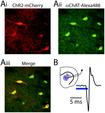
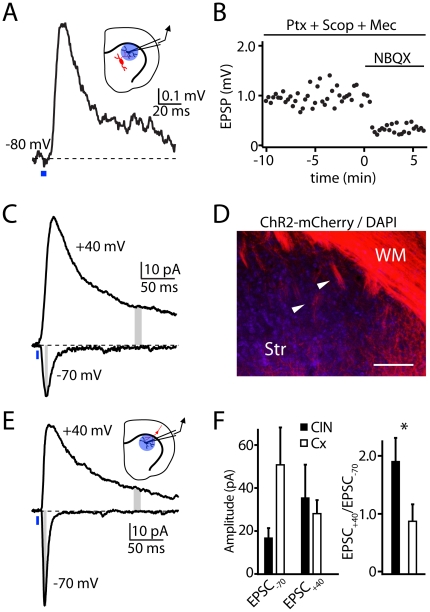
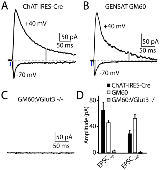
Verification
Claims the paper makes
The claims and statistics of this paper haven’t been checked yet.
Run the check — free, no account needed. It reads the open-access text and checks the sources it cites.
Claims other papers make about this one
Each is checked against this paper’s own text: does it say what the citing paper says it does?
In the citing paper
“Glutamate release onto SPNs activates postsynaptic ionotropic glutamate receptors with distinct AMPA/NMDA ratios compared with cortico-striatal synapses, suggesting distinct plasticity rules from these disparate inputs.60”
From Synaptic and circuit functions of multitransmitter neurons in the mammalian brain (2023) · cites this paper as [60]
In the citing paper
“Striatal cholinergic interneurons (CINs) cotransmit Ach and glutamate locally onto spiny projection neurons (SPNs) 60 and dopaminergic axonal terminals.[61][62][63]”
From Synaptic and circuit functions of multitransmitter neurons in the mammalian brain (2023) · cites this paper as [60]
Self-check
Percentages, sums and reported differences re-computed from the paper’s own numbers.
The self-check needs the paper’s full text.
Citations
Cites
Phasic Firing in Dopaminergic Neurons Is Sufficient for Behavioral Conditioning
Tsai, H.-C., Zhang, F., Adamantidis, A., Stuber, G. D., Bonci, A., de Lecea, L., & Deisseroth, K. (2009) · Science · doi:10.1126/science.1168878 · Cited by 1,266 · cited 1×
“Intracranial injection of adeno-associated virus encoding a Credependent (double floxed inverted) Channelrhodopsin2-mCherry fusion protein (DFI-ChR2-mCherry) [25,26] into the striatum of these mice enabled the CIN-specific expression of the lightactivated excitatory ChR2, which was clearly observed 7–14 days after inj…”
BackgroundStriatal Plasticity and Basal Ganglia Circuit Function
Kreitzer, A. C., & Malenka, R. C. (2008) · Neuron · doi:10.1016/j.neuron.2008.11.005 · Cited by 1,071 · cited 1×
“Synaptic plasticity in the striatum is thought to be critical for normal motor learning, and many forms are dependent on activation of both NMDARs and metabotropic glutamate receptors [33].”
BackgroundGong, S., Doughty, M., Harbaugh, C. R., Cummins, A., Hatten, M. E., Heintz, N., & Gerfen, C. R. (2007) · The Journal of Neuroscience · doi:10.1523/jneurosci.2707-07.2007 · Cited by 977 · cited 10×
“To examine this possibility, we utilized mice expressing Cre-recombinase under control of the endogenous choline acetyltransferase locus and conditionally expressed light-activated Channelrhodopsin2 in CINs.”
MethodInfluentialOttersen, O. P., & Storm‐Mathisen, J. (1984) · The Journal of Comparative Neurology · doi:10.1002/cne.902290308 · Cited by 848 · cited 1×
“brainstem and mesencephalon [1] and revealed the presence of”
BackgroundOptogenetic interrogation of neural circuits: technology for probing mammalian brain structures
Zhang, F., Gradinaru, V., Adamantidis, A. R., Durand, R., Airan, R. D., de Lecea, L., & Deisseroth, K. (2010) · Nature Protocols · doi:10.1038/nprot.2009.226 · Cited by 692 · cited 1×
“The virus carried either a double-floxed-inverted Channelrhodopsin2-mCherry construct under control of the Ef1a promoter (CINs) or non-conditional construct coding for Channelrhodopsin2-mCherry under the synapsin promoter (corticostriatal) [34].”
Rossi, J., Balthasar, N., Olson, D., Scott, M., Berglund, E., Lee, C. E., Choi, M. J., Lauzon, D., Lowell, B. B., & Elmquist, J. K. (2011) · Cell Metabolism · doi:10.1016/j.cmet.2011.01.010 · Cited by 672
Self-citationPatneau, D. K., & Mayer, M. (1990) · Journal of Neuroscience · doi:10.1523/jneurosci.10-07-02385.1990 · Cited by 653 · cited 2×
“2C), estimating the contribution of AMPARs.”
BackgroundA third vesicular glutamate transporter expressed by cholinergic and serotoninergic neurons
Gras, C., Herzog, E., Bellenchi, G. C., Bernard, V., Ravassard, P., Pohl, M., Gasnier, B., Giros, B., & El Mestikawy, S. (2002) · The Journal of Neuroscience · doi:10.1523/jneurosci.22-13-05442.2002 · Cited by 639 · cited 4×
“the vesicular glutamate transporter VGluT3, which co-localizes with the vesicular ACh transporter in the same presynaptic terminals [6,7,9].”
BackgroundInfluentialAtasoy, D., Aponte, Y., Su, H. H., & Sternson, S. M. (2008) · Journal of Neuroscience · doi:10.1523/jneurosci.1954-08.2008 · Cited by 635 · cited 1×
“Intracranial injection of adeno-associated virus encoding a Credependent (double floxed inverted) Channelrhodopsin2-mCherry fusion protein (DFI-ChR2-mCherry) [25,26] into the striatum of these mice enabled the CIN-specific expression of the lightactivated excitatory ChR2, which was clearly observed 7–14 days after inj…”
BackgroundCoincident but Distinct Messages of Midbrain Dopamine and Striatal Tonically Active Neurons
Morris, G., Arkadir, D., Nevet, A., Vaadia, E., & Bergman, H. (2004) · Neuron · doi:10.1016/j.neuron.2004.06.012 · Cited by 574
Cited by
Sorted by citations
Cachope, R., Mateo, Y., Mathur, B. N., Irving, J. M., Wang, H., Morales, M., Lovinger, D. M., & Cheer, J. F. (2012) · Cell Reports · doi:10.1016/j.celrep.2012.05.011 · Cited by 529
“This establishes even broader implications, given that glutamate released from cholinergic interneurons mediates not only excitation of medium spiny neurons, as previously described (Higley et al., 2011), but also shapes accumbal dopamine release.”
InfluentialNeurotransmitter Corelease: Mechanism and Physiological Role
Hnasko, T. S., & Edwards, R. H. (2012) · Annual Review of Physiology · doi:10.1146/annurev-physiol-020911-153315 · Cited by 284
“Consistent with VGLUT3 expression by these cells, the response was abolished in VGLUT3 KO mice (110).”
Stouffer, M. A., Woods, C. A., Patel, J. C., Lee, C. R., Witkovsky, P., Bao, L., Machold, R. P., Jones, K. T., de Vaca, S. C., Reith, M. E. A., et al. (2015) · Nature Communications · doi:10.1038/ncomms9543 · Cited by 288
“However, the effect of insulin on evoked [DA] o was absent throughout the striatal complex in forebrain ChAT KO mice, demonstrating that insulin-mediated enhancement of DA release requires striatal ACh, but not co-released transmitters from ChIs, such as glutamate 36 .”
Striatal Local Circuitry: A New Framework for Lateral Inhibition
Burke, D. A., Rotstein, H. G., & Alvarez, V. A. (2017) · Neuron · doi:10.1016/j.neuron.2017.09.019 · Cited by 287
Striatal cholinergic interneuron regulation and circuit effects
Lim, S. A. O., Kang, U. J., & McGehee, D. S. (2014) · Frontiers in Synaptic Neuroscience · doi:10.3389/fnsyn.2014.00022 · Cited by 244
“Striatal ChIs also co-release glutamate with ACh (Higley et al., 2011).”
BackgroundGABAergic RIP-Cre Neurons in the Arcuate Nucleus Selectively Regulate Energy Expenditure
Kong, D., Tong, Q., Ye, C., Koda, S., Fuller, P. M., Krashes, M. J., Vong, L., Ray, R. S., Olson, D. P., & Lowell, B. B. (2012) · Cell · doi:10.1016/j.cell.2012.09.020 · Cited by 215
“ChR2 and CRACM Viral Channelrhodopsin-2 expression and activation were similarly performed as previously described (Atasoy et al., 2008; Haubensak et al., 2010; Higley et al., 2011; Zhang et al., 2007).”
Dual-transmitter neurons: functional implications of co-release and co-transmission
Vaaga, C. E., Borisovska, M., & Westbrook, G. L. (2014) · Current Opinion in Neurobiology · doi:10.1016/j.conb.2014.04.010 · Cited by 221
Cholinergic modulation of hippocampal network function
Ruivo, L. M. T.-G., & Mellor, J. R. (2013) · Frontiers in Synaptic Neuroscience · doi:10.3389/fnsyn.2013.00002 · Cited by 214
“…in CA1 pyramidal neurons (Gu and Yakel, 2011). α4β2 receptors are expressed on interneuron axons terminating on excitatory and inhibitory neurons (Hill et al., 1993; Alkondon et al., 1999; Alkondon and Albuquerque, 2001; Graham et al., 2003; Bell et al., 2011). α3β4 receptors are expressed at…”
BackgroundCorelease of acetylcholine and GABA from cholinergic forebrain neurons
Saunders, A., Granger, A. J., & Sabatini, B. L. (2015) · eLife · doi:10.7554/elife.06412 · Cited by 200
“In the central brain, selective activation of striatal cholinergic interneurons results in cholinergic and glutamatergic responses (Gras et al., 2008; Higley et al., 2011; Nelson et al., 2014).”
BackgroundSelf-citationJiang, L., Kundu, S., Lederman, J., López‐Hernández, G. Y., Ballinger, E. C., Wang, S., Talmage, D. A., & Role, L. W. (2016) · Neuron · doi:10.1016/j.neuron.2016.04.028 · Cited by 186
Local control of striatal dopamine release
Cachope, R., & Cheer, J. F. (2014) · Frontiers in Behavioral Neuroscience · doi:10.3389/fnbeh.2014.00188 · Cited by 191
“Also, following Sabatini’s group’s report regarding the ability of CINs to evoke glutamatergic responses (Higley et al., 2011), we showed that AChevoked DA release is sensitive to AMPA blockers (Cachope et al., 2012).”
BackgroundEndocannabinoid modulation of dopamine neurotransmission
Covey, D. P., Mateo, Y., Sulzer, D., Cheer, J. F., & Lovinger, D. M. (2017) · Neuropharmacology · doi:10.1016/j.neuropharm.2017.04.033 · Cited by 197
“For example, M5 mACh-Rs potentiate glutamate released from VGluT2-expressing DA terminals (Shin et al., 2015), and striatal CINs, which express VGluT3, also release glutamate in the striatum (Higley et al., 2011; Nelson et al., 2014).”
BackgroundNicotine aversion: Neurobiological mechanisms and relevance to tobacco dependence vulnerability
Fowler, C. D., & Kenny, P. J. (2013) · Neuropharmacology · doi:10.1016/j.neuropharm.2013.09.008 · Cited by 176
“In addition to releasing acetylcholine, these interneurons may also corelease glutamate (Higley et al., 2011).”
Sgambato, V., & Cenci, M. A. (2011) · Progress in Neurobiology · doi:10.1016/j.pneurobio.2011.10.005 · Cited by 175
“Intriguingly, a recent study has revealed that striatal cholinergic interneurons express VGLUT3 and can elicit fast glutamatergic responses in the medium-spiny neurons (Higley et al., 2011).”
BackgroundGonzales, K. K., & Smith, Y. (2015) · Annals of the New York Academy of Sciences · doi:10.1111/nyas.12762 · Cited by 175
Cortical Control of Striatal Dopamine Transmission via Striatal Cholinergic Interneurons
Kosillo, P., Zhang, Y.-F., Threlfell, S., & Cragg, S. J. (2016) · Cerebral Cortex · doi:10.1093/cercor/bhw252 · Cited by 169
“2012); and therefore, the glutamate dependence of DA release driven by Pf or mCtx does not result from glutamate potentially co-released by ChIs (Higley et al. 2011).”
BackgroundBasal forebrain cholinergic signalling: development, connectivity and roles in cognition
Ananth, M. R., Rajebhosale, P., Kim, R., Talmage, D. A., & Role, L. W. (2023) · Nature Reviews Neuroscience · doi:10.1038/s41583-023-00677-x · Cited by 148
Dopaminergic modulation of striatal neurons, circuits, and assemblies
Surmeier, D. J., Carrillo‐Reid, L., & Bargas, J. (2011) · Neuroscience · doi:10.1016/j.neuroscience.2011.08.051 · Cited by 141
Cholinergic circuits in cognitive flexibility
Prado, V. F., Janíčková, H., Al‐Onaizi, M., & Prado, M. A. M. (2016) · Neuroscience · doi:10.1016/j.neuroscience.2016.09.013 · Cited by 158
“In addition to VAChT, striatal CINs also express the vesicular glutamate transporter 3 (VGLUT3) in synaptic vesicles (Fremeau et al., 2002; Gras et al., 2002; Takamori et al., 2002); which give them the ability to corelease ACh and glutamate (Higley et al., 2011).”
BackgroundInfluentialDoig, N. M., Magill, P. J., Apicella, P., Bolam, J. P., & Sharott, A. (2014) · Journal of Neuroscience · doi:10.1523/jneurosci.4627-13.2014 · Cited by 138
Lozovaya, N., Eftekhari, S., Cloarec, R., Gouty‐Colomer, L., Dufour, A., Riffault, B., Billon-Grand, M., Pons-Bennaceur, A., Oumar, N., Burnashev, N., et al. (2018) · Nature Communications · doi:10.1038/s41467-018-03802-y · Cited by 143
Regulation of cholinergic activity by the vesicular acetylcholine transporter
Prado, V. F., Roy, A., Kolisnyk, B., Gros, R., & Prado, M. A. M. (2013) · Biochemical Journal · doi:10.1042/bj20121662 · Cited by 137
Kaufman, A. M., Milnerwood, A. J., Sepers, M. D., Coquinco, A., She, K., Wang, L., Lee, H., Craig, A. M., Cynader, M. S., & Raymond, L. A. (2012) · Journal of Neuroscience · doi:10.1523/jneurosci.4129-11.2012 · Cited by 135
Matikainen-Ankney, B. A., Kezunovic, N., Mesias, R. E., Tian, Y., Williams, F. M., Huntley, G. W., & Benson, D. L. (2016) · Journal of Neuroscience · doi:10.1523/jneurosci.3314-15.2016 · Cited by 139
“The vast majority of extrinsic glutamatergic inputs to striatum derive from neocortex and thalamus (Wilson et al., 1983; Gerfen, 1992), with a smaller glutamatergic contribution via corelease from intrinsic cholinergic neurons (Higley et al., 2011).”
BackgroundGeneral Principles of Neuronal Co-transmission: Insights From Multiple Model Systems
Svensson, E., Apergis‐Schoute, J., Burnstock, G., Nusbaum, M. P., Parker, D., & Schiöth, H. B. (2019) · Frontiers in Neural Circuits · doi:10.3389/fncir.2018.00117 · Cited by 134
Cholinergic modulation of striatal microcircuits
Abudukeyoumu, N., Hernández-Flores, T., Garcia-Munoz, M., & Arbuthnott, G. W. (2018) · European Journal of Neuroscience · doi:10.1111/ejn.13949 · Cited by 144
“…approach, there are two studies: one reports that optical stimulation of ChIs induces in MSNs two glutamate-dependent responses (Higley et al., 2011) and another reports that ACh release following synchronous ChIs triggers an action potential-independent presynaptic release of…”
Maurice, N., Liberge, M., Jaouen, F., Ztaou, S., Hanini, M., Camon, J., Deisseroth, K. A., Amalric, M., Goff, L. K., & Beurrier, C. (2015) · Cell Reports · doi:10.1016/j.celrep.2015.09.034 · Cited by 125
Aroniadou‐Anderjaska, V., Apland, J. P., Figueiredo, T. H., Furtado, M. D. A., & Braga, M. F. M. (2020) · Neuropharmacology · doi:10.1016/j.neuropharm.2020.108298 · Cited by 131
Pistillo, F., Clementi, F., Zoli, M., & Gotti, C. (2014) · Progress in Neurobiology · doi:10.1016/j.pneurobio.2014.10.002 · Cited by 120
“2B) (Higley et al., 2011; Lenz and Lobo, 2013).”
Tanimura, A., Du, Y., Kondapalli, J., Wokosin, D. L., & Surmeier, D. J. (2019) · Neuron · doi:10.1016/j.neuron.2018.12.004 · Cited by 111
“Because ChIs can release glutamate (Higley et al., 2011; Nelson et al., 2014), one interpretation of this result is that ChIs are directly activating postsynaptic iSPN glutamate receptors.”
Mateo, Y., Johnson, K. A., Covey, D. P., Atwood, B. K., Wang, H., Zhang, S., Gildish, I., Cachope, R., Bellocchio, L., Guzmán, M., et al. (2017) · Neuron · doi:10.1016/j.neuron.2017.11.012 · Cited by 111
Dual-transmitter systems regulating arousal, attention, learning and memory
Ma, S., Hangya, B., Leonard, C. S., Wisden, W., & Gundlach, A. L. (2017) · Neuroscience & Biobehavioral Reviews · doi:10.1016/j.neubiorev.2017.07.009 · Cited by 107
“It has been shown that two separate cholinergic systems, the tonically-active interneurons of the striatum (Gras et al., 2002; Guzman et al., 2011; Higley et al., 2011) and the cholinergic cells of the medial habenula (Ren et al., 2011) can release both ACh and glutamate.”
New Insights Into Cholinergic Neuron Diversity
Ahmed, N. Y., Knowles, R., & Dehorter, N. (2019) · Frontiers in Molecular Neuroscience · doi:10.3389/fnmol.2019.00204 · Cited by 108
“They also drive spontaneous activation of SPNs via muscarinic (Mamaligas and Ford, 2016) and glutamatergic transmission (Higley et al., 2011).”
BackgroundCholinergic modulation of synaptic integration and dendritic excitability in the striatum
Oldenburg, I. A., & Ding, J. (2011) · Current Opinion in Neurobiology · doi:10.1016/j.conb.2011.04.004 · Cited by 103
Cotransmission of acetylcholine and GABA
Granger, A. J., Mulder, N., Saunders, A. B., & Sabatini, B. L. (2015) · Neuropharmacology · doi:10.1016/j.neuropharm.2015.07.031 · Cited by 101
“…field potentials or single unit spiking in cortex following activation of BF axons (Pinto et al., 2013), or recording postsynaptic currents in conditions that would explicitly preclude observing GABAergic responses by including GABA receptor antagonists (Higley et al., 2011; Yang et al., 2014).”
BackgroundXu, J., Liu, R.-J., Fahey, S., Frick, L. R., Leckman, J. F., Vaccarino, F. M., Duman, R. S., Williams, K. B., Swedo, S. E., & Pittenger, C. (2020) · American Journal of Psychiatry · doi:10.1176/appi.ajp.2020.19070698 · Cited by 102
“The CINs interact with other neurotransmitter systems; they co-release glutamate (68) and drive dopamine and GABA release from dopamine terminals (69–71).”
Striatal cholinergic neurotransmission requires VGLUT3
Nelson, A. B., Bussert, T. G., Kreitzer, A. C., & Seal, R. P. (2014) · Journal of Neuroscience · doi:10.1523/jneurosci.0901-14.2014 · Cited by 99
Kondabolu, K., Roberts, E. A., Bucklin, M. E., McCarthy, M. M., Kopell, N., & Han, X. (2016) · Proceedings of the National Academy of Sciences · doi:10.1073/pnas.1605658113 · Cited by 98
“Glutamate, which is also coreleased from SChIs, may be responsible for the increase in high gamma oscillations during SChI stimulation (37).”
Multiplexed neurochemical signaling by neurons of the ventral tegmental area
Barker, D. J., Root, D. H., Zhang, S., & Morales, M. (2016) · Journal of Chemical Neuroanatomy · doi:10.1016/j.jchemneu.2015.12.016 · Cited by 105
“…are multiplexed throughout the brain (Trudeau, 2004; Gillespie et al., 2005; Zhou et al., 2005; Gras et al., 2008; Noh et al., 2010; Higley et al., 2011; Tritsch et al., 2012; Hnasko and Edwards, 2012; Münster-Wandowski et al., 2013; Nelson et al., 2014; Root et al., 2014a; Shabel…”
BackgroundOptogenetic insights into striatal function and behavior
Lenz, J. D., & Lobo, M. K. (2013) · Behavioural Brain Research · doi:10.1016/j.bbr.2013.04.018 · Cited by 95
“[84] Higley MJ, Gittis AH, Oldenburg IA, Balthasar N, Seal RP, Edwards RH, et al.”
BackgroundDisrupted striatal neuron inputs and outputs in Huntington's disease
Reiner, A. J., & Deng, Y. (2018) · CNS Neuroscience & Therapeutics · doi:10.1111/cns.12844 · Cited by 93
Guzmán, M. S., Jaeger, X. D., Raulic, S., Souza, I. A., Li, A. X., Schmid, S., Menon, R. S., Gainetdinov, R. R., Caron, M. G., Bartha, R., et al. (2011) · PLoS Biology · doi:10.1371/journal.pbio.1001194 · Cited by 99
“predominant activity over NMDA receptors [27].”
BackgroundHeterogeneity in striatal dopamine circuits: Form and function in dynamic reward seeking
Collins, A. L., & Saunders, B. T. (2020) · Journal of Neuroscience Research · doi:10.1002/jnr.24587 · Cited by 93
“For example, CINs release GABA (Saunders, Granger, & Sabatini, 2015) and glutamate (Fremeau et al., 2002; Gras et al., 2002, 2008; Higley et al., 2011) in addition to acetylcholine.”
BackgroundStriatal cholinergic interneurons and Parkinson's disease
Tanimura, A., Pancani, T., Lim, S. A. O., Tubert, C., Melendez-Zaidi, A. E., Shen, W., & Surmeier, D. J. (2017) · European Journal of Neuroscience · doi:10.1111/ejn.13638 · Cited by 94
“In particular, using optogenetic approaches it was shown that ChIs are capable of releasing glutamate (Higley et al., 2011; Nelson et al., 2014).”
Laszlovszky, T., Schlingloff, D., Hegedüs, P., Freund, T. F., Gulyás, A. I., Kepecs, Á., & Hangya, B. (2020) · Nature Neuroscience · doi:10.1038/s41593-020-0648-0 · Cited by 98
“Ethics oversight Adult (over 2 months old) ChAT-Cre (N = 15, 14/15 male, Higley et al., 2011), ChAT-ChR2 (N = 3, 3/3 male, Zhao et al., 2011) and PV-Cre (n = 4, 4/4 male) mice were used for behavioral recording experiments under the protocol approved by Cold Spring Harbor Laboratory Institutional…”
Sun, Y.-G., Pita‐Almenar, J. D., Wu, C., Renger, J. J., Uebele, V. N., Lu, H., & Beierlein, M. (2013) · Journal of Neuroscience · doi:10.1523/jneurosci.3177-12.2013 · Cited by 79
“Recent studies have demonstrated the synaptic release of glutamate from cholinergic neurons (Higley et al., 2011; Ren et al., 2011).”
BackgroundPatel, J. C., Rossignol, E., Rice, M. E., & Machold, R. P. (2012) · Nature Communications · doi:10.1038/ncomms2144 · Cited by 80
Protons Regulate Vesicular Glutamate Transporters through an Allosteric Mechanism
Eriksen, J., Chang, R., McGregor, M., Silm, K., Suzuki, T., & Edwards, R. H. (2016) · Neuron · doi:10.1016/j.neuron.2016.03.026 · Cited by 76
Trudeau, L., & Mestikawy, S. E. (2018) · Frontiers in Neural Circuits · doi:10.3389/fncir.2018.00113 · Cited by 83
“…DA neurons (Sulzer et al., 1998; Bourque and Trudeau, 2000; Hnasko et al., 2010; Tecuapetla et al., 2010; Chuhma et al., 2014; Trudeau et al., 2014), ACh (Huh et al., 2008; Higley et al., 2011; Nelson et al., 2014; Frahm et al., 2015) and GABA neurons (Shabel et al., 2014; Root et al., 2018).”
Basal forebrain subcortical projections
Agostinelli, L. J., Geerling, J. C., & Scammell, T. E. (2019) · Brain Structure and Function · doi:10.1007/s00429-018-01820-6 · Cited by 77
Fasano, C., Bourque, M.-J., Lapointe, G., Leo, D., Thibault, D., Haber, M., Kortleven, C., DesGroseillers, L., Murai, K. K., & Trudeau, L.-É. (2013) · Neuropharmacology · doi:10.1016/j.neuropharm.2012.11.030 · Cited by 67
Upmanyu, N., Jin, J., Emde, H. V. D., Ganzella, M., Bösche, L., Malviya, V. N., Zhuleku, E., Politi, A. Z., Ninov, M., Silbern, I., et al. (2022) · Neuron · doi:10.1016/j.neuron.2022.02.008 · Cited by 69
“, 2009), or with acetylcholine in striatal interneurons and medial habenula projections (Frahm et al., 2015; Gras et al., 2008; Higley et al., 2011; Ren et al., 2011).”
BackgroundLuchicchi, A., Bloem, B., Viaña, J. N. M., Mansvelder, H. D., & Role, L. W. (2014) · Frontiers in Synaptic Neuroscience · doi:10.3389/fnsyn.2014.00024 · Cited by 74
Developmental specification of forebrain cholinergic neurons
Allaway, K. C., & Machold, R. (2017) · Developmental Biology · doi:10.1016/j.ydbio.2016.11.007 · Cited by 68
“However, some of these populations (e.g., striatal cholinergic interneurons) have also been found to express the vesicular glutamate transporter VGLUT3 (slc17a8) and co-release glutamate (Higley et al., 2011), an indication of additional functional heterogeneity.”
Spontaneous Synaptic Activation of Muscarinic Receptors by Striatal Cholinergic Neuron Firing
Mamaligas, A. A., & Ford, C. (2016) · Neuron · doi:10.1016/j.neuron.2016.06.021 · Cited by 70
“, 2012) and the co-release of glutamate from cholinergic interneurons (Higley et al., 2011; Nelson et al., 2014a) onto MSNs.”
Gonzalez, D., Tomasek, M. P., Hays, S. A., Sridhar, V., Ammanuel, S. G., Chang, C.-W., Pawlowski, K. S., Huber, K. M., & Gibson, J. R. (2019) · Journal of Neuroscience · doi:10.1523/jneurosci.0886-19.2019 · Cited by 64
“Serotonergic and cholinergic neurons have been reported to release glutamate, but VGlut3 is the transporter in these neurons (Higley et al., 2011; Wang et al., 2019).”
BackgroundSakae, D. Y., Marti, F., Lecca, S., Vorspan, F., Martín‐García, E., Morel, L. J., Henrion, A., Cuesta, J., Besnard, A., Heck, N., et al. (2015) · Molecular Psychiatry · doi:10.1038/mp.2015.104 · Cited by 68
Regulation of the Hippocampal Network by VGLUT3-Positive CCK- GABAergic Basket Cells
Fasano, C., Rocchetti, J., Pietrajtis, K., Zander, J.-F., Manseau, F., Sakae, D. Y., Marcus-Sells, M., Ramet, L., Morel, L. J., Carrel, D., et al. (2017) · Frontiers in Cellular Neuroscience · doi:10.3389/fncel.2017.00140 · Cited by 67
Crofton, E. J., Nenov, M. N., Zhang, Y., Scala, F., Page, S. A., McCue, D. L., Li, D., Hommel, J. D., Laezza, F., & Green, T. A. (2017) · Neuropharmacology · doi:10.1016/j.neuropharm.2017.01.020 · Cited by 64
“…role of TANs in cocaine addiction with immunotoxin mediated cell ablation resulting in increased sensitivity to cocaine (Hikida et al., 2001) and preventing behavioral abnormalities associated with cocaine induced by centrally active acetylcholinesterase inhibitors in the NAc (Hikida et al., 2003).”
BackgroundMuñoz, B., Fritz, B. M., Yin, F., & Atwood, B. K. (2018) · Nature Communications · doi:10.1038/s41467-018-03683-1 · Cited by 63
Divito, C. B., Steece‐Collier, K., Case, D. T., Williams, S. G., Stancati, J. A., Zhi, L., Rubio, M. E., Sortwell, C. E., Collier, T. J., Sulzer, D., et al. (2015) · Journal of Neuroscience · doi:10.1523/jneurosci.2124-15.2015 · Cited by 62
“Therefore, deletion of VGLUT3 from these cells not only abolishes the glutamate signaling (Higley et al., 2011), but also markedly reduces cholinergic transmission (Nelson et al., 2014).”
BackgroundDopaminergic Regulation of Striatal Interneurons in Reward and Addiction: Focus on Alcohol
Clarke, R. B. C., & Adermark, L. (2015) · Neural Plasticity · doi:10.1155/2015/814567 · Cited by 68
“Tonically active cholinergic interneurons modulate dopamine output through activation of both muscarinic and nicotinic cholinergic receptors on dopamine terminals [125, 139–141] and through the corelease of glutamate [142, 143].”
BackgroundSynaptic and circuit functions of multitransmitter neurons in the mammalian brain
Wallace, M. L., & Sabatini, B. L. (2023) · Neuron · doi:10.1016/j.neuron.2023.06.003 · Cited by 59
Self-citationLee, J., Finkelstein, J., Choi, J. Y., & Witten, I. B. (2016) · Neuron · doi:10.1016/j.neuron.2016.05.001 · Cited by 59
“, 2001), and their activity modulates the activity of themedium spiny neurons (MSNs), the output neurons in the striatum (English et al., 2012; Oldenburg and Ding, 2011; Witten et al., 2010; Nelson et al., 2014; Higley et al., 2011).”
BackgroundOptogenetic interrogations of the neural circuits underlying addiction
Britt, J. P., & Bonci, A. (2013) · Current Opinion in Neurobiology · doi:10.1016/j.conb.2013.01.010 · Cited by 64
Ma, T., Huang, Z., Xie, X., Cheng, Y., Zhuang, X., Childs, M., Gangal, H., Wang, X., Smith, L., Smith, R., et al. (2021) · Journal of Clinical Investigation · doi:10.1172/jci154969 · Cited by 53
Paradoxical network excitation by glutamate release from VGluT3+ GABAergic interneurons
Pelkey, K. A., Calvigioni, D., Fang, C., Vargish, G., Ekins, T., Auville, K., Wester, J. C., Lai, M., Mackenzie-Gray Scott, C., Yuan, X., et al. (2020) · eLife · doi:10.7554/elife.51996 · Cited by 46
“In many of these systems, VGluT3 imparts unique synaptic properties such as glutamate corelease and vesicular synergy (Amilhon et al., 2010; Gras et al., 2008; Higley et al., 2011; Nelson et al., 2014; Noh et al., 2010; Sakae et al., 2015; Varga et al., 2009; Wang et al., 2019).”
BackgroundPoppi, L. A., Ho-Nguyen, K.-T., Shi, A., Daut, C. T., & Tischfield, M. A. (2021) · Cells · doi:10.3390/cells10040907 · Cited by 47
“To add to the complexity, ChINs can also co-release GABA [141] and glutamate [68,142], in addition to ACh.”
BackgroundIn vivo optogenetic control of striatal and thalamic neurons in non-human primates
Galvan, A., Hu, X., Smith, Y., & Wichmann, T. (2012) · PLoS ONE · doi:10.1371/journal.pone.0050808 · Cited by 45
“The value of using the far more specific optogenetic techniques to study the physiology of the basal ganglia is well established in rodent studies [12,13,14,15,16,17].”
BackgroundWalker, L. C., Berizzi, A. E., Chen, N. A., Rueda, P., Perreau, V. M., Huckstep, K., Srisontiyakul, J., Govitrapong, P., Jia, X., Lindsley, C. W., et al. (2020) · Biological Psychiatry · doi:10.1016/j.biopsych.2020.02.019 · Cited by 45
Luquin, E., Huerta, I., Aymerich, M. S., & Mengual, E. (2018) · Frontiers in Neuroanatomy · doi:10.3389/fnana.2018.00034 · Cited by 46
Lu, J., Cheng, Y., Wang, X., Woodson, K., Kemper, W. C., Disney, E., & Wang, J. (2019) · Neuropsychopharmacology · doi:10.1038/s41386-019-0332-9 · Cited by 42
Stone, T. W. (2021) · Neuroscience · doi:10.1016/j.neuroscience.2021.06.007 · Cited by 49
Adler, A., Katabi, S., Finkes, I., Prut, Y., & Bergman, H. (2013) · Frontiers in Systems Neuroscience · doi:10.3389/fnsys.2013.00047 · Cited by 41
“Thus, they can modulate (in conjunction with the dopaminergic and other modulators of the striatum) the efficacy of the cortico and thalamo-striatal synapses, rather than directly affecting their target neurons’ ongoing discharge (Kreitzer, 2009; Higley et al., 2011).”
BackgroundOmiya, Y., Uchigashima, M., Konno, K., Yamasaki, M., Miyazaki, T., Yoshida, T., Kusumi, I., & Watanabe, M. (2015) · Journal of Neuroscience · doi:10.1523/jneurosci.4681-14.2015 · Cited by 42
“Both CCK and CCK2R are densely expressed in the BLA (Larsson and Rehfeld, 1979; Vanderhaeghen et al., 1985; Honda et al., 1993; Mascagni and McDonald, 2003), and rich CCK2R mRNA expression in BA pyramidal cells was shown in the present study.”
BackgroundPomierny, B., Krzyżanowska, W., Broniowska, Ż., Strach, B., Bystrowska, B., Starek‐Świechowicz, B., Maciejska, A., Skórkowska, A., Wesołowska, J., Walczak, M., et al. (2019) · Toxicological Sciences · doi:10.1093/toxsci/kfz160 · Cited by 42
Lv, X., Dickerson, J. W., Rook, J. M., Lindsley, C. W., Conn, P. J., & Xiang, Z. (2017) · Neuropharmacology · doi:10.1016/j.neuropharm.2017.03.017 · Cited by 40
“A previous study indicated that striatal cholinergic interneurons could co-release glutamate, which activates NMDA and AMPA/ kainate glutamate receptors in MSNs (Higley et al., 2011).”
InfluentialZimmermann, J., Herman, M. A., & Rosenmund, C. (2015) · Frontiers in Synaptic Neuroscience · doi:10.3389/fnsyn.2015.00016 · Cited by 37
Skirzewski, M., Princz‐Lebel, O., German‐Castelan, L., Crooks, A. M., Kim, K., Tarnow, S. H., Reichelt, A. C., Memar, S., Palmer, D. D., Li, Y., et al. (2022) · Nature Communications · doi:10.1038/s41467-022-35601-x · Cited by 41
“Please, improve statistics/analysis section including these and other relevant details for better understanding of the data analysis.”
Kljakic, O., Janíčková, H., Prado, V. F., & Prado, M. A. M. (2017) · Journal of Neurochemistry · doi:10.1111/jnc.14003 · Cited by 39
Crittenden, J. R., Lacey, C. J., Weng, F., Garrison, C. E., Gibson, D. J., Lin, Y., & Graybiel, A. M. (2017) · Frontiers in Neuroanatomy · doi:10.3389/fnana.2017.00020 · Cited by 42
“Analyses of SPN responses to ChI stimulation have shown that there are multiple inhibitory post-synaptic currents with fast, slow and very slow time-courses (English et al., 2012), and small excitatory post-synaptic currents (Higley et al., 2011).”
BackgroundNucleus accumbens in the pathogenesis of major depressive disorder: A brief review
Yajie, J., Zou, M., Wang, Y., & Wang, Y. (2023) · Brain Research Bulletin · doi:10.1016/j.brainresbull.2023.03.004 · Cited by 41
“At the circuit level, activation of TANs has been shown to elicit both fast glutamatergic transmission (Higley et al., 2011) and GABAergic inhibition of MSNs, the latter coincident with synchronous cholinergic activation and sufficient to pause MSNs firing (English et al.”
A distinct set of synaptic‐like microvesicles in atroglial cells contain VGLUT3
Ormel, L., Stensrud, M. J., Chaudhry, F. A., & Gundersen, V. (2012) · Glia · doi:10.1002/glia.22348 · Cited by 38
“Indeed, it has recently been reported that glutamate is released from cholinergic neurons to give fast glutamatergic postsynaptic responses (Higley et al., 2011; Ren et al., 2011).”
BackgroundStensrud, M. J., Chaudhry, F. A., Leergaard, T. B., Bjaalie, J. G., & Gundersen, V. (2013) · The Journal of Comparative Neurology · doi:10.1002/cne.23331 · Cited by 35
“Recently, VGLUT3dependent glutamate receptor responses were detected in the striatum (Higley et al., 2011), indicative of glutamate release from cholinergic neurons.”
BackgroundDescending projections from the basal forebrain to the orexin neurons in mice
Agostinelli, L. J., Ferrari, L. L., Mahoney, C. E., Mochizuki, T., Lowell, B. B., Arrigoni, E., & Scammell, T. E. (2016) · The Journal of Comparative Neurology · doi:10.1002/cne.24158 · Cited by 38
“The ChAT-Cre mice have been validated in prior studies (Higley et al., 2011; Rossi et al., 2011).”
BackgroundMethodDopaminergic modulation of basal ganglia output through coupled excitation–inhibition
Budzillo, A., Duffy, A., Miller, K. E., Fairhall, A. L., & Perkel, D. J. (2017) · Proceedings of the National Academy of Sciences · doi:10.1073/pnas.1611146114 · Cited by 35
“It is not the cholinergic neuron type, which also releases glutamate in mammals (40, 46), but may nonetheless have similar function.”
Backgroundd’Incamps, B. L., Bhumbra, G. S., Foster, J. D., Beato, M., & Ascher, P. (2017) · Scientific Reports · doi:10.1038/s41598-017-04266-8 · Cited by 38
Neale, S. A., Copeland, C. S., & Salt, T. E. (2013) · Neurochemistry International · doi:10.1016/j.neuint.2013.10.001 · Cited by 35
Nasirova, N., Quina, L. A., Agosto‐Marlin, I. M., Ramirez, J., Lambe, E. K., & Turner, E. E. (2019) · The Journal of Comparative Neurology · doi:10.1002/cne.24753 · Cited by 33
“…cholinergic neurons can exhibit glutamatergic transmission dependent on the glutamate transporter VGluT3 (Boulland et al., 2004; Gras et al., 2002; Higley et al., 2011), which is atypical in that it is gener- ally coexpressed in neurons with other neurotransmitters (El…”
BackgroundGalaj, E., Kipp, B. T., Floresco, S., & Savage, L. M. (2019) · Neuroscience · doi:10.1016/j.neuroscience.2019.01.062 · Cited by 37
“Cholinergic interneurons in the NAc control local circuits (Cachope et al. 2012; Higley et al. 2011; Threlfell et al. 2012), modulate the activity of GABAergic projection neurons (Witten et al.”
BackgroundMultiplexed neurotransmission emulated for emotion control
Ni, Y., Ma, M., Wei, H., Gong, J., Han, H., Liu, L., Xu, Z., & Xu, W. (2021) · Nano Energy · doi:10.1016/j.nanoen.2021.106038 · Cited by 34
A Feud that Wasn't: Acetylcholine Evokes Dopamine Release in the Striatum
Surmeier, D. J., & Graybiel, A. M. (2012) · Neuron · doi:10.1016/j.neuron.2012.06.028 · Cited by 32
“They linked this to the recently described corelease of glutamate and AChbyChIs (Higley et al., 2011).”
BackgroundRole of the atypical vesicular glutamate transporter VGLUT3 in l-DOPA-induced dyskinesia
Gangarossa, G., Guzmán, M. S., Prado, V. F., Prado, M. A. M., Daumas, S., Mestikawy, S. E., & Valjent, E. (2015) · Neurobiology of Disease · doi:10.1016/j.nbd.2015.12.010 · Cited by 35
Xie, Y., Heida, T., Stegenga, J., Zhao, Y., Moser, A., Tronnier, V. M., Feuerstein, T. J., & Hofmann, U. (2014) · European Journal of Neuroscience · doi:10.1111/ejn.12736 · Cited by 30
“DBS is currently proposed as an effective surgical intervention for various dysfunctional syndromes, such as epilepsy, depression and obsessive-compulsive disorder (OCD) (Tronnier & Fogel, 2000; Gross, 2004; Huff et al., 2010).”
BackgroundProtonic conductor: better understanding neural resting and action potential
Lee, J. W. (2020) · Journal of Neurophysiology · doi:10.1152/jn.00281.2020 · Cited by 28
Morgenstern, N. A., Isidro, A. F., Israely, I., & Costa, R. M. (2022) · Science Advances · doi:10.1126/sciadv.abh4315 · Cited by 30
“However, in the DLS, ChIs corelease glutamate together with acetylcholine ( 63 ).”
Cholinergic activity reflects reward expectations and predicts behavioral responses
Hegedüs, P., Sviatkó, K., Király, B., Martínez‐Bellver, S., & Hangya, B. (2022) · iScience · doi:10.1016/j.isci.2022.105814 · Cited by 28
Cholinergic modulation of striatal nitric oxide‐producing interneurons
Melendez-Zaidi, A. E., Lakshminarasimhah, H., & Surmeier, D. J. (2019) · European Journal of Neuroscience · doi:10.1111/ejn.14528 · Cited by 25
“It is also worth noting that strong optogenetic stimulation of ChIs did evoke an excitatory response in some LTSIs (Figure 5d; Higley et al., 2011).”
BackgroundSynapse-specific expression of mu opioid receptor long-term depression in the dorsomedial striatum
Muñoz, B., Haggerty, D. L., & Atwood, B. K. (2020) · Scientific Reports · doi:10.1038/s41598-020-64203-0 · Cited by 27
Xu, J., Frankovich, J., Liu, R.-J., Thienemann, M., Silverman, M. A., Farhadian, B., Willett, T. A., Manko, C., Columbo, L. A., Leibold, C. M., et al. (2024) · Brain Behavior and Immunity · doi:10.1016/j.bbi.2024.07.044 · Cited by 28
Metadata
- Authors
- Higley, M. J., Gittis, A. H., Oldenburg, I. A., Balthasar, N., Seal, R. P., Edwards, R. H., Lowell, B. B., Kreitzer, A. C., & Sabatini, B. L.
- Affiliations
- Howard Hughes Medical Institute (US)
- University of California, San Francisco (US)
- Beth Israel Deaconess Medical Center (US)
- Corresponding author
- Sabatini, Bernardo L.
- ORCID
- Venue
- PLoS ONE
- Publisher
- Public Library of Science (PLoS)
- Published
- 2011-04-22
- Volume
- 6
- Issue
- 4
- Pages
- e19155
- ISSN
- 1932-6203 (ISSN-L)
- PMC
- PMC3081336
- OpenAlex
- W2137833675
- Cited by
- 175
- References
- 35
- Citations by year
- 2026: 4 · 2025: 6 · 2024: 8 · 2023: 10 · 2022: 14 · 2021: 11 · 2020: 14 · 2019: 10 · 2018: 12 · 2017: 16 · 2016: 14 · 2015: 16 · 2014: 12 · 2013: 10 · 2012: 13
- FWCI
- 5.30
- Type
- journal article
- Language
- en
- Open access
- gold
- doi.org
- Locations
- PLoS ONE (publishedVersion, cc-by)
- Europe PMC (PubMed Central) (publishedVersion, cc-by)
- PubMed Central (publishedVersion, cc-by)
- PubMed (publishedVersion)
- Explore Bristol Research (publishedVersion)
- Figshare (submittedVersion, cc-by)
- Digital Access to Scholarship at Harvard (DASH) (Harvard University) (submittedVersion, cc-by)
- KiltHub Repository (submittedVersion, other-oa)
- DOAJ (DOAJ: Directory of Open Access Journals) (submittedVersion)
- eScholarship (California Digital Library) (submittedVersion)
- eScholarship (California Digital Library) (submittedVersion, cc-by)
- Bristol Research (University of Bristol) (submittedVersion)
- KiltHub Repository (cc-by)
- Funders
- Howard Hughes Medical Institute
- W. M. Keck Foundation
- Parkinson's Disease Foundation
- National Alliance for Research on Schizophrenia and Depression
- National Institutes of Health (DK057521, DK046200, DA10154, NS046579)
- National Institute of Mental Health (F32MH068085, MH50712, R01MH050712, R37MH050712, F31MH093026)
- National Institute on Drug Abuse (P01DA010154, DA-10154)
- National Institute of Diabetes and Digestive and Kidney Diseases (P30DK057521, R01DK075632, P30DK046200, DK075632)
- National Institute of Neurological Disorders and Stroke (NS064984, R01NS046579, F32NS063663, R01NS064984, R37NS046579, NS063663, NS046579)
- Topics
- Life Sciences › Neuroscience › Cellular and Molecular Neuroscience › Neuroscience and Neuropharmacology Research
- Life Sciences › Neuroscience › Cellular and Molecular Neuroscience › Neurotransmitter Receptor Influence on Behavior
- Health Sciences › Medicine › Clinical Neurology › Deep Brain Stimulation and Movement Disorders
- Keywords
- cholinergic interneurons, striatum, VGLUT3, glutamatergic neurotransmission, medium spiny neurons, NMDA receptor, choline acetyltransferase, channelrhodopsin-2, optogenetic stimulation
- MeSH terms
- Channelrhodopsins, Central Nervous System Stimulants, Animals, Corpus Striatum, Evoked Potentials, Female, Interneurons, Male, Mecamylamine, Picrotoxin, Receptors, Cholinergic, Scopolamine, Receptors, N-Methyl-D-Aspartate, Amino Acid Transport Systems, Acidic, Mice
About these checks
Registry, retraction, Cites and statistics results are computed, not judged, with one exception: a printed reference that no index lists is read once by a model before it counts as a citation error. Claims are extracted by a model and kept only when their quote appears verbatim in the text; each cited source is then checked, with a model’s help, for whether it says what the claim relies on. Those verdicts can be wrong — read the claim’s quote and the cited source before relying on one.
Statistics and arithmetic are recomputed, not judged.
Confidence bands are shown only for fields calibrated on our annotated test set. None is calibrated yet, so no band is shown.
What counts as a claim ›How we measure confidence ›How we check this ›
Are you an author, or do you see a problem with this page? Request a review
