

The DOI matches its Crossref record; 83 of the 84 references it cites were found in a registry or an index.
Protein 3D Structure Computed from Evolutionary Sequence Variation
Open accessAbstract
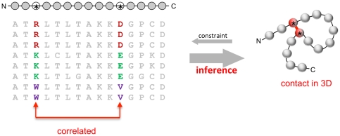
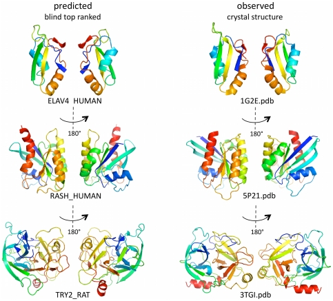
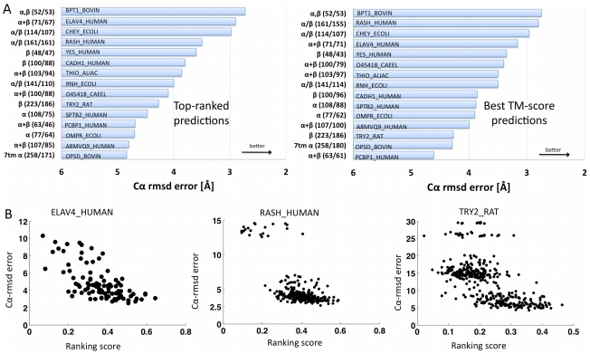
The evolutionary trajectory of a protein through sequence space is constrained by its function. Collections of sequence homologs record the outcomes of millions of evolutionary experiments in which the protein evolves according to these constraints. Deciphering the evolutionary record held in these sequences and exploiting it for predictive and engineering purposes presents a formidable challenge. The potential benefit of solving this challenge is amplified by the advent of inexpensive high-throughput genomic sequencing.In this paper we ask whether we can infer evolutionary constraints from a set of sequence homologs of a protein. The challenge is to distinguish true co-evolution couplings from the noisy set of observed correlations. We address this challenge using a maximum entropy model of the protein sequence, constrained by the statistics of the multiple sequence alignment, to infer residue pair couplings. Surprisingly, we find that the strength of these inferred couplings is an excellent predictor of residue-residue proximity in folded structures. Indeed, the top-scoring residue couplings are sufficiently accurate and well-distributed to define the 3D protein fold with remarkable accuracy.We quantify this observation by computing, from sequence alone, all-atom 3D structures of fifteen test proteins from different fold classes, ranging in size from 50 to 260 residues, including a G-protein coupled receptor. These blinded inferences are de novo, i.e., they do not use homology modeling or sequence-similar fragments from known structures. The co-evolution signals provide sufficient information to determine accurate 3D protein structure to 2.7-4.8 Å C(α)-RMSD error relative to the observed structure, over at least two-thirds of the protein (method called EVfold, details at http://EVfold.org). This discovery provides insight into essential interactions constraining protein evolution and will facilitate a comprehensive survey of the universe of protein structures, new strategies in protein and drug design, and the identification of functional genetic variants in normal and disease genomes.
Overview
The claims and statistics of this paper haven’t been checked yet.
Run the check — free, no account needed. It reads the open-access text and checks the sources it cites.
Integrity signals
Registry
Cites
Statistics
Figures
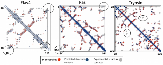
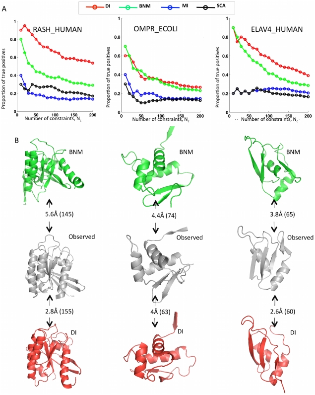
![Figure 3 Progress in contact prediction using the maximum entropy method. Extraction of evolutionary information about residue coupling and predicted contacts from multiple sequence alignments works much better using the global statistical model (right, Direct Information, DI, Equation 3) than the local statistical model (left, Mutual Information, MI, Equation 1). Predicted contacts for DI (red lines connecting the residues predicted to be coupled from sequence information) are better positioned in the experimentally observed structure (grey ribbon diagram), than those for MI (left, blue lines), shown here for the RAS protein (upper) and ELAV4 protein (lower). The DI residue pairs are also more evenly distributed along the chain and overlap more accurately with the contacts in the observed structure (red stars [predicted, grey circles [observed] in contact map; center, upper right triangle) than those using MI (blue [predicted], grey circles [observed]; center, lower left triangle). Details of contact maps for all proteins comparing predicted and observed contacts are in Figures S1 and S2, Text S1.](/paper-figures/ad0edff5338c41479018a65a69fdb4cd/protein-3d-structure-computed-from-evolutionary-sequence-variation-figure-3.jpg)
Verification
Claims the paper makes
The claims and statistics of this paper haven’t been checked yet.
Run the check — free, no account needed. It reads the open-access text and checks the sources it cites.
Claims other papers make about this one
Each is checked against this paper’s own text: does it say what the citing paper says it does?
In the citing paper
“The premise of methods to infer structure based on evolutionary couplings is that, because amino acids exist and evolve in the context of 3D structure, they are not free to evolve independently, but instead co-evolve in patterns reflective of the underlying structure.”
From Predicting multiple conformations via sequence clustering and AlphaFold2 (2024) · cites this paper as [9]
In the citing paper
“One of the major insights leveraged by AlphaFold 1,2 comes from the use of evolutionary covariations that can be extracted from large-scale multiple sequence alignments.[49][50][51][52][53][54]”
From AlphaFold and Implications for Intrinsically Disordered Proteins (2021) · cites this paper as [50]
Self-check
Percentages, sums and reported differences re-computed from the paper’s own numbers.
The self-check needs the paper’s full text.
Citations
Cites
The PyMOL Molecular Graphics System
DeLano, W. L. (2002) · Medical Entomology and Zoology · Cited by 18,147
Crystallography & NMR System: A New Software Suite for Macromolecular Structure Determination
Brünger, A. T., Adams, P. D., Clore, G. M., DeLano, W. L., Gros, P., Grosse‐Kunstleve, R. W., Jiang, J., Kuszewski, J. J., Nilges, M., Pannu, N., et al. (1998) · Acta Crystallographica Section D Biological Crystallography · doi:10.1107/s0907444998003254 · Cited by 15,723
The Pfam Protein Families Database
Bateman, A. G. (2002) · Nucleic Acids Research · doi:10.1093/nar/30.1.276 · Cited by 14,075
I-TASSER: a unified platform for automated protein structure and function prediction
Roy, A., Küçükural, A., & Zhang, Y. (2010) · Nature Protocols · doi:10.1038/nprot.2010.5 · Cited by 6,212
The Pfam protein families database
Finn, R. D., Mistry, J., Tate, J., Coggill, P., Heger, A., Pollington, J. E., Gavin, O. L., Gunasekaran, P., Ceric, G., Forslund, K., et al. (2010) · Nucleic Acids Research · doi:10.1093/nar/gkp985 · Cited by 2,694
Scoring function for automated assessment of protein structure template quality
Zhang, Y., & Skolnick, J. (2004) · Proteins: Structure, Function, and Bioinformatics · doi:10.1002/prot.20264 · Cited by 2,392
Atomic-level characterization of the structural dynamics of proteins
Shaw, D. E., Maragakis, P., Lindorff-Larsen, K., Piana, S., Dror, R. O., Eastwood, M. P., Bank, J. A., Jumper, J. M., Salmon, J. K., Shan, Y., et al. (2010) · Science · doi:10.1126/science.1187409 · Cited by 1,923
Weak pairwise correlations imply strongly correlated network states in a neural population
Schneidman, E., Berry, M. J., Segev, R., & Bialek, W. (2006) · Nature · doi:10.1038/nature04701 · Cited by 1,745
Holm, L., & Sander, C. (1996) · Science · doi:10.1126/science.273.5275.595 · Cited by 1,497
Self-citationMorcos, F., Pagnani, A., Lunt, B., Bertolino, A., Marks, D. S., Sander, C., Zecchina, R., Onuchic, J. N., Hwa, T., & Weigt, M. (2011) · Proceedings of the National Academy of Sciences · doi:10.1073/pnas.1111471108 · Cited by 1,434
Self-citationLGA: a method for finding 3D similarities in protein structures
Zemla, A. (2003) · Nucleic Acids Research · doi:10.1093/nar/gkg571 · Cited by 1,069
Identification of direct residue contacts in protein–protein interaction by message passing
Weigt, M., White, R. A., Szurmant, H., Hoch, J. A., & Hwa, T. (2009) · Proceedings of the National Academy of Sciences · doi:10.1073/pnas.0805923106 · Cited by 1,025
The Sorcerer II Global Ocean Sampling Expedition: Expanding the Universe of Protein Families
Yooseph, S., Sutton, G., Rusch, D. B., Halpern, A. L., Williamson, S. J., Remington, K., Eisen, J. A., Heidelberg, K. B., Manning, G., Li, W., et al. (2007) · PLoS Biology · doi:10.1371/journal.pbio.0050016 · Cited by 925
High-dimensional Ising model selection using ℓ1-regularized logistic regression
Ravikumar, P., Wainwright, M. J., & Lafferty, J. D. (2010) · The Annals of Statistics · doi:10.1214/09-aos691 · Cited by 896
Toward High-Resolution de Novo Structure Prediction for Small Proteins
Bradley, P. H., Misura, K. M. S., & Baker, D. A. (2005) · Science · doi:10.1126/science.1113801 · Cited by 865
Correlated mutations and residue contacts in proteins
Göbel, U., Sander, C., Schneider, R., & Valencia, A. (1994) · Proteins Structure Function and Bioinformatics · doi:10.1002/prot.340180402 · Cited by 863
Self-citationPathway and mechanism of drug binding to G-protein-coupled receptors
Dror, R. O., Pan, A. C., Arlow, D. H., Borhani, D. W., Maragakis, P., Shan, Y., Xu, H., & Shaw, D. E. (2011) · Proceedings of the National Academy of Sciences · doi:10.1073/pnas.1104614108 · Cited by 737
Protein Sectors: Evolutionary Units of Three-Dimensional Structure
Halabi, N. M., Rivoire, O., Leibler, S., & Ranganathan, R. (2009) · Cell · doi:10.1016/j.cell.2009.07.038 · Cited by 697
A large‐scale experiment to assess protein structure prediction methods
Moult, J., Pedersen, J. T., Judson, R., & Fidelis, K. (1995) · Proteins: Structure, Function, and Bioinformatics · doi:10.1002/prot.340230303 · Cited by 555
Engineering of stable and fast-folding sequences of model proteins.
Shakhnovich, E. I., & Gutin, A. M. (1993) · Proceedings of the National Academy of Sciences · doi:10.1073/pnas.90.15.7195 · Cited by 516
Structure prediction for CASP8 with all‐atom refinement using Rosetta
Raman, S., Vernon, R. M., Thompson, J., Tyka, M. D., Sadreyev, R. I., Pei, J., Kim, D. E., Kellogg, E. H., DiMaio, F., Lange, O. F., et al. (2009) · Proteins Structure Function and Bioinformatics · doi:10.1002/prot.22540 · Cited by 511
Conformation of twisted β-pleated sheets in proteins
Chothia, C. (1973) · Journal of Molecular Biology · doi:10.1016/0022-2836(73)90022-3 · Cited by 501
Dunn, S. D., Wahl, L. M., & Gloor, G. B. (2007) · Bioinformatics · doi:10.1093/bioinformatics/btm604 · Cited by 490
A decade of CASP: progress, bottlenecks and prognosis in protein structure prediction
Moult, J. (2005) · Current Opinion in Structural Biology · doi:10.1016/j.sbi.2005.05.011 · Cited by 485
Rewiring the Specificity of Two-Component Signal Transduction Systems
Skerker, J. M., Perchuk, B. S., Siryaporn, A., Lubin, E. A., Ashenberg, O., Goulian, M., & Laub, M. T. (2008) · Cell · doi:10.1016/j.cell.2008.04.040 · Cited by 464
MaxSub: an automated measure for the assessment of protein structure prediction quality
Siew, N., Elofsson, A., Rychlewski, L., & Fischer, D. (2000) · Bioinformatics · doi:10.1093/bioinformatics/16.9.776 · Cited by 401
Maximum entropy models for antibody diversity
Mora, T., Walczak, A. M., Bialek, W., & Callan, C. G. (2010) · Proceedings of the National Academy of Sciences · doi:10.1073/pnas.1001705107 · Cited by 328
Critical assessment of methods of protein structure prediction—Round VII
Moult, J., Fidelis, K. A., Kryshtafovych, A., Rost, B., Hubbard, T., & Tramontano, A. L. (2007) · Proteins Structure Function and Bioinformatics · doi:10.1002/prot.21767 · Cited by 321
Determinants of protein function revealed by combinatorial entropy optimization
Reva, B. A., Antipin, Y., & Sander, C. (2007) · Genome biology · doi:10.1186/gb-2007-8-11-r232 · Cited by 307
Self-citationShindyalov, I. N., Kolchanov, N. A., & Sander, C. (1994) · "Protein Engineering, Design and Selection" · doi:10.1093/protein/7.3.349 · Cited by 297
Self-citationAltschuh, D., Lesk, A. M., Bloomer, A. C., & Klug, A. (1987) · Journal of Molecular Biology · doi:10.1016/0022-2836(87)90352-4 · Cited by 284
Recovery of protein structure from contact maps
Vendruscolo, M., Kussell, E. L., & Domany, E. (1997) · Folding and Design · doi:10.1016/s1359-0278(97)00041-2 · Cited by 270
NMR Structure Determination for Larger Proteins Using Backbone-Only Data
Raman, S., Lange, O. F., Rossi, P., Tyka, M. D., Wang, X., Aramini, J. M., Liu, G., Ramelot, T. A., Eletsky, A., Szyperski, T., et al. (2010) · Science · doi:10.1126/science.1183649 · Cited by 268
Lezon, T. R., Banavar, J. R., Cieplak, M., Maritan, A., & Fedoroff, N. V. (2006) · Proceedings of the National Academy of Sciences · doi:10.1073/pnas.0609152103 · Cited by 266
Natural-like function in artificial WW domains
Russ, W. P., Lowery, D. M., Mishra, P., Yaffe, M. B., & Ranganathan, R. (2005) · Nature · doi:10.1038/nature03990 · Cited by 257
Correlations Among Amino Acid Sites in bHLH Protein Domains: An Information Theoretic Analysis
Atchley, W. R., Wollenberg, K. R., Fitch, W. M., Terhalle, W. F., & Dress, A. (2000) · Molecular Biology and Evolution · doi:10.1093/oxfordjournals.molbev.a026229 · Cited by 256
MONSSTER: a method for folding globular proteins with a small number of distance restraints
Skolnick, J., Koliński, A., & Ortíz, Á. R. (1997) · Journal of Molecular Biology · doi:10.1006/jmbi.1996.0720 · Cited by 255
Crystal structure of bovine trypsinogen at 1·8 Å resolution
Fehlhammer, H., Bode, W., & Huber, R. (1977) · Journal of Molecular Biology · doi:10.1016/s0022-2836(77)80062-4 · Cited by 245
How frequent are correlated changes in families of protein sequences?
Neher, E. (1994) · Proceedings of the National Academy of Sciences · doi:10.1073/pnas.91.1.98 · Cited by 240
Influence of conservation on calculations of amino acid covariance in multiple sequence alignments
Fodor, A. A., & Aldrich, R. W. (2004) · Proteins Structure Function and Bioinformatics · doi:10.1002/prot.20098 · Cited by 238
Joost, P., & Methner, A. (2002) · Genome biology · doi:10.1186/gb-2002-3-11-research0063 · Cited by 219
Structure prediction for CASP7 targets using extensive all-atom refinement with Rosetta@home
Das, R., Qian, B., Raman, S., Vernon, R. M., Thompson, J., Bradley, P. H., Khare, S. D., Tyka, M. D., Bhat, D., Chivian, D. C., et al. (2007) · Proteins Structure Function and Bioinformatics · doi:10.1002/prot.21636 · Cited by 209
Prediction of contact maps with neural networks and correlated mutations
Fariselli, P., Olmea, O., Valencia, A., & Casadio, R. (2001) · Protein Engineering Design and Selection · doi:10.1093/protein/14.11.835 · Cited by 206
Disentangling Direct from Indirect Co-Evolution of Residues in Protein Alignments
Burger, L., & van Nimwegen, E. (2009) · PLoS Computational Biology · doi:10.1371/journal.pcbi.1000633 · Cited by 193
Accurate prediction of protein–protein interactions from sequence alignments using a Bayesian method
Burger, L., & van Nimwegen, E. (2008) · Molecular Systems Biology · doi:10.1038/msb4100203 · Cited by 191
Efficient Learning in Boltzmann Machines Using Linear Response Theory
Kappen, H. J., & Rodríguez, F. B. (1998) · Neural Computation · doi:10.1162/089976698300017386 · Cited by 187
Wu, S., & Zhang, Y. (2008) · Bioinformatics · doi:10.1093/bioinformatics/btn069 · Cited by 186
Critical assessment of methods of protein structure prediction—Round VIII
Moult, J., Fidelis, K. A., Kryshtafovych, A., Rost, B., & Tramontano, A. L. (2009) · Proteins Structure Function and Bioinformatics · doi:10.1002/prot.22589 · Cited by 186
TASSER: An automated method for the prediction of protein tertiary structures in CASP6
Zhang, Y., Arakaki, A. K., & Skolnick, J. (2005) · Proteins Structure Function and Bioinformatics · doi:10.1002/prot.20724 · Cited by 185
Rosetta predictions in CASP5: Successes, failures, and prospects for complete automation
Bradley, P. H., Chivian, D. C., Meiler, J., Misura, K. M. S., Rohl, C. A., Schief, W. R., Wedemeyer, W. J., Schueler‐Furman, O., Murphy, P. M., Schonbrun, J., et al. (2003) · Proteins Structure Function and Bioinformatics · doi:10.1002/prot.10552 · Cited by 181
Progress over the first decade of CASP experiments
Kryshtafovych, A., Venclovas, Č., Fidelis, K. A., & Moult, J. (2005) · Proteins Structure Function and Bioinformatics · doi:10.1002/prot.20740 · Cited by 179
Duan, Y., Wang, L., & Kollman, P. A. (1998) · Proceedings of the National Academy of Sciences · doi:10.1073/pnas.95.17.9897 · Cited by 163
High-resolution protein complexes from integrating genomic information with molecular simulation
Schug, A. H., Weigt, M., Onuchic, J. N., Hwa, T., & Szurmant, H. (2009) · Proceedings of the National Academy of Sciences · doi:10.1073/pnas.0912100106 · Cited by 157
Pathway and Mechanism of Drug Binding to G-Protein-Coupled Receptors
Dror, R. O., Pan, A. C., Arlow, D. H., Borhani, D. W., Maragakis, P., Shan, Y., Xu, H., & Shaw, D. E. (2012) · Biophysical Journal · doi:10.1016/j.bpj.2011.11.2241 · Cited by 155
Compensating changes in protein multiple sequence alignments
Taylor, W. R., & Hatrick, K. (1994) · Protein Engineering Design and Selection · doi:10.1093/protein/7.3.341 · Cited by 152
Statistical physics of pairwise probability models
Roudi, Y. (2009) · Frontiers in Computational Neuroscience · doi:10.3389/neuro.10.022.2009 · Cited by 143
Small-correlation expansions for the inverse Ising problem
Sessak, V., & Monasson, R. (2009) · Journal of Physics A Mathematical and Theoretical · doi:10.1088/1751-8113/42/5/055001 · Cited by 142
Constraint satisfaction problems and neural networks: A statistical physics perspective
Mézard, M., & Mora, T. (2009) · Journal of Physiology-Paris · doi:10.1016/j.jphysparis.2009.05.013 · Cited by 138
Correlated mutations in models of protein sequences: phylogenetic and structural effects
Lapedes, A. S., Giraud, B., Liu, L., & Stormo, G. D. (1999) · Institute of Mathematical Statistics Lecture Notes - Monograph Series · doi:10.1214/lnms/1215455556 · Cited by 124
Protein knots and fold complexity: Some new twists
Taylor, W. R. (2007) · Computational Biology and Chemistry · doi:10.1016/j.compbiolchem.2007.03.002 · Cited by 118
An Evolutionary-Network Model Reveals Stratified Interactions in the V3 Loop of the HIV-1 Envelope
Poon, A. F. Y., Lewis, F. I., Pond, S. L. K., & Frost, S. D. W. (2007) · PLoS Computational Biology · doi:10.1371/journal.pcbi.0030231 · Cited by 106
Sampling Bottlenecks in De novo Protein Structure Prediction
Kim, D. E., Blum, B., Bradley, P. H., & Baker, D. A. (2009) · Journal of Molecular Biology · doi:10.1016/j.jmb.2009.07.063 · Cited by 101
Coordinated amino acid changes in homologous protein families
Altschuh, D., Vernet, T., Berti, P. J., Moras, D., & Nagai, K. (1988) · Protein Engineering Design and Selection · doi:10.1093/protein/2.3.193 · Cited by 96
Protein knot server: detection of knots in protein structures
Kolesov, G., Virnau, P., Mehran Kardar, A., & Mirny, L. A. (2007) · Nucleic Acids Research · doi:10.1093/nar/gkm312 · Cited by 94
Ranking predicted protein structures with support vector regression
Qiu, J., Sheffler, W., Baker, D. A., & Noble, W. S. (2007) · Proteins Structure Function and Bioinformatics · doi:10.1002/prot.21809 · Cited by 90
All-Atom Ab Initio Folding of a Diverse Set of Proteins
Yang, J. S., Chen, W. W., Skolnick, J., & Shakhnovich, E. I. (2007) · Structure · doi:10.1016/j.str.2006.11.010 · Cited by 86
Antibody structure, prediction and redesign
Morea, V., Tramontano, A. L., Rustici, M., Chothia, C., & Lesk, A. M. (1997) · Biophysical Chemistry · doi:10.1016/s0301-4622(96)02266-1 · Cited by 82
Spidermonkey: rapid detection of co-evolving sites using Bayesian graphical models
Poon, A. F. Y., Lewis, F. I., Frost, S. D. W., & Pond, S. L. K. (2008) · Bioinformatics · doi:10.1093/bioinformatics/btn313 · Cited by 78
Structural genomics is the largest contributor of novel structural leverage
Nair, R. P., Liu, J., Soong, T.-T., Acton, T., Everett, J. K., Kouranov, A., Fiser, A., Godzik, A., Jaroszewski, L., Orengo, C. A., et al. (2009) · Journal of Structural and Functional Genomics · doi:10.1007/s10969-008-9055-6 · Cited by 77
Improving Protein Structure Prediction Using Multiple Sequence-Based Contact Predictions
Wu, S., Szilágyi, A., & Zhang, Y. (2011) · Structure · doi:10.1016/j.str.2011.05.004 · Cited by 70
Mimicking the folding pathway to improve homology-free protein structure prediction
DeBartolo, J., Colubri, A., Jha, A., Fitzgerald, J. E., Freed, K. F., & Sosnick, T. R. (2009) · Proceedings of the National Academy of Sciences · doi:10.1073/pnas.0811363106 · Cited by 65
Optimal contact definition for reconstruction of Contact Maps
Duarte, J. M., Sathyapriya, R., Stehr, H., Filippis, I., & Lappé, M. (2010) · BMC Bioinformatics · doi:10.1186/1471-2105-11-283 · Cited by 62
Using inferred residue contacts to distinguish between correct and incorrect protein models
Miller, C. S., & Eisenberg, D. S. (2008) · Bioinformatics · doi:10.1093/bioinformatics/btn248 · Cited by 62
A New Approach to an Old Problem
Fineman, M. S., & Augsburger, J. J. (1999) · Survey of Ophthalmology · doi:10.1016/s0039-6257(99)00038-7 · Cited by 62
Havel, T. F., Kuntz, I. D., & Crippen, G. M. (1983) · Journal of Theoretical Biology · doi:10.1016/0022-5193(83)90112-1 · Cited by 57
Ortíz, Á. R., Koliński, A., & Skolnick, J. (1998) · Proceedings of the National Academy of Sciences · doi:10.1073/pnas.95.3.1020 · Cited by 56
Defining an Essence of Structure Determining Residue Contacts in Proteins
Sathyapriya, R., Duarte, J. M., Stehr, H., Filippis, I., & Lappé, M. (2009) · PLoS Computational Biology · doi:10.1371/journal.pcbi.1000584 · Cited by 44
Contact patterns between helices and strands of sheet define protein folding patterns
Kamat, A. P., & Lesk, A. M. (2007) · Proteins Structure Function and Bioinformatics · doi:10.1002/prot.21241 · Cited by 36
Maximum Entropy Reconstructions of Dynamic Signaling Networks from Quantitative Proteomics Data
Locasale, J. W., & Wolf‐Yadlin, A. (2009) · PLoS ONE · doi:10.1371/journal.pone.0006522 · Cited by 36
GIRAUD, B. G., Heumann, J. M., & Lapedes, A. S. (1999) · Physical review. E, Statistical physics, plasmas, fluids, and related interdisciplinary topics · doi:10.1103/physreve.59.4983 · Cited by 29
Ten years of predictions … and counting
Cozzetto, D., Matteo, A. D., & Tramontano, A. L. (2005) · FEBS Journal · doi:10.1111/j.1742-4658.2005.04549.x · Cited by 15
Miyano, M., Kumasaka, T., Hori, T., & Yamamoto, M. (2001) · PubMed · Cited by 2
Research works printed but not linked: 3
- KT Simons, 1999, Ab initio protein structure prediction of CASP III targets using ROSETTA., Proteins, Suppl 3, 171, 10.1002/(SICI)1097-0134(1999)37:3+<171::AID-PROT21>3.0.CO;2-Z · DOI
- MMT Mezard, 2009, Constraint satisfaction problems and neural networks: A statistical physics perspective., Journal of Physiology, 103, 107
- TR Lezon, 2006, Using the principle of entropy maximization to infer genetic interaction networks from gene expression patterns., Proceedings of the National Academy of Sciences of the United States of America, 103, 19033, 10.1073/pnas.0609152103 · DOI
Cited by
Sorted by citations
Highly accurate protein structure prediction with AlphaFold
Jumper, J., Evans, R., Pritzel, A., Green, T., Figurnov, M., Ronneberger, O., Tunyasuvunakool, K., Bates, R., Žídek, A., Potapenko, A., et al. (2021) · Nature · doi:10.1038/s41586-021-03819-2 · Cited by 43,764
Rives, A., Meier, J., Sercu, T., Goyal, S., Lin, Z., Liu, J., Guo, D., Ott, M., Zitnick, C. L., Ma, J., et al. (2021) · Proceedings of the National Academy of Sciences · doi:10.1073/pnas.2016239118 · Cited by 3,286
ProtTrans: Toward Understanding the Language of Life Through Self-Supervised Learning
Elnaggar, A., Heinzinger, M., Dallago, C., Rehawi, G., Wang, Y., Jones, L., Gibbs, T., Feher, T., Angerer, C., Steinegger, M., et al. (2022) · IEEE Transactions on Pattern Analysis and Machine Intelligence · doi:10.1109/tpami.2021.3095381 · Cited by 2,329
Opportunities and obstacles for deep learning in biology and medicine
Ching, T., Himmelstein, D. S., Beaulieu-Jones, B. K., Kalinin, A. A., Do, B. T., Way, G. P., Ferrero, E., Agapow, P.-M., Zietz, M., Hoffman, M. M., et al. (2018) · Journal of The Royal Society Interface · doi:10.1098/rsif.2017.0387 · Cited by 2,310
HHblits: lightning-fast iterative protein sequence searching by HMM-HMM alignment
Remmert, M., Biegert, A., Hauser, A., & Söding, J. (2012) · Nature Methods · doi:10.1038/nmeth.1818 · Cited by 2,288
Metadata
- Authors
- Marks, D. S., Colwell, L. J., Sheridan, R., Hopf, T. A., Pagnani, A., Zecchina, R., & Sander, C.
- Affiliations
- Harvard University (US)
- MRC Laboratory of Molecular Biology (GB)
- Memorial Sloan Kettering Cancer Center (US)
- Italian institute for Genomic Medicine (IT)
- Politecnico di Torino (IT)
- Corresponding author
- Marks, Debora S.
- Venue
- PLoS ONE
- Publisher
- Public Library of Science (PLoS)
- Published
- 2011-12-07
- Volume
- 6
- Issue
- 12
- Pages
- e28766
- ISSN
- 1932-6203 (ISSN-L)
- PMC
- PMC3233603
- OpenAlex
- W2061042699
- Cited by
- 1,144
- References
- 84
- Citations by year
- 2026: 61 · 2025: 60 · 2024: 47 · 2023: 72 · 2022: 96 · 2021: 104 · 2020: 90 · 2019: 110 · 2018: 91 · 2017: 107 · 2016: 71 · 2015: 74 · 2014: 72 · 2013: 56 · 2012: 29
- FWCI
- 20.79
- Type
- journal article
- Language
- en
- Open access
- gold
- doi.org
- Locations
- PLoS ONE (publishedVersion, cc-by)
- Europe PMC (PubMed Central) (publishedVersion, cc-by)
- PubMed Central (publishedVersion, cc-by)
- PubMed (publishedVersion)
- PORTO@Iris (submittedVersion, cc-by)
- DOAJ (DOAJ: Directory of Open Access Journals) (submittedVersion, cc-by-sa)
- Figshare (submittedVersion, cc-by)
- PORTO Publications Open Repository TOrino (Politecnico di Torino) (submittedVersion, cc-by)
- Funders
- Memorial Sloan-Kettering Cancer Center
- Studienstiftung des Deutschen Volkes
- National Institutes of Health (NIH U54, CA143798)
- Engineering and Physical Sciences Research Council (EP/H028064/1)
- National Cancer Institute (U54CA143798)
- National Institute of General Medical Sciences (R01GM106303)
- Topics
- Life Sciences › Biochemistry, Genetics and Molecular Biology › Molecular Biology › Protein Structure and Dynamics
- Life Sciences › Biochemistry, Genetics and Molecular Biology › Structural Biology › Enzyme Structure and Function
- Life Sciences › Biochemistry, Genetics and Molecular Biology › Molecular Biology › RNA and protein synthesis mechanisms
- Keywords
- protein structure prediction, coevolution, maximum entropy model, multiple sequence alignment, de novo structure prediction, protein evolution, G protein-coupled receptors, protein engineering
- MeSH terms
- Animals, Humans, Protein Conformation, Proteins, Rhodopsin, Trypsin, Genetic Variation, Drug Design, Reproducibility of Results, Models, Statistical, Genome, Protein Structure, Tertiary, Evolution, Molecular, Entropy, Computational Biology, Catalytic Domain, Imaging, Three-Dimensional
About these checks
Registry, retraction, Cites and statistics results are computed, not judged, with one exception: a printed reference that no index lists is read once by a model before it counts as a citation error. Claims are extracted by a model and kept only when their quote appears verbatim in the text; each cited source is then checked, with a model’s help, for whether it says what the claim relies on. Those verdicts can be wrong — read the claim’s quote and the cited source before relying on one.
Statistics and arithmetic are recomputed, not judged.
Confidence bands are shown only for fields calibrated on our annotated test set. None is calibrated yet, so no band is shown.
What counts as a claim ›How we measure confidence ›How we check this ›
Are you an author, or do you see a problem with this page? Request a review
