

The DOI matches its Crossref record; 83 of the 84 references it cites were found in a registry or an index.
Protein 3D Structure Computed from Evolutionary Sequence Variation
Open accessAbstract
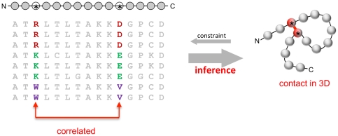
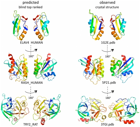
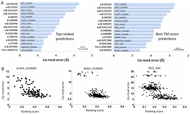
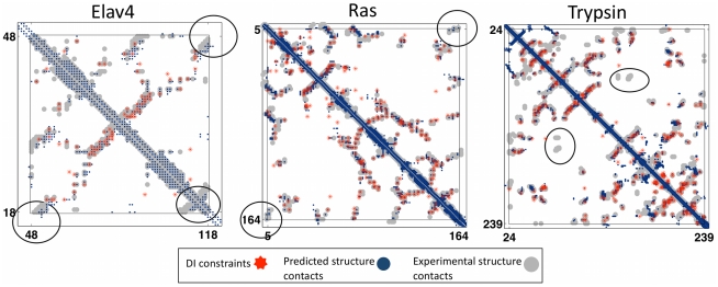
The evolutionary trajectory of a protein through sequence space is constrained by its function. Collections of sequence homologs record the outcomes of millions of evolutionary experiments in which the protein evolves according to these constraints. Deciphering the evolutionary record held in these sequences and exploiting it for predictive and engineering purposes presents a formidable challenge. The potential benefit of solving this challenge is amplified by the advent of inexpensive high-throughput genomic sequencing.In this paper we ask whether we can infer evolutionary constraints from a set of sequence homologs of a protein. The challenge is to distinguish true co-evolution couplings from the noisy set of observed correlations. We address this challenge using a maximum entropy model of the protein sequence, constrained by the statistics of the multiple sequence alignment, to infer residue pair couplings. Surprisingly, we find that the strength of these inferred couplings is an excellent predictor of residue-residue proximity in folded structures. Indeed, the top-scoring residue couplings are sufficiently accurate and well-distributed to define the 3D protein fold with remarkable accuracy.We quantify this observation by computing, from sequence alone, all-atom 3D structures of fifteen test proteins from different fold classes, ranging in size from 50 to 260 residues, including a G-protein coupled receptor. These blinded inferences are de novo, i.e., they do not use homology modeling or sequence-similar fragments from known structures. The co-evolution signals provide sufficient information to determine accurate 3D protein structure to 2.7-4.8 Å C(α)-RMSD error relative to the observed structure, over at least two-thirds of the protein (method called EVfold, details at http://EVfold.org). This discovery provides insight into essential interactions constraining protein evolution and will facilitate a comprehensive survey of the universe of protein structures, new strategies in protein and drug design, and the identification of functional genetic variants in normal and disease genomes.
Overview
The claims and statistics of this paper haven’t been checked yet.
Run the check — free, no account needed. It reads the open-access text and checks the sources it cites.
Integrity signals
Registry
Cites
Statistics
Figures
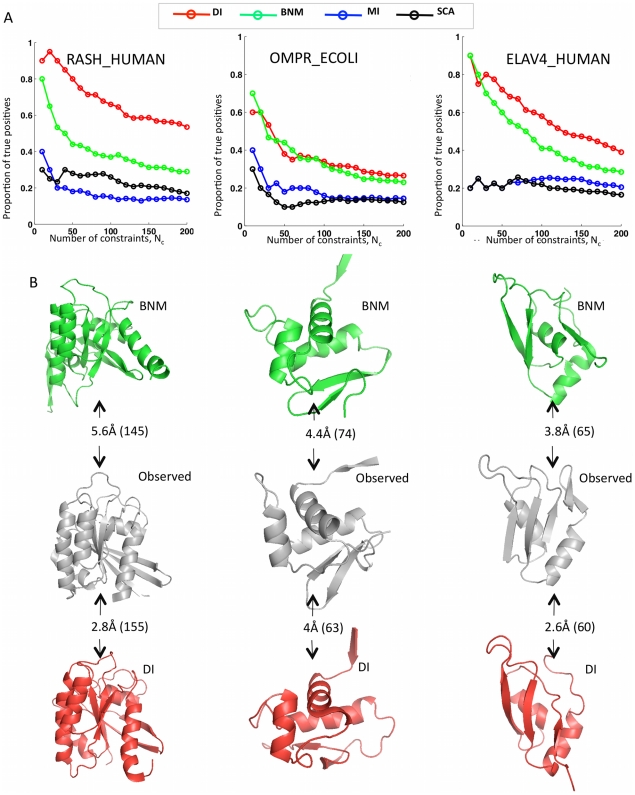
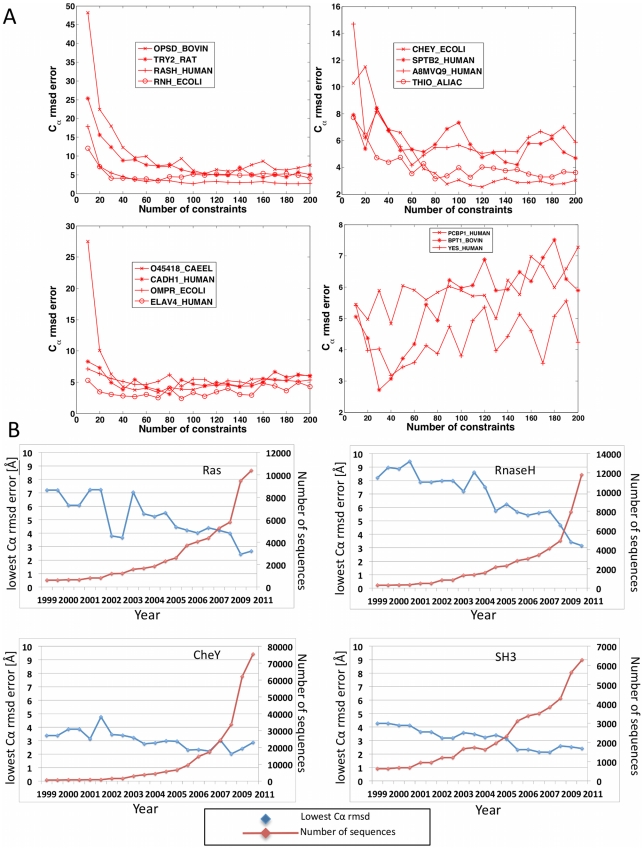
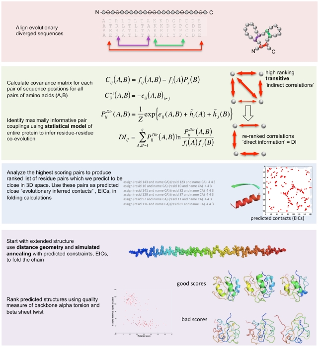
![Figure 3 Progress in contact prediction using the maximum entropy method. Extraction of evolutionary information about residue coupling and predicted contacts from multiple sequence alignments works much better using the global statistical model (right, Direct Information, DI, Equation 3) than the local statistical model (left, Mutual Information, MI, Equation 1). Predicted contacts for DI (red lines connecting the residues predicted to be coupled from sequence information) are better positioned in the experimentally observed structure (grey ribbon diagram), than those for MI (left, blue lines), shown here for the RAS protein (upper) and ELAV4 protein (lower). The DI residue pairs are also more evenly distributed along the chain and overlap more accurately with the contacts in the observed structure (red stars [predicted, grey circles [observed] in contact map; center, upper right triangle) than those using MI (blue [predicted], grey circles [observed]; center, lower left triangle). Details of contact maps for all proteins comparing predicted and observed contacts are in Figures S1 and S2, Text S1.](/paper-figures/ad0edff5338c41479018a65a69fdb4cd/protein-3d-structure-computed-from-evolutionary-sequence-variation-figure-3.jpg)
Verification
Claims the paper makes
The claims and statistics of this paper haven’t been checked yet.
Run the check — free, no account needed. It reads the open-access text and checks the sources it cites.
Claims other papers make about this one
Each is checked against this paper’s own text: does it say what the citing paper says it does?
In the citing paper
“The premise of methods to infer structure based on evolutionary couplings is that, because amino acids exist and evolve in the context of 3D structure, they are not free to evolve independently, but instead co-evolve in patterns reflective of the underlying structure.”
From Predicting multiple conformations via sequence clustering and AlphaFold2 (2024) · cites this paper as [9]
In the citing paper
“One of the major insights leveraged by AlphaFold 1,2 comes from the use of evolutionary covariations that can be extracted from large-scale multiple sequence alignments.[49][50][51][52][53][54]”
From AlphaFold and Implications for Intrinsically Disordered Proteins (2021) · cites this paper as [50]
Self-check
Percentages, sums and reported differences re-computed from the paper’s own numbers.
The self-check needs the paper’s full text.
Citations
Cites
The PyMOL Molecular Graphics System
DeLano, W. L. (2002) · Medical Entomology and Zoology · Cited by 18,147
Crystallography & NMR System: A New Software Suite for Macromolecular Structure Determination
Brünger, A. T., Adams, P. D., Clore, G. M., DeLano, W. L., Gros, P., Grosse‐Kunstleve, R. W., Jiang, J., Kuszewski, J. J., Nilges, M., Pannu, N., et al. (1998) · Acta Crystallographica Section D Biological Crystallography · doi:10.1107/s0907444998003254 · Cited by 15,723
The Pfam Protein Families Database
Bateman, A. G. (2002) · Nucleic Acids Research · doi:10.1093/nar/30.1.276 · Cited by 14,075
I-TASSER: a unified platform for automated protein structure and function prediction
Roy, A., Küçükural, A., & Zhang, Y. (2010) · Nature Protocols · doi:10.1038/nprot.2010.5 · Cited by 6,212
The Pfam protein families database
Finn, R. D., Mistry, J., Tate, J., Coggill, P., Heger, A., Pollington, J. E., Gavin, O. L., Gunasekaran, P., Ceric, G., Forslund, K., et al. (2010) · Nucleic Acids Research · doi:10.1093/nar/gkp985 · Cited by 2,694
Scoring function for automated assessment of protein structure template quality
Zhang, Y., & Skolnick, J. (2004) · Proteins: Structure, Function, and Bioinformatics · doi:10.1002/prot.20264 · Cited by 2,392
Atomic-level characterization of the structural dynamics of proteins
Shaw, D. E., Maragakis, P., Lindorff-Larsen, K., Piana, S., Dror, R. O., Eastwood, M. P., Bank, J. A., Jumper, J. M., Salmon, J. K., Shan, Y., et al. (2010) · Science · doi:10.1126/science.1187409 · Cited by 1,923
Weak pairwise correlations imply strongly correlated network states in a neural population
Schneidman, E., Berry, M. J., Segev, R., & Bialek, W. (2006) · Nature · doi:10.1038/nature04701 · Cited by 1,745
Holm, L., & Sander, C. (1996) · Science · doi:10.1126/science.273.5275.595 · Cited by 1,497
Self-citationMorcos, F., Pagnani, A., Lunt, B., Bertolino, A., Marks, D. S., Sander, C., Zecchina, R., Onuchic, J. N., Hwa, T., & Weigt, M. (2011) · Proceedings of the National Academy of Sciences · doi:10.1073/pnas.1111471108 · Cited by 1,434
Self-citation
Research works printed but not linked: 3
- KT Simons, 1999, Ab initio protein structure prediction of CASP III targets using ROSETTA., Proteins, Suppl 3, 171, 10.1002/(SICI)1097-0134(1999)37:3+<171::AID-PROT21>3.0.CO;2-Z · DOI
- MMT Mezard, 2009, Constraint satisfaction problems and neural networks: A statistical physics perspective., Journal of Physiology, 103, 107
- TR Lezon, 2006, Using the principle of entropy maximization to infer genetic interaction networks from gene expression patterns., Proceedings of the National Academy of Sciences of the United States of America, 103, 19033, 10.1073/pnas.0609152103 · DOI
Cited by
Sorted by citations
Highly accurate protein structure prediction with AlphaFold
Jumper, J., Evans, R., Pritzel, A., Green, T., Figurnov, M., Ronneberger, O., Tunyasuvunakool, K., Bates, R., Žídek, A., Potapenko, A., et al. (2021) · Nature · doi:10.1038/s41586-021-03819-2 · Cited by 43,764
Rives, A., Meier, J., Sercu, T., Goyal, S., Lin, Z., Liu, J., Guo, D., Ott, M., Zitnick, C. L., Ma, J., et al. (2021) · Proceedings of the National Academy of Sciences · doi:10.1073/pnas.2016239118 · Cited by 3,286
ProtTrans: Toward Understanding the Language of Life Through Self-Supervised Learning
Elnaggar, A., Heinzinger, M., Dallago, C., Rehawi, G., Wang, Y., Jones, L., Gibbs, T., Feher, T., Angerer, C., Steinegger, M., et al. (2022) · IEEE Transactions on Pattern Analysis and Machine Intelligence · doi:10.1109/tpami.2021.3095381 · Cited by 2,329
Opportunities and obstacles for deep learning in biology and medicine
Ching, T., Himmelstein, D. S., Beaulieu-Jones, B. K., Kalinin, A. A., Do, B. T., Way, G. P., Ferrero, E., Agapow, P.-M., Zietz, M., Hoffman, M. M., et al. (2018) · Journal of The Royal Society Interface · doi:10.1098/rsif.2017.0387 · Cited by 2,310
HHblits: lightning-fast iterative protein sequence searching by HMM-HMM alignment
Remmert, M., Biegert, A., Hauser, A., & Söding, J. (2012) · Nature Methods · doi:10.1038/nmeth.1818 · Cited by 2,288
JPred4: a protein secondary structure prediction server
Drozdetskiy, A., Cole, C., Procter, J. B., & Barton, G. J. (2015) · Nucleic Acids Research · doi:10.1093/nar/gkv332 · Cited by 1,714
The Protein-Folding Problem, 50 Years On
Dill, K. A., & MacCallum, J. L. (2012) · Science · doi:10.1126/science.1219021 · Cited by 1,699
Accurate De Novo Prediction of Protein Contact Map by Ultra-Deep Learning Model
Wang, S., Sun, S., Li, Z., Zhang, R., & Xu, J. (2017) · PLOS Computational Biology · doi:10.1371/journal.pcbi.1005324 · Cited by 931
Perspective: Coarse-grained models for biomolecular systems
Noid, W. G. (2013) · The Journal of Chemical Physics · doi:10.1063/1.4818908 · Cited by 907
Advances in protein structure prediction and design
Kuhlman, B., & Bradley, P. (2019) · Nature Reviews Molecular Cell Biology · doi:10.1038/s41580-019-0163-x · Cited by 806
Disease variant prediction with deep generative models of evolutionary data
Frazer, J., Notin, P., Dias, M., Gomez, A., Min, J. K., Brock, K., Gal, Y., & Marks, D. S. (2021) · Nature · doi:10.1038/s41586-021-04043-8 · Cited by 791
Self-citationMutation effects predicted from sequence co-variation
Hopf, T. A., Ingraham, J. B., Poelwijk, F. J., Schärfe, C. P. I., Springer, M., Sander, C., & Marks, D. S. (2017) · Nature Biotechnology · doi:10.1038/nbt.3769 · Cited by 775
Self-citationPerformance and Its Limits in Rigid Body Protein-Protein Docking
Desta, I., Porter, K. A., Xia, B., Kozakov, D., & Vajda, S. (2020) · Structure · doi:10.1016/j.str.2020.06.006 · Cited by 709
AlphaFold and Implications for Intrinsically Disordered Proteins
Ruff, K. M., & Pappu, R. V. (2021) · Journal of Molecular Biology · doi:10.1016/j.jmb.2021.167208 · Cited by 635
Improved contact prediction in proteins: using pseudolikelihoods to infer Potts models
Ekeberg, M., Lövkvist, C., Lan, Y., Weigt, M., & Aurell, E. (2013) · Physical Review E · doi:10.1103/physreve.87.012707 · Cited by 661
Kamisetty, H., Ovchinnikov, S., & Baker, D. A. (2013) · Proceedings of the National Academy of Sciences · doi:10.1073/pnas.1314045110 · Cited by 672
Computational methods in drug discovery
Leelananda, S. P., & Lindert, S. (2016) · Beilstein Journal of Organic Chemistry · doi:10.3762/bjoc.12.267 · Cited by 675
Ovchinnikov, S., Kamisetty, H., & Baker, D. (2014) · eLife · doi:10.7554/elife.02030 · Cited by 629
Protein structure prediction from sequence variation
Marks, D. S., Hopf, T. A., & Sander, C. (2012) · Nature Biotechnology · doi:10.1038/nbt.2419 · Cited by 622
Self-citationEmerging methods in protein co-evolution
de Juan, D., Pazos, F., & Valencia, A. (2013) · Nature Reviews Genetics · doi:10.1038/nrg3414 · Cited by 607
Modeling aspects of the language of life through transfer-learning protein sequences
Heinzinger, M., Elnaggar, A., Wang, Y., Dallago, C., Nechaev, D., Matthes, F., & Rost, B. (2019) · BMC Bioinformatics · doi:10.1186/s12859-019-3220-8 · Cited by 606
Modeling the language of life – Deep Learning Protein Sequences
Heinzinger, M., Elnaggar, A., Wang, Y., Dallago, C., Nechaev, D., Matthes, F., & Rost, B. (2019) · doi:10.1101/614313 · Cited by 606
Deep generative models of genetic variation capture the effects of mutations
Riesselman, A. J., Ingraham, J. B., & Marks, D. S. (2018) · Nature Methods · doi:10.1038/s41592-018-0138-4 · Cited by 601
Self-citationZheng, W. H., Zhang, C., Li, Y., Pearce, R., Bell, E. W., & Zhang, Y. (2021) · Cell Reports Methods · doi:10.1016/j.crmeth.2021.100014 · Cited by 588
Epistasis in protein evolution
Starr, T. N., & Thornton, J. W. (2016) · Protein Science · doi:10.1002/pro.2897 · Cited by 598
Predicting multiple conformations via sequence clustering and AlphaFold2
Wayment-Steele, H. K., Ojoawo, A., Otten, R., Apitz, J. M., Pitsawong, W., Hömberger, M., Ovchinnikov, S., Colwell, L., & Kern, D. (2024) · Nature · doi:10.1038/s41586-023-06832-9 · Cited by 545
Self-citationZhou, X., Zheng, W. H., Li, Y., Pearce, R., Zhang, C., Bell, E. W., Zhang, G., & Zhang, Y. (2022) · Nature Protocols · doi:10.1038/s41596-022-00728-0 · Cited by 538
Protein structure determination using metagenome sequence data
Ovchinnikov, S., Park, H., Varghese, N., Huang, P.-S., Pavlopoulos, G. A., Kim, D. E., Kamisetty, H., Kyrpides, N. C., & Baker, D. (2017) · Science · doi:10.1126/science.aah4043 · Cited by 520
Sequence co-evolution gives 3D contacts and structures of protein complexes
Hopf, T. A., Schärfe, C. P. I., Rodrigues, J. P. G. L. M., Green, A. G., Kohlbacher, O., Sander, C., Bonvin, A. M. J. J., & Marks, D. S. (2014) · eLife · doi:10.7554/elife.03430 · Cited by 515
Self-citationThree-Dimensional Structures of Membrane Proteins from Genomic Sequencing
Hopf, T. A., Colwell, L. J., Sheridan, R., Rost, B., Sander, C., & Marks, D. S. (2012) · Cell · doi:10.1016/j.cell.2012.04.012 · Cited by 509
Self-citationAhdritz, G., Bouatta, N., Floristean, C., Kadyan, S., Xia, Q., Gerecke, W., O’Donnell, T. J., Berenberg, D., Fisk, I., Zanichelli, N., et al. (2024) · Nature Methods · doi:10.1038/s41592-024-02272-z · Cited by 439
Geometric deep learning of RNA structure
Townshend, R. J. L., Eismann, S., Watkins, A. M., Rangan, R., Karelina, M., Das, R., & Dror, R. O. (2021) · Science · doi:10.1126/science.abe5650 · Cited by 424
CCMpred—fast and precise prediction of protein residue–residue contacts from correlated mutations
Seemayer, S., Gruber, M., & Söding, J. (2014) · Bioinformatics · doi:10.1093/bioinformatics/btu500 · Cited by 416
Distance-based protein folding powered by deep learning
Xu, J. (2019) · Proceedings of the National Academy of Sciences · doi:10.1073/pnas.1821309116 · Cited by 401
Rives, A., Meier, J., Sercu, T., Goyal, S., Lin, Z., Liu, J., Guo, D., Ott, M., Zitnick, C. L., Ma, J., et al. (2019) · doi:10.1101/622803 · Cited by 3,280
“Contacts can be inferred through evolutionary correlations between residues in a sequence family (Marks et al., 2011; Anishchenko et al., 2017).”
BackgroundProtTrans: Towards Cracking the Language of Life’s Code Through Self-Supervised LearningPreprint
Elnaggar, A., Heinzinger, M., Dallago, C., Rehawi, G., Wang, Y., Jones, L., Gibbs, T., Feher, T., Angerer, C., Steinegger, M., et al. (2020) · bioRxiv (Cold Spring Harbor Laboratory) · doi:10.1101/2020.07.12.199554 · Cited by 424
Expanding functional protein sequence spaces using generative adversarial networks
Repecka, D., Jauniskis, V., Karpus, L., Rembeza, E., Rokaitis, I., Zrimec, J., Poviloniene, S., Laurynenas, A., Viknander, S., Abuajwa, W., et al. (2021) · Nature Machine Intelligence · doi:10.1038/s42256-021-00310-5 · Cited by 388
Bouchard, J. J., Otero, J. H., Scott, D. C., Szulc, E., Martin, E. W., Sabri, N., Granata, D., Marzahn, M. R., Lindorff‐Larsen, K., Salvatella, X., et al. (2018) · Molecular Cell · doi:10.1016/j.molcel.2018.08.027 · Cited by 387
Currin, A., Swainston, N., Day, P. J., & Kell, D. B. (2014) · Chemical Society Reviews · doi:10.1039/c4cs00351a · Cited by 393
End-to-End Differentiable Learning of Protein Structure
AlQuraishi, M. (2019) · Cell Systems · doi:10.1016/j.cels.2019.03.006 · Cited by 371
An evolution-based model for designing chorismate mutase enzymes
Russ, W. P., Figliuzzi, M., Stocker, C., Barrat-Charlaix, P., Socolich, M., Kast, P., Hilvert, D., Monasson, R., Cocco, S., Weigt, M., et al. (2020) · Science · doi:10.1126/science.aba3304 · Cited by 313
TMC1 Forms the Pore of Mechanosensory Transduction Channels in Vertebrate Inner Ear Hair Cells
Pan, B., Akyuz, N., Liu, X.-P., Asai, Y., Nist-Lund, C. A., Kurima, K., Derfler, B. H., György, B., Limapichat, W., Walujkar, S., et al. (2018) · Neuron · doi:10.1016/j.neuron.2018.07.033 · Cited by 356
Jones, D. T., Singh, T., Kościółek, T., & Tetchner, S. (2014) · Bioinformatics · doi:10.1093/bioinformatics/btu791 · Cited by 345
A Comprehensive Biophysical Description of Pairwise Epistasis throughout an Entire Protein Domain
Olson, C. A., Wu, N. C., & Sun, R. (2014) · Current Biology · doi:10.1016/j.cub.2014.09.072 · Cited by 340
Transformer protein language models are unsupervised structure learners
Rao, R., Meier, J., Sercu, T., Ovchinnikov, S., & Rives, A. (2020) · doi:10.1101/2020.12.15.422761 · Cited by 341
Protein design and variant prediction using autoregressive generative models
Shin, J.-E., Riesselman, A. J., Kollasch, A. W., McMahon, C., Simon, E., Sander, C., Manglik, A., Kruse, A. C., & Marks, D. S. (2021) · Nature Communications · doi:10.1038/s41467-021-22732-w · Cited by 320
Self-citationγ-Aminobutyric acid (GABA) signalling in plants
Ramesh, S. A., Tyerman, S. D., Gilliham, M., & Xu, B. (2016) · Cellular and Molecular Life Sciences · doi:10.1007/s00018-016-2415-7 · Cited by 326
AlQuraishi, M. (2019) · Bioinformatics · doi:10.1093/bioinformatics/btz422 · Cited by 333
Structure and conformational states of the bovine mitochondrial ATP synthase by cryo-EM
Zhou, A., Rohou, A. L., Schep, D. G., Bason, J. V., Montgomery, M. G., Walker, J. E., Grigorieff, N., & Rubinstein, J. L. (2015) · eLife · doi:10.7554/elife.10180 · Cited by 298
Should evolutionary geneticists worry about higher-order epistasis?
Weinreich, D. M., Lan, Y., Wylie, C. S., & Heckendorn, R. B. (2013) · Current Opinion in Genetics & Development · doi:10.1016/j.gde.2013.10.007 · Cited by 277
Network Topology Inference from Spectral Templates
Segarra, S., Marques, A. G., Mateos, G., & Ribeiro, A. (2017) · IEEE Transactions on Signal and Information Processing over Networks · doi:10.1109/tsipn.2017.2731051 · Cited by 286
Network deconvolution as a general method to distinguish direct dependencies in networks
Feizi, S., Marbach, D., Médard, M., & Kellis, M. (2013) · Nature Biotechnology · doi:10.1038/nbt.2635 · Cited by 287
The EVcouplings Python framework for coevolutionary sequence analysis
Hopf, T. A., Green, A. G., Schubert, B., Mersmann, S., Schärfe, C., Ingraham, J., Tóth-Petróczy, Á., Brock, K. P., Riesselman, A. J., Palmedo, P., et al. (2018) · Bioinformatics · doi:10.1093/bioinformatics/bty862 · Cited by 286
Self-citationDeep learning methods in protein structure prediction
Torrisi, M., Pollastri, G., & Le, Q. (2020) · Computational and Structural Biotechnology Journal · doi:10.1016/j.csbj.2019.12.011 · Cited by 297
Inverse statistical problems: from the inverse Ising problem to data science
Nguyen, H. C., Zecchina, R., & Berg, J. (2017) · Advances In Physics · doi:10.1080/00018732.2017.1341604 · Cited by 319
Self-citationThe Causes and Consequences of Genetic Interactions (Epistasis)
Domingo, J., Baeza-Centurion, P., & Lehner, B. (2019) · Annual Review of Genomics and Human Genetics · doi:10.1146/annurev-genom-083118-014857 · Cited by 242
Protein Contact Networks: An Emerging Paradigm in Chemistry
Paola, L. D., Ruvo, M. D., Paci, P., Santoni, D., & Giuliani, A. (2012) · Chemical Reviews · doi:10.1021/cr3002356 · Cited by 270
Generative models for graph-based protein design
Ingraham, J., Garg, V., Barzilay, R., & Jaakkola, T. (2019) · DSpace@MIT (Massachusetts Institute of Technology) · Cited by 258
Controlling Allosteric Networks in Proteins
Dokholyan, N. V. (2016) · Chemical Reviews · doi:10.1021/acs.chemrev.5b00544 · Cited by 260
SPEACH_AF: Sampling protein ensembles and conformational heterogeneity with Alphafold2
Stein, R. A., & Mchaourab, H. S. (2022) · PLOS Computational Biology · doi:10.1371/journal.pcbi.1010483 · Cited by 258
A Guide to In Silico Drug Design
Chang, Y., Hawkins, B. A., Du, J. J., Groundwater, P. W., Hibbs, D. E., & Lai, F. (2022) · Pharmaceutics · doi:10.3390/pharmaceutics15010049 · Cited by 296
Elnaggar, A., Heinzinger, M., Dallago, C., Rihawi, G., Wang, Y., Jones, L., Gibbs, T., Fehér, T., Angerer, C. M., Steinegger, M., et al. (2020) · arXiv (Cornell University) · doi:10.48550/arxiv.2007.06225 · Cited by 364
Thermodynamics and signatures of criticality in a network of neurons
Tkačik, G., Mora, T., Marre, O., Amodei, D., Palmer, S. E., Berry, M. J., & Bialek, W. (2015) · Proceedings of the National Academy of Sciences · doi:10.1073/pnas.1514188112 · Cited by 271
Machine learning in protein structure prediction
AlQuraishi, M. (2021) · Current Opinion in Chemical Biology · doi:10.1016/j.cbpa.2021.04.005 · Cited by 263
Searching for Collective Behavior in a Large Network of Sensory Neurons
Tkačik, G., Marre, O., Amodei, D., Schneidman, E., Bialek, W., & Berry, M. J. (2014) · PLoS Computational Biology · doi:10.1371/journal.pcbi.1003408 · Cited by 252
Inverse statistical physics of protein sequences: a key issues review
Cocco, S., Feinauer, C., Figliuzzi, M., Monasson, R., & Weigt, M. (2017) · Reports on Progress in Physics · doi:10.1088/1361-6633/aa9965 · Cited by 260
Protein interaction networks revealed by proteome coevolution
Cong, Q., Anishchenko, I., Ovchinnikov, S., & Baker, D. (2019) · Science · doi:10.1126/science.aaw6718 · Cited by 241
Sixty-five years of the long march in protein secondary structure prediction: the final stretch?
Yang, Y., Gao, J., Wang, J., Heffernan, R., Hanson, J., Paliwal, K. K., & Zhou, Y. (2016) · Briefings in Bioinformatics · doi:10.1093/bib/bbw129 · Cited by 240
Genomics-aided structure prediction
Sulkowska, J. I., Morcos, F., Weigt, M., Terence Hwa, B., & Onuchic, J. N. (2012) · Proceedings of the National Academy of Sciences · doi:10.1073/pnas.1207864109 · Cited by 226
Araya, C. L., Fowler, D. M., Chen, W., Muniez, I., Kelly, J. W., & Fields, S. (2012) · Proceedings of the National Academy of Sciences · doi:10.1073/pnas.1209751109 · Cited by 230
LDAF1 and Seipin Form a Lipid Droplet Assembly Complex
Chung, J., Wu, X., Lambert, T. J., Lai, Z. W., Walther, T. C., & Farese, R. V. (2019) · Developmental Cell · doi:10.1016/j.devcel.2019.10.006 · Cited by 229
Modeling protein–protein and protein–peptide complexes: CAPRI 6th edition
Lensink, M. F., Velankar, S., & Wodak, S. J. (2017) · Proteins Structure Function and Bioinformatics · doi:10.1002/prot.25215 · Cited by 222
Deep Learning in Protein Structural Modeling and Design
Gao, W., Mahajan, S. P., Sulam, J., & Gray, J. J. (2020) · Patterns · doi:10.1016/j.patter.2020.100142 · Cited by 227
Hopf, T. A., Ingraham, J., Poelwijk, F. J., Springer, M., Sander, C., & Marks, D. S. (2015) · arXiv (Cornell University) · doi:10.48550/arxiv.1510.04612 · Cited by 249
Self-citationLarge-scale determination of previously unsolved protein structures using evolutionary information
Ovchinnikov, S., Kinch, L., Park, H., Liao, Y., Pei, J., Kim, D. E., Kamisetty, H., Grishin, N. V., & Baker, D. (2015) · eLife · doi:10.7554/elife.09248 · Cited by 222
3D clusters of somatic mutations in cancer reveal numerous rare mutations as functional targets
Gao, J., Chang, M. T., Johnsen, H. C., Gao, S. P., Sylvester, B. E., Sümer, S. O., Zhang, H., Solit, D. B., Taylor, B. S., Schultz, N. D., et al. (2017) · Genome Medicine · doi:10.1186/s13073-016-0393-x · Cited by 220
Self-citationEvol and ProDy for bridging protein sequence evolution and structural dynamics
Bakan, A., Dutta, A., Mao, W., Liu, Y., Chennubhotla, C., Lezon, T. R., & Bahar, İ. (2014) · Bioinformatics · doi:10.1093/bioinformatics/btu336 · Cited by 214
Information Processing in Living Systems
Tkačik, G., & Bialek, W. (2016) · Annual Review of Condensed Matter Physics · doi:10.1146/annurev-conmatphys-031214-014803 · Cited by 224
Sledzieski, S., Singh, R., Cowen, L., & Berger, B. (2021) · Cell Systems · doi:10.1016/j.cels.2021.08.010 · Cited by 235
Generating functional protein variants with variational autoencoders
Hawkins‐Hooker, A., Depardieu, F., Baur, S., Couairon, G., Chen, A., & Bikard, D. (2021) · PLoS Computational Biology · doi:10.1371/journal.pcbi.1008736 · Cited by 217
Toward optimal fragment generations for ab initio protein structure assembly
Xu, D. H., & Zhang, Y. (2012) · Proteins Structure Function and Bioinformatics · doi:10.1002/prot.24179 · Cited by 206
Amino acid coevolution induces an evolutionary Stokes shift
Pollock, D. D., Thiltgen, G., & Goldstein, R. A. (2012) · Proceedings of the National Academy of Sciences · doi:10.1073/pnas.1120084109 · Cited by 193
Protein–protein interaction prediction with deep learning: A comprehensive review
Soleymani, F., Paquet, E., Viktor, H. L., Michalowski, W., & Spinello, D. (2022) · Computational and Structural Biotechnology Journal · doi:10.1016/j.csbj.2022.08.070 · Cited by 209
Tamir, S., Paddock, M. L., Darash-Yahana-Baram, M., Holt, S. H., Sohn, Y. S., Agranat-Tamir, L., Michaeli, D., Stofleth, J. T., Lipper, C. H., Morcos, F., et al. (2014) · Biochimica et Biophysica Acta (BBA) - Molecular Cell Research · doi:10.1016/j.bbamcr.2014.10.014 · Cited by 198
Cryo–electron microscopy structure of the lipid droplet–formation protein seipin
Sui, X., Arlt, H., Brock, K. P., Lai, Z. W., DiMaio, F., Marks, D. S., Liao, M., Farese, R. V., & Walther, T. C. (2018) · The Journal of Cell Biology · doi:10.1083/jcb.201809067 · Cited by 188
Self-citationApplications of targeted proteomics in systems biology and translational medicine
Ebhardt, H. A., Root, A., Sander, C., & Aebersold, R. (2015) · PROTEOMICS · doi:10.1002/pmic.201500004 · Cited by 184
Self-citationOrigins of coevolution between residues distant in protein 3D structures
Anishchenko, I., Ovchinnikov, S., Kamisetty, H., & Baker, D. (2017) · Proceedings of the National Academy of Sciences · doi:10.1073/pnas.1702664114 · Cited by 185
Ekeberg, M., Hartonen, T., & Aurell, E. M. (2014) · Journal of Computational Physics · doi:10.1016/j.jcp.2014.07.024 · Cited by 184
Jones, D. T., & Kandathil, S. M. (2018) · Bioinformatics · doi:10.1093/bioinformatics/bty341 · Cited by 183
The landscape of tolerated genetic variation in humans and primates
(2023) · doi:10.1126/science.abn8197 · Cited by 182
Greener, J. G., Kandathil, S. M., & Jones, D. T. (2019) · Nature Communications · doi:10.1038/s41467-019-11994-0 · Cited by 178
Assessment of contact predictions in CASP12: Co‐evolution and deep learning coming of age
Schaarschmidt, J., Monastyrskyy, B., Kryshtafovych, A., & Bonvin, A. M. J. J. (2018) · Proteins: Structure, Function, and Bioinformatics · doi:10.1002/prot.25407 · Cited by 177
Nugent, T., & Jones, D. T. (2012) · Proceedings of the National Academy of Sciences · doi:10.1073/pnas.1120036109 · Cited by 177
Cryo-EM structure of the human L-type amino acid transporter 1 in complex with glycoprotein CD98hc
Lee, Y., Wiriyasermkul, P., Jin, C., Quan, L., Ohgaki, R., Okuda, S., Kusakizako, T., Nishizawa, T., Oda, K., Ishitani, R., et al. (2019) · Nature Structural & Molecular Biology · doi:10.1038/s41594-019-0237-7 · Cited by 175
ECNet is an evolutionary context-integrated deep learning framework for protein engineering
Luo, Y., Jiang, G., Yu, T., Liu, Y., Vo, L. T., Ding, H., Su, Y., Qian, W. W., Zhao, H., & Peng, J. (2021) · Nature Communications · doi:10.1038/s41467-021-25976-8 · Cited by 174
Structures of the human mitochondrial ribosome in native states of assembly
Brown, A., Rathore, S., Kimanius, D., Aibara, S., Bai, X., Rorbach, J., Amunts, A., & Ramakrishnan, V. (2017) · Nature Structural & Molecular Biology · doi:10.1038/nsmb.3464 · Cited by 172
Machine learning for protein folding and dynamics
Noé, F., Fabritiis, G. D., & Clementi, C. (2019) · Current Opinion in Structural Biology · doi:10.1016/j.sbi.2019.12.005 · Cited by 177
Coevolutionary signals across protein lineages help capture multiple protein conformations
Morcos, F., Jana, B., Terence Hwa, B., & Onuchic, J. N. (2013) · Proceedings of the National Academy of Sciences · doi:10.1073/pnas.1315625110 · Cited by 170
Kell, D. B., & Goodacre, R. (2013) · Drug Discovery Today · doi:10.1016/j.drudis.2013.07.014 · Cited by 175
Biophysical and Mechanistic Models for Disease-Causing Protein Variants
Stein, A., Fowler, D. M., Hartmann‐Petersen, R., & Lindorff‐Larsen, K. (2019) · Trends in Biochemical Sciences · doi:10.1016/j.tibs.2019.01.003 · Cited by 167
Metadata
- Authors
- Marks, D. S., Colwell, L. J., Sheridan, R., Hopf, T. A., Pagnani, A., Zecchina, R., & Sander, C.
- Affiliations
- Harvard University (US)
- MRC Laboratory of Molecular Biology (GB)
- Memorial Sloan Kettering Cancer Center (US)
- Italian institute for Genomic Medicine (IT)
- Politecnico di Torino (IT)
- Corresponding author
- Marks, Debora S.
- Venue
- PLoS ONE
- Publisher
- Public Library of Science (PLoS)
- Published
- 2011-12-07
- Volume
- 6
- Issue
- 12
- Pages
- e28766
- ISSN
- 1932-6203 (ISSN-L)
- PMC
- PMC3233603
- OpenAlex
- W2061042699
- Cited by
- 1,144
- References
- 84
- Citations by year
- 2026: 61 · 2025: 60 · 2024: 47 · 2023: 72 · 2022: 96 · 2021: 104 · 2020: 90 · 2019: 110 · 2018: 91 · 2017: 107 · 2016: 71 · 2015: 74 · 2014: 72 · 2013: 56 · 2012: 29
- FWCI
- 20.79
- Type
- journal article
- Language
- en
- Open access
- gold
- doi.org
- Locations
- PLoS ONE (publishedVersion, cc-by)
- Europe PMC (PubMed Central) (publishedVersion, cc-by)
- PubMed Central (publishedVersion, cc-by)
- PubMed (publishedVersion)
- PORTO@Iris (submittedVersion, cc-by)
- DOAJ (DOAJ: Directory of Open Access Journals) (submittedVersion, cc-by-sa)
- Figshare (submittedVersion, cc-by)
- PORTO Publications Open Repository TOrino (Politecnico di Torino) (submittedVersion, cc-by)
- Funders
- Memorial Sloan-Kettering Cancer Center
- Studienstiftung des Deutschen Volkes
- National Institutes of Health (NIH U54, CA143798)
- Engineering and Physical Sciences Research Council (EP/H028064/1)
- National Cancer Institute (U54CA143798)
- National Institute of General Medical Sciences (R01GM106303)
- Topics
- Life Sciences › Biochemistry, Genetics and Molecular Biology › Molecular Biology › Protein Structure and Dynamics
- Life Sciences › Biochemistry, Genetics and Molecular Biology › Structural Biology › Enzyme Structure and Function
- Life Sciences › Biochemistry, Genetics and Molecular Biology › Molecular Biology › RNA and protein synthesis mechanisms
- Keywords
- protein structure prediction, coevolution, maximum entropy model, multiple sequence alignment, de novo structure prediction, protein evolution, G protein-coupled receptors, protein engineering
- MeSH terms
- Animals, Humans, Protein Conformation, Proteins, Rhodopsin, Trypsin, Genetic Variation, Drug Design, Reproducibility of Results, Models, Statistical, Genome, Protein Structure, Tertiary, Evolution, Molecular, Entropy, Computational Biology, Catalytic Domain, Imaging, Three-Dimensional
About these checks
Registry, retraction, Cites and statistics results are computed, not judged, with one exception: a printed reference that no index lists is read once by a model before it counts as a citation error. Claims are extracted by a model and kept only when their quote appears verbatim in the text; each cited source is then checked, with a model’s help, for whether it says what the claim relies on. Those verdicts can be wrong — read the claim’s quote and the cited source before relying on one.
Statistics and arithmetic are recomputed, not judged.
Confidence bands are shown only for fields calibrated on our annotated test set. None is calibrated yet, so no band is shown.
What counts as a claim ›How we measure confidence ›How we check this ›
Are you an author, or do you see a problem with this page? Request a review
