

The DOI matches its Crossref record; 60 of the 60 references it cites were found in a registry or an index.
Commensal Akkermansia muciniphila Exacerbates Gut Inflammation in Salmonella Typhimurium-Infected Gnotobiotic Mice
Open accessAbstract
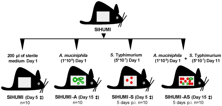
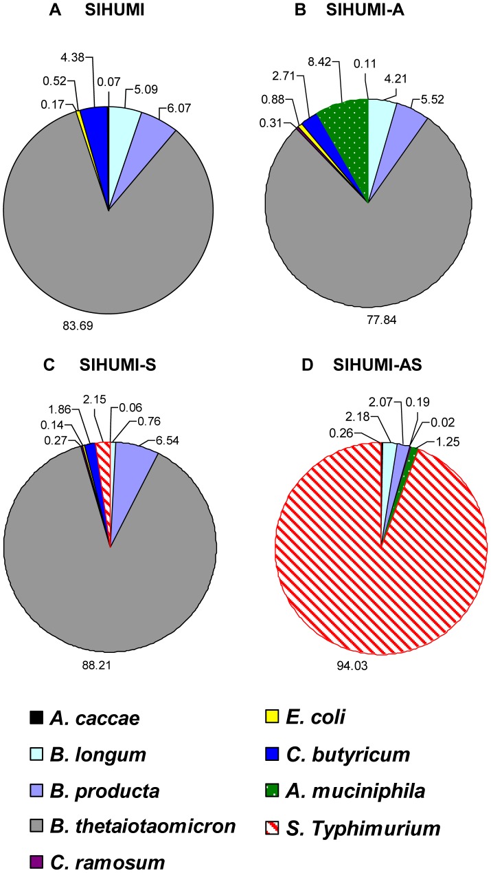
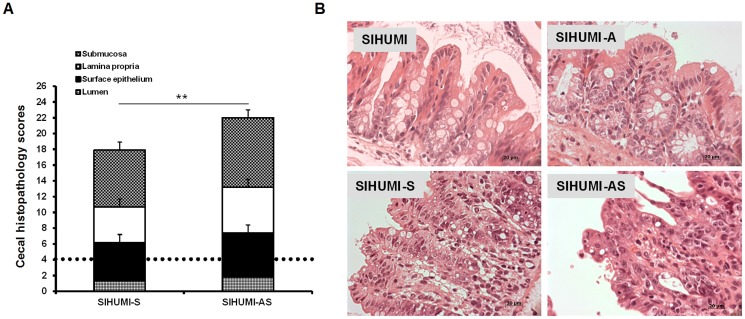
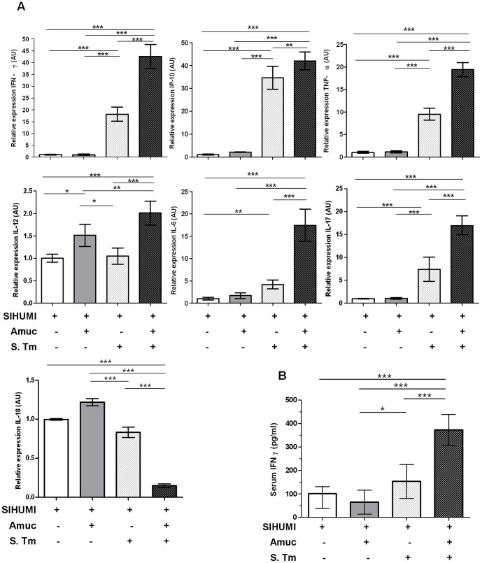
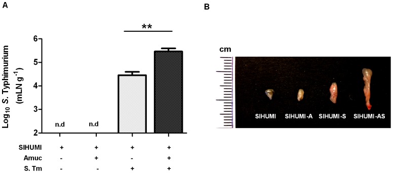
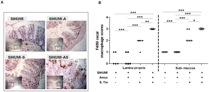
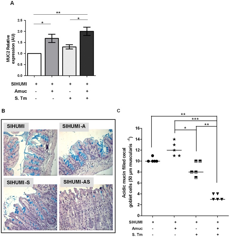
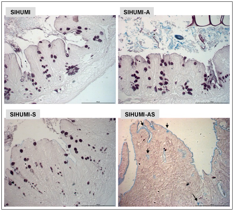
Excessive mucin degradation by intestinal bacteria may contribute to inflammatory bowel diseases because access of luminal antigens to the intestinal immune system is facilitated. This study investigated how the presence of a mucin degrading commensal bacterium affects the severity of an intestinal Salmonella enterica Typhimurium-induced gut inflammation. Using a gnotobiotic C3H mouse model with a background microbiota of eight bacterial species (SIHUMI) the impact of the mucin-degrading commensal bacterium Akkermansia muciniphila (SIHUMI-A) on inflammatory and infectious symptoms caused by S. Typhimurium was investigated. Presence of A. muciniphila in S. Typhimurium-infected SIHUMI mice caused significantly increased histopathology scores and elevated mRNA levels of IFN-γ, IP-10, TNF-α, IL-12, IL-17 and IL-6 in cecal and colonic tissue. The increase in pro-inflammatory cytokines was accompanied by 10-fold higher S. Typhimurium cell numbers in mesenteric lymph nodes of SIHUMI mice associated with A. muciniphila and S. Typhimurium (SIHUMI-AS) compared to SIHUMI mice with S. Typhimurium only (SIHUMI-S). The number of mucin filled goblet cells was 2- to 3-fold lower in cecal tissue of SIHUMI-AS mice compared to SIHUMI-S, SIHUMI-A or SIHUMI mice. Reduced goblet cell numbers significantly correlated with increased IFN-γ mRNA levels (r(2) = -0.86, ***P<0.001) in all infected mice. In addition, loss of cecal mucin sulphation was observed in SIHUMI mice containing both A. muciniphila and S. Typhimurium compared to other mouse groups. Concomitant presence of A. muciniphila and S. Typhimurium resulted in a drastic change in microbiota composition of SIHUMI mice: the proportion of B. thetaiotaomicron in SIHUMI-AS mice was 0.02% of total bacteria compared to 78%-88% in the other mouse groups and the proportion of S. Typhimurium was 94% in SIHUMI-AS mice but only 2.2% in the SIHUMI-S mice. These results indicate that A. muciniphila exacerbates S. Typhimurium-induced intestinal inflammation by its ability to disturb host mucus homeostasis.
Overview
The claims and statistics of this paper haven’t been checked yet.
Run the check — free, no account needed. It reads the open-access text and checks the sources it cites.
Integrity signals
Registry
Cites
Statistics
Figures
Verification
Claims the paper makes
The claims and statistics of this paper haven’t been checked yet.
Run the check — free, no account needed. It reads the open-access text and checks the sources it cites.
Claims other papers make about this one
Each is checked against this paper’s own text: does it say what the citing paper says it does?
The cited source doesn’t support the claim as written.
In the citing paper
“It has been reported that the abundance of g_Akkermansia has a negative correlation with the value of body mass index (BMI) [59] and that g_Akkermansia has a protective effect for the mucus layer in pro-inflammation [60].”
From Preventive Effects and Mechanisms of Garlic on Dyslipidemia and Gut Microbiome Dysbiosis (2019) · cites this paper as [60]
Checked by a reasoning model — may be wrong.
Experimental — accuracy not yet measuredIn the citing paper
“The Gram-negative bacteria, Akkermansia, can degrade mucin, cause intestinal inflammation and permeability, and increase endotoxemia and systemic inflammation, eventually promoting neuropathology [53,54,55].”
From Neuroprotective Effects of Sodium Butyrate by Restoring Gut Microbiota and Inhibiting TLR4 Signaling in Mice with MPTP-Induced Parkinson’s Disease (2023) · cites this paper as [55]
In the citing paper
“its mucin degrading activity might also damage the gut-barrier triggering inflammation and promoting gut permeability [83,84]”
From The Interplay between Gut Microbiota and Parkinson’s Disease: Implications on Diagnosis and Treatment (2022) · cites this paper as [84]
Self-check
Percentages, sums and reported differences re-computed from the paper’s own numbers.
The self-check needs the paper’s full text.
Citations
Cites
Gut microbiota in health and disease
Sekirov, I., Russell, S. L., Antunes, L. C. M., & Finlay, B. B. (2010) · Physiological Reviews · doi:10.1152/physrev.00045.2009 · Cited by 4,182
Akkermansia muciniphila gen. nov., sp. nov., a human intestinal mucin-degrading bacterium
Derrien, M., Vaughan, E. E., Plugge, C. M., & de Vos, W. M. (2004) · International Journal of Systematic and Evolutionary Microbiology · doi:10.1099/ijs.0.02873-0 · Cited by 2,094 · cited 1×
“muciniphila is known for its ability to degrade mucins [16,17].”
BackgroundMechanisms of Disease: pathogenesis of Crohn's disease and ulcerative colitis
Sartor, R. B. (2006) · Nature Clinical Practice Gastroenterology & Hepatology · doi:10.1038/ncpgasthep0528 · Cited by 1,806
Intestinal Goblet Cells and Mucins in Health and Disease: Recent Insights and Progress
Kim, Y. S., & Ho, S. B. (2010) · Current Gastroenterology Reports · doi:10.1007/s11894-010-0131-2 · Cited by 1,531 · cited 1×
“muciniphila is the main intestinal representative of this phylum [2], suggesting that A.”
BackgroundMucin dynamics and enteric pathogens
McGuckin, M. A., Lindén, S. K., Sutton, P., & Florin, T. H. (2011) · Nature Reviews Microbiology · doi:10.1038/nrmicro2538 · Cited by 1,428 · cited 1×
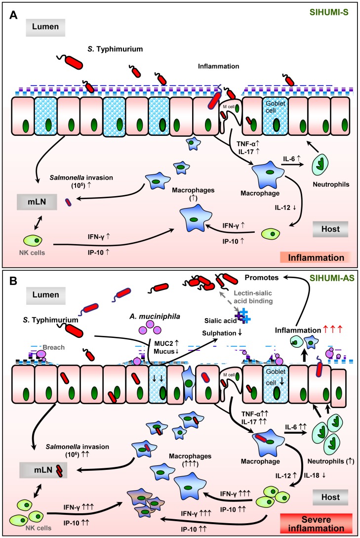
“(q increased; Q decreased; grey dotted line: assumed processes including lectin-sialic acid binding [56], M-cells for pathogen transit [43,58,59]; black line: supported by data of the present study).”
ResultGut inflammation provides a respiratory electron acceptor for Salmonella
Winter, S., Thiennimitr, P., Winter, M. G., Butler, B. P., Huseby, D. L., Crawford, R. W., Russell, J. M., Bevins, C. L., Adams, L. G., Tsolis, R. M., et al. (2010) · Nature · doi:10.1038/nature09415 · Cited by 1,271 · cited 1×
“[21,22] and therefore represents a highly suitable model for”
BackgroundStecher, B., Robbiani, R., Walker, A. W., Westendorf, A. M., Barthel, M., Kremer, M., Chaffron, S., Macpherson, A. J., Buer, J., Parkhill, J., et al. (2007) · PLoS Biology · doi:10.1371/journal.pbio.0050244 · Cited by 1,048 · cited 4×
“Typhimurium-induced gut inflammation [14,50] or in inflamed IL-10 mice [51].”
BackgroundInfluentialHost-Derived Nitrate Boosts Growth of E. coli in the Inflamed Gut
Winter, S. E., Winter, M. G., Xavier, M. N., Thiennimitr, P., Poon, V., Keestra, A. M., Laughlin, R. C., Gomez, G., Wu, J., Lawhon, S. D., et al. (2013) · Science · doi:10.1126/science.1232467 · Cited by 1,019 · cited 2×
“Typhimurium-induced gut inflammation [14,50] or in inflamed IL-10 mice [51].”
BackgroundHelicobacter pylori SabA Adhesin in Persistent Infection and Chronic Inflammation
Mahdavi, J., Sondén, B., Hurtig, M., Olfat, F. O., Forsberg, L., Roche, N., Ångström, J., Larsson, T. P., Teneberg, S., Karlsson, K., et al. (2002) · Science · doi:10.1126/science.1069076 · Cited by 935 · cited 1×
“Interestingly, changes in intestinal mucin composition characterized by a lower degree of sulphation [7,46] and a higher degree of sialylation have previously been reported to occur in UC, Crohn’s Disease (CD) and gastric ulcer caused by Helicobacter pylori-induced inflammation [7,46,47].”
BackgroundCollado, M. C., Derrien, M., Isolauri, E., de Vos, W. M., & Salminen, S. (2007) · Applied and Environmental Microbiology · doi:10.1128/aem.01477-07 · Cited by 706
Cited by
Sorted by citations
Items 401–500 of 525
Qu, D., Gu, Z., Feng, S., Yu, L., Tian, F., Zhang, H., Chen, W., & Zhai, Q. (2023) · Food Science and Human Wellness · doi:10.26599/fshw.2022.9250100 · Cited by 5
Akkermansia muciniphila in infectious disease: A new target for this next-generation probiotic?
Keane, J. M., Cazzaniga, M., & Gahan, C. G. M. (2024) · Science Progress · doi:10.1177/00368504241231159 · Cited by 4
Shin, J., Ahn, Y. J., Chung, W., Lim, M. Y., Hong, S. P., Kim, J., Park, M. H., & Nam, Y. (2021) · Frontiers in Nutrition · doi:10.3389/fnut.2021.743620 · Cited by 4
“exacerbated gut inflammation in an animal infection model based on their ability to degrade mucins (51).”
BackgroundCross-feeding interactions of gut symbionts driven by human milk oligosaccharidesand mucins
Chia, L. W. (2018) · doi:10.18174/444432 · Cited by 4
“However, it cannot be excluded that under certain conditions, Akkermansia muciniphila could modulate the host response differently, as shown by its exacerbation of gut inflammation in the Salmonella Typhimurium-infected gnotobiotic mice (Ganesh et al 2013).”
BackgroundYang, X., Dai, J., Yang, Q., Wei, H., Song, H., Liu, L., Huang, Y., Wang, L., Chen, P., Chen, X., et al. (2025) · Food Bioscience · doi:10.1016/j.fbio.2025.106930 · Cited by 3
Immunomodulatory effect of Qihuang Biwen decoction and its postbiotic product
Li, J., Wang, M., Liu, K., Liang, Y., Wang, H., Yang, L., Huo, Z., Zhang, Z., & Zhang, C. (2025) · World Journal of Microbiology and Biotechnology · doi:10.1007/s11274-025-04569-3 · Cited by 3
“Previous studies found that monoclonal Akkermansia in germ-free IL10 −/− mice led to increased inflammation in colitis, and the presence of Akkermansia also aggravated Salmonella -induced colitis(Ganesh et al. 2013; Seregin et al. 2017).”
Campanale, A., Mir, H.-D., Dumais, E., Inserra, A., Flamand, N., Chakravarty, M. M., Gantois, I., Siddiqui, N., Sonenberg, N., Gobbi, G., et al. (2025) · Progress in Neuro-Psychopharmacology and Biological Psychiatry · doi:10.1016/j.pnpbp.2025.111575 · Cited by 3
Ahmadi, S., Sedaghat, F. R., Mohammadzadeh-Asl, Y., Khabbaz, A., Hosseinzadeh, N., Poortahmasebi, V., Hasani, R., Hosseini, S., Yazdchi, M., Mehdizadehfar, E., et al. (2026) · Antonie van Leeuwenhoek · doi:10.1007/s10482-026-02304-y · Cited by 3
Liechty, Z., Baldwin, A., Isidean, S. D., Suvarnapunya, A. E., Frenck, R. W., Porter, C. K., & Goodson, M. S. (2025) · mSphere · doi:10.1128/msphere.00906-24 · Cited by 3
Cytochrome P450 Cyp2s1 regulation of the intestinal metabolome and microbiome
Dylgjeri, S., Bartoszek, E. M., Hruz, P. L., Melhem, H., & Niess, J. H. (2025) · Mucosal Immunology · doi:10.1016/j.mucimm.2025.10.007 · Cited by 3
Biochemical Features of Beneficial Microbes: Foundations for Therapeutic Microbiology
Engevik, M. A., & Versalovic, J. A. (2018) · ASM Press eBooks · doi:10.1128/9781555819705.ch1 · Cited by 2
Yasuzawa, T., Nishi, R., Ishitani, S., Matsuo, O., & Ueshima, S. (2022) · Nutrients · doi:10.3390/nu14224743 · Cited by 3
“It was reported that the excessive degradation of mucin by Akkermansia muciniphila exacerbated intestinal inflammation [24].”
BackgroundLü, F., MacPherson, C. W., Tremblay, J., Iskandar, M. M., & Kubow, S. J. (2023) · Frontiers in Nutrition · doi:10.3389/fnut.2023.1130841 · Cited by 3
From Microbes to Immunity: A Comprehensive Review of Microbiome Modulation
Shahid, A., Fatima, M., Khan, M. S. I., Ali, U., Fareed, S. Z., & Qureshi, M. A. (2023) · Journal of Health and Rehabilitation Research · doi:10.61919/jhrr.v3i2.238 · Cited by 3
Chung, A., & Schunter, C. (2024) · Reviews in Fish Biology and Fisheries · doi:10.1007/s11160-024-09907-6 · Cited by 3
Specific targeting of intestinal Prevotella copri by a Listeria monocytogenes bacteriocinPreprint
Rolhion, N., Chassaing, B., Nahori, M., Bodt, J. D., Moura, A., Lecuit, M., Dussurget, O., Berard, M., Marzorati, M., Fehlner-Peach, H., et al. (2019) · bioRxiv (Cold Spring Harbor Laboratory) · doi:10.1101/680801 · Cited by 3
Leleiwi, I., Rodríguez-Ramos, J. A., Shaffer, M., Sabag-Daigle, A., Kokkinias, K., Flynn, R. M., Daly, R. A., Kop, L. F. M., Solden, L., Ahmer, B. M. M., et al. (2022) · bioRxiv (Cold Spring Harbor Laboratory) · doi:10.1101/2022.10.24.513540 · Cited by 3
Jagadeesan, G., Das, T. K., Mendoza, J. M., Alrousan, G., Blasco-Conesa, M. P., Thangaraj, P., & Ganesh, B. P. (2025) · International Journal of Molecular Sciences · doi:10.3390/ijms26073140 · Cited by 2
“Sections of the brain (10 µ m), pancreas (5 µ m), and ileum (5 µ m) were prepared and stained with hematoxylin and eosin (H&E) [62,63].”
Self-citationShehata, F., Dwyer, K. M., Axtens, M. J., McGee, S. L., & Rivera, L. R. (2025) · Metabolites · doi:10.3390/metabo15110692 · Cited by 3
Huang, B., Guo, S., Lin, X., Li, Y., Ma, H., Zhang, J., & Chen, Q. (2025) · World Journal of Diabetes · doi:10.4239/wjd.v16.i12.111771 · Cited by 2
Zhang, S., Sun, Y., Li, Y., Chen, C., & Shaoping, N. (2026) · Food Research International · doi:10.1016/j.foodres.2026.118617 · Cited by 2
Zhao, X., Mahsa, G. C., Xiao, L., Fan, X., Jiang, W., Jia, X., Ma, K., Zhang, C., Geng, S., Ji, F., et al. (2026) · Food Bioscience · doi:10.1016/j.fbio.2026.108631 · Cited by 2
Franklin, S., Sahasrabhojane, P. V., Hayase, T., Hayase, E., Chang, C.-C., Senapati, J., Desikan, S. P., Kadia, T. M., Lorenzi, P. L., Jenq, R. R., et al. (2026) · mSystems · doi:10.1128/msystems.01343-25 · Cited by 2
“…conditions (i.e., chemotherapy-induced epithelial injury and broad-spectrum antibiotic exposure), excessive expansion of mucin-degrading bacteria may become maladaptive, leading to degradation of the mucosal barrier, potentially promoting pathogen translocation and febrile responses (37, 38).”
Hansen, A. K. (2021) · Laboratory animal science and medicine · doi:10.1007/978-3-030-66147-2_4 · Cited by 2
Zhou, K., Zhang, Q., Yuan, Z., Yan, Y., Zhao, Q., & Wang, J. (2024) · Frontiers in Psychiatry · doi:10.3389/fpsyt.2024.1368942 · Cited by 2
Xia, S., Kiss, G. B., Megens, H., Groenen, M. A. M., Zwaan, B. J., Bijma, P., & Pannebakker, B. A. (2025) · G3 Genes Genomes Genetics · doi:10.1093/g3journal/jkaf285 · Cited by 2
Host mucin glycosylation and gut symbiosis
Juge, N., Latousakis, D., & Crost, E. H. (2024) · Elsevier eBooks · doi:10.1016/b978-0-12-819655-7.00018-7 · Cited by 2
Biagioli, M., Giorgio, C. D., Massa, C., Marchianò, S., Bellini, R., Bordoni, M., Urbani, G., Roselli, R., Lachi, G., Morretta, E., et al. (2024) · bioRxiv (Cold Spring Harbor Laboratory) · doi:10.1101/2024.04.08.588556 · Cited by 2
Gut Microbiome in Microbial Pathogenicity
Misra, P., & Singh, S. (2020) · Springer eBooks · doi:10.1007/978-981-15-8562-3_1 · Cited by 1
The effect of Akkermansia in patients with pregnancy induced hypertension
Yao, C., Yang, Y., Qiu, M., Jin, D., & Huang, J. (2025) · BMC Microbiology · doi:10.1186/s12866-025-04087-0 · Cited by 1
“According to Ganesh and colleagues, the degree of inflammation may influence the differential effects of Akkermansia on intestinal ecology [79].”
Bugs and babies: How gut microbiota affect infertility? A narrative Review
Nabeh, O. A., Menshawey, R., Menshawey, E., & Moubarak, E. S. (2025) · Urologia Journal · doi:10.1177/03915603251358953 · Cited by 1
“46 Furthermore, subjects who are either obese or overweight were reported to exhibit a lower abundance of Firmicutes (butyrate producer) and increased Bacteroidetes .”
Growth rates and metabolic traits differ by diarrhoeal manifestation in Campylobacter jejuni strains
Bosquez, J. M., Parker, C. T., Pascoe, B., & Cooper, K. K. (2025) · Journal of Medical Microbiology · doi:10.1099/jmm.0.002053 · Cited by 1
“Mucin degradation by A. muciniphila is known to support intestinal barrier function [67] and provide physiological benefits that prevent disease [67–69].”
Rondon‐Lorefice, I., Lopez, J. I., Ugalde‐Olano, A., Zufiaurre, M., Astobiza, I., Martín-Martín, N., Bozal‐Basterra, L., Garcia‐Longarte, S., Zabala‐Letona, A., Rey, S., et al. (2025) · Molecular Oncology · doi:10.1002/1878-0261.70164 · Cited by 1
Zhang, C., Li, Z., Cao, S., Liu, X., Tian, Y., Xu, J., Cheng, M., Liu, G., Zhong, H., Zhang, P., et al. (2025) · Clinical Nutrition ESPEN · doi:10.1016/j.clnesp.2025.11.002 · Cited by 1
Husník, R., Fletcher, J. M., Pilla, R., Ingram, D. K., Gaschen, F. P., Roth, G., Chen, C.-C., & Suchodolski, J. S. (2025) · Microbiology Research · doi:10.3390/microbiolres16080190 · Cited by 1
Reghu, G., Fathima, A., Swaminathan, N., Sakhuja, R., & Jamma, T. (2026) · Steroids · doi:10.1016/j.steroids.2026.109749 · Cited by 1
Brent, A. E., Chan, R., Sirkay, V., Morton, B. R., & Mansfield, J. H. (2026) · Developmental Biology · doi:10.1016/j.ydbio.2026.02.008 · Cited by 1
Lei, M., Xu, H., Jin, X., Chen, X., Chen, K., Yang, Z., Xie, Y., Li, D., Ao, M., Zhu, Y. Z., et al. (2026) · Foods · doi:10.3390/foods15091587 · Cited by 1
“Although Akkermansia is generally considered beneficial as a mucin-degrading bacterium, its excessive expansion under conditions of impaired intestinal barrier function may aggravate mucus erosion and metabolic disturbance in aged hosts [44,45].”
Zhang, W., Wu, S., Shen, L., Peng, P., Li, R., Zheng, Q., Jia, X. M., Liu, Q., & Liu, Y. (2026) · Frontiers in Cellular and Infection Microbiology · doi:10.3389/fcimb.2026.1813261 · Cited by 1
Lakey, B. D., Wozniak, K. J., Britton, R. A., & Tabor, J. J. (2026) · Gut Microbes · doi:10.1080/19490976.2026.2691334 · Cited by 1
“62 Beyond host glycan composition, these metabolic constraints may also structure interactions between A. muciniphila and co-occurring taxa, reinforcing its role in metabolic exchange within the gut microbiota.”
Peng, Y., Huang, Y., Li, L., Li, X., Zhang, J., Shu, Z., Li, X., Hu, C., & Lan, S. (2023) · Italian Journal of Animal Science · doi:10.1080/1828051x.2023.2276261 · Cited by 1
“A. muciniphila can stimulate mucin synthesis and maintain the intestinal barrier to some extent (Derrien et al. 2017; Zhang et al. 2019; Ansaldo et al. 2019), although its adverse effects on the host have also been reported (Ganesh et al. 2013).”
BackgroundAkkermansia mucinifila and the part that it plays in metabolic disturbance
Fadieienko, G. D., & Gridnyev, O. (2020) · Ukrainian Therapeutical Journal · doi:10.30978/utj2020-4-55 · Cited by 1
Lakshmanan, A. P., Murugesan, S., & Bangarusamy, D. K. (2022) · Springer eBooks · doi:10.1007/978-981-16-6226-3_4 · Cited by 1
Characterizing the Role of the Gut Microbiome in Colorectal Cancer.
Zackular, J. P. (2014) · Deep Blue (University of Michigan) · Cited by 1
Wang, Y., Morishima, T., Sezaki, M., Nakato, G., Fukuda, S., Li, Y., & Takizawa, H. (2022) · bioRxiv (Cold Spring Harbor Laboratory) · doi:10.1101/2022.01.03.474846 · Cited by 1
Longley, C. M., Xu, X., Messier, J. E., Cai, Z., Park, J. W., Chen, H., Reznik, D. L., Jadi, M. P., & Xue, M. (2022) · bioRxiv (Cold Spring Harbor Laboratory) · doi:10.1101/2022.09.23.509267 · Cited by 1
Chatrizeh, M., Tian, J., Rogers, M. B., Feturi, F. G., Wu, G. G., Firek, B., Nikonov, R., Cass, L., Sheppeck, A., Ramos-Jiménez, R. G., et al. (2025) · medRxiv · doi:10.1101/2025.03.19.25323813 · Cited by 1
Lavrador, A. S., Afonso, I., Chaínho, P., Costa, A. C., Medeiros, J. P., Parente, M. I., Parretti, P., Ramalhosa, P., Sousa, R., Vieira, P. E., et al. (2025) · bioRxiv (Cold Spring Harbor Laboratory) · doi:10.1101/2025.06.23.660658 · Cited by 1
The Role of the Gut Microbiota in Colonization Resistance against Clostridium difficile.
Schubert, A. M. (2015) · Deep Blue (University of Michigan)
PROMOTION OF COLON HEALTH BY STRAWBERRY AND CRANBERRY
Han, Y. (2020) · ScholarWorks@UMassAmherst (University of Massachusetts Amherst) · doi:10.7275/bcy8-p973
Efficiency of Akkermansia muciniphila in type 2 diabetes and obesity
Rostami, F., Shalibeik, S., & Esfahani, B. N. (2020) · Journal of Current Biomedical Reports · doi:10.52547/jcbior.1.1.38
“This protection is reflected by less subcutaneous and internal fat mass as well as by increased markers of fatty acid oxidation (Acox1, Cpt1a, Acacb) in adipose tissue [30, 31].”
BackgroundResultKweon, M., Kim, S., Kim, T.-Y., Kim, Y., Lee, Y., Lee, S.-H., & Kim, M. K. (2020) · doi:10.21203/rs.3.rs-29112/v1
“muciniphila and reduced microbiome diversity leads to a deleterious modification of the gut environment [49].”
BackgroundBescucci, D. M. (2020) · ERA: Education and Research Archive (University of Alberta) · doi:10.7939/r3-ytng-wt48
Deeper than you think: partisan-dependent brain responsePreprint
Katabi, N., Simon, H., Yakim, S., Ravreby, I., & Yeshurun, Y. (2021) · bioRxiv (Cold Spring Harbor Laboratory) · doi:10.1101/2021.11.23.469735
Quigley, E. M. M. (2020) · Faculty Opinions – Post-Publication Peer Review of the Biomedical Literature · doi:10.3410/f.731115520.793569663
Nájera-cortazar, L. A., Keen, A., Kitching, T., Stokes, D. C., & Goodman, S. J. (2022) · bioRxiv (Cold Spring Harbor Laboratory) · doi:10.1101/2022.08.09.503385
Microorganisms in Pathogenesis and Management of Multiple Sclerosis (MS)
Ortíz, G. G., Bitzer-Quintero, O. K., Niño, C. L. C., Panduro-Cerda, A., Mireles-Ramírez, M. A., Ramírez-Jirano, L. J., Torres-Sánchez, E. D., González-Usigli, H., Torres-Mendoza, B. M., & Delgado-Lara, D. L. C. (2022) · Springer eBooks · doi:10.1007/978-981-19-4800-8_8
Tang, Z.-W., Zhang, C.-E., Ma, F.-Z., Cui, Y.-T., Ye, R.-H., Pu, S.-B., & Ma, Z. (2023) · SSRN Electronic Journal · doi:10.2139/ssrn.4632823
Meekers, D., Turk, L. A., & Olutola, O. (2023) · medRxiv · doi:10.1101/2023.11.26.23298739
Next-generation probiotics: Health-promoting bacteria of the human gut
Golić, N., Đokić, J., Tolinački, M., & Živković, M. (2023) · Arhiv za farmaciju · doi:10.5937/arhfarm73-46921
“At other hand Ganesh et al., published that treatment with A. muciniphila aggravates IBD (45).”
BackgroundInfluentialMicrobiome in multiple sclerosis
Baranzini, S. E. (2024) · Elsevier eBooks · doi:10.1016/b978-0-12-823848-6.00012-9
Campanari, D. D. (2024) · Digital Library of Theses and Dissertations (Universidade de São Paulo) · doi:10.11606/t.17.2024.tde-13052024-155734
A role for class I PAKs in the regulation of the excitability of the actin cytoskeletonPreprint
Tyler, J. J., Davidson, A. C., Poxon, M. E., Llanses, M., Hume, P., King, J., & Koronakis, V. E. (2024) · bioRxiv (Cold Spring Harbor Laboratory) · doi:10.1101/2024.10.16.618394
Growth Rates and Metabolic Traits Differ by Diarrheal Manifestation in Campylobacter jejuni StrainsPreprint
Bosquez, J. M., Parker, C. T., Pascoe, B., & Cooper, K. K. (2025) · bioRxiv (Cold Spring Harbor Laboratory) · doi:10.1101/2025.04.24.650550
Peng, X.-R., Jia, Z., Zhou, M., Lu, M., Li, Y., & Chen, D. (2025) · Asian Journal of Surgery · doi:10.1016/j.asjsur.2025.04.098
Targeting the homeodomain of ceramide-synthase can ameliorate insulin resistancePreprint
Vera-Granda, C., Sociale, M., Beckschäfer, K., Kanevskij, I., Gerding, M., Eckardt, F., Rindsfuesser, H., Thiele, C., Ziegler, A. B., Bülow, M. H., et al. (2025) · bioRxiv (Cold Spring Harbor Laboratory) · doi:10.1101/2025.07.24.666377
Yousfi, F. Z., Farid, B., Fatna, M., Yasmine, K., Walid, M. M., Belarbi, L., Boukraa, D., Khaled, R., Ali, K., Ech‐Chergui, A. N., et al. (2025) · Vegetos · doi:10.1007/s42535-025-01345-4
Radovic, L. K., Orr, R., Lane, M. M., Jacka, F. N., Jabeen, T., Young, L. M., Saunders, D., Mahoney, S., Turner, M., Bryan, M., et al. (2025) · medRxiv · doi:10.1101/2025.10.30.25339194
Impact of gut microbiome on outcomes following endoscopic interventions in gastrointestinal disease
Agrawal, H., Agarwal, N., & Gupta, N. (2025) · World Journal of Gastrointestinal Endoscopy · doi:10.4253/wjge.v17.i11.110207
Tan, H., Chen, M., Yao, H., Li, S., & Nie, S. (2025) · Journal of Agricultural and Food Chemistry · doi:10.1021/acs.jafc.5c09097 · Cited by 1
Yuan, Z., Kang, Y., Mo, C., Huang, S., Qin, F., Zhang, J., Wang, F., Jiang, J., Yang, X., Liang, H., et al. (2024) · Figshare · doi:10.6084/m9.figshare.26660816
Leleiwi, I., Rodriguez-Ramos, J., Shaffer, M., Sabag-Daigle, A., Kokkinias, K., Flynn, R. M., Daly, R. A., Kop, L. F. M., Solden, L. M., Ahmer, B. M. M., et al. (2023) · Figshare · doi:10.6084/m9.figshare.23001947
Yuan, Z., Kang, Y., Mo, C., Huang, S., Qin, F., Zhang, J., Wang, F., Jiang, J., Yang, X., Liang, H., et al. (2024) · Figshare · doi:10.6084/m9.figshare.26660798
Conesa, M. P. B., Blixt, F. W., Peesh, P., Khan, R., Korf, J. M., Lee, J., Jagadeesan, G., Andersohn, A. P., Das, T. K., Tan, C., et al. (2024) · Figshare · doi:10.6084/m9.figshare.26624434
Self-citationYuan, Z., Kang, Y., Mo, C., Huang, S., Qin, F., Zhang, J., Wang, F., Jiang, J., Yang, X., Liang, H., et al. (2024) · Figshare · doi:10.6084/m9.figshare.26660807
Yuan, Z., Kang, Y., Mo, C., Huang, S., Qin, F., Zhang, J., Wang, F., Jiang, J., Yang, X., Liang, H., et al. (2024) · Figshare · doi:10.6084/m9.figshare.26660804
Additional file 2 of Direct impact of commonly used dietary emulsifiers on human gut microbiota
Naimi, S., Viennois, É., Gewirtz, A. T., & Chassaing, B. (2021) · Figshare · doi:10.6084/m9.figshare.14267040
Yuan, X., Xue, H., Xu, X., Jiao, X., Pan, Z., & Zhang, Y. (2022) · Figshare · doi:10.6084/m9.figshare.19068744
Wade, H., Pan, K., Duan, Q., Kaluzny, S., Pandey, E., Fatumoju, L., Saraswathi, V., Wu, R. R., Harris, E. N., & Su, Q. (2024) · Figshare · doi:10.6084/m9.figshare.26595476
Conesa, M. P. B., Blixt, F. W., Peesh, P., Khan, R., Korf, J. M., Lee, J., Jagadeesan, G., Andersohn, A. P., Das, T. K., Tan, C., et al. (2024) · Figshare · doi:10.6084/m9.figshare.26624443
Self-citationBescucci, D. M., Clarke, S. T., Brown, C. L. J., Boras, V. F., Montina, T., Uwiera, R. R. E., & Inglis, G. D. (2020) · Figshare · doi:10.6084/m9.figshare.13237446
Yuan, Z., Kang, Y., Mo, C., Huang, S., Qin, F., Zhang, J., Wang, F., Jiang, J., Yang, X., Liang, H., et al. (2024) · Figshare · doi:10.6084/m9.figshare.26660819
Kaczmarczyk, M., Löber, U., Adamek, K., Węgrzyn, D., Skonieczna‐Żydecka, K., Malinowski, D., Łoniewski, I., Markó, L., Ulas, T., Forslund, S. K., et al. (2021) · Figshare · doi:10.6084/m9.figshare.14509527
Conesa, M. P. B., Blixt, F. W., Peesh, P., Khan, R., Korf, J. M., Lee, J., Jagadeesan, G., Andersohn, A. P., Das, T. K., Tan, C., et al. (2024) · Figshare · doi:10.6084/m9.figshare.26624440
Self-citationConesa, M. P. B., Blixt, F. W., Peesh, P., Khan, R., Korf, J. M., Lee, J., Jagadeesan, G., Andersohn, A. P., Das, T. K., Tan, C., et al. (2024) · Figshare · doi:10.6084/m9.figshare.26624446
Self-citationConesa, M. P. B., Blixt, F. W., Peesh, P., Khan, R., Korf, J. M., Lee, J., Jagadeesan, G., Andersohn, A. P., Das, T. K., Tan, C., et al. (2024) · Figshare · doi:10.6084/m9.figshare.26624449
Self-citation
Metadata
- Authors
- Ganesh, B. P., Klopfleisch, R., Loh, G., & Blaut, M.
- Affiliations
- German Institute of Human Nutrition (DE)
- Freie Universität Berlin (DE)
- Corresponding author
- Loh, Gunnar
- Venue
- PLoS ONE
- Publisher
- Public Library of Science (PLoS)
- Published
- 2013-09-10
- Volume
- 8
- Issue
- 9
- Pages
- e74963
- ISSN
- 1932-6203 (ISSN-L)
- PMC
- PMC3769299
- OpenAlex
- W2085991415
- Cited by
- 522
- References
- 60
- Citations by year
- 2026: 27 · 2025: 47 · 2024: 69 · 2023: 56 · 2022: 59 · 2021: 50 · 2020: 46 · 2019: 41 · 2018: 37 · 2017: 27 · 2016: 36 · 2015: 13 · 2014: 11 · 2013: 2
- FWCI
- 9.26
- Type
- journal article
- Language
- en
- Open access
- gold
- doi.org
- Locations
- PLoS ONE (publishedVersion, cc-by)
- Europe PMC (PubMed Central) (publishedVersion, other-oa)
- PubMed Central (publishedVersion, cc-by)
- PubMed (publishedVersion)
- DOAJ (DOAJ: Directory of Open Access Journals) (submittedVersion, cc-by-sa)
- Figshare (submittedVersion, cc-by)
- Refubium (Universitätsbibliothek der Freien Universität Berlin) (submittedVersion, cc-by)
- Universitätsbibliothek der FU Berlin Hochschulschriftenstelle u. Dokumentenserver (cc-by)
- Funders
- Deutsche Forschungsgemeinschaft
- Topics
- Life Sciences › Biochemistry, Genetics and Molecular Biology › Molecular Biology › Gut microbiota and health
- Health Sciences › Medicine › Gastroenterology › Inflammatory Bowel Disease
- Health Sciences › Medicine › Gastroenterology › Gastrointestinal motility and disorders
- Keywords
- Akkermansia muciniphila, Salmonella typhimurium, intestinal inflammation, mucin degradation, commensal bacteria, gnotobiotic mice, goblet cells, Bacteroides thetaiotaomicron, inflammatory cytokines, interferon-gamma, gut microbiota, Salmonella enteritis
- MeSH terms
- Animals, Germ-Free Life, Inflammation, Interferon-gamma, Interleukin-1, Intestines, Lymph Nodes, Macrophages, Mice, Inbred C3H, RNA, Messenger, Salmonella Infections, Salmonella typhimurium, Symbiosis, Tumor Necrosis Factor-alpha, Interleukin-6, Interleukin-12, Mice, Chemokine CXCL10, Mucin-2, Verrucomicrobia
About these checks
Registry, retraction, Cites and statistics results are computed, not judged, with one exception: a printed reference that no index lists is read once by a model before it counts as a citation error. Claims are extracted by a model and kept only when their quote appears verbatim in the text; each cited source is then checked, with a model’s help, for whether it says what the claim relies on. Those verdicts can be wrong — read the claim’s quote and the cited source before relying on one.
Statistics and arithmetic are recomputed, not judged.
Confidence bands are shown only for fields calibrated on our annotated test set. None is calibrated yet, so no band is shown.
What counts as a claim ›How we measure confidence ›How we check this ›
Are you an author, or do you see a problem with this page? Request a review
