

The DOI matches its Crossref record; 60 of the 60 references it cites were found in a registry or an index.
Commensal Akkermansia muciniphila Exacerbates Gut Inflammation in Salmonella Typhimurium-Infected Gnotobiotic Mice
Open accessAbstract
Excessive mucin degradation by intestinal bacteria may contribute to inflammatory bowel diseases because access of luminal antigens to the intestinal immune system is facilitated. This study investigated how the presence of a mucin degrading commensal bacterium affects the severity of an intestinal Salmonella enterica Typhimurium-induced gut inflammation. Using a gnotobiotic C3H mouse model with a background microbiota of eight bacterial species (SIHUMI) the impact of the mucin-degrading commensal bacterium Akkermansia muciniphila (SIHUMI-A) on inflammatory and infectious symptoms caused by S. Typhimurium was investigated. Presence of A. muciniphila in S. Typhimurium-infected SIHUMI mice caused significantly increased histopathology scores and elevated mRNA levels of IFN-γ, IP-10, TNF-α, IL-12, IL-17 and IL-6 in cecal and colonic tissue. The increase in pro-inflammatory cytokines was accompanied by 10-fold higher S. Typhimurium cell numbers in mesenteric lymph nodes of SIHUMI mice associated with A. muciniphila and S. Typhimurium (SIHUMI-AS) compared to SIHUMI mice with S. Typhimurium only (SIHUMI-S). The number of mucin filled goblet cells was 2- to 3-fold lower in cecal tissue of SIHUMI-AS mice compared to SIHUMI-S, SIHUMI-A or SIHUMI mice. Reduced goblet cell numbers significantly correlated with increased IFN-γ mRNA levels (r(2) = -0.86, ***P<0.001) in all infected mice. In addition, loss of cecal mucin sulphation was observed in SIHUMI mice containing both A. muciniphila and S. Typhimurium compared to other mouse groups. Concomitant presence of A. muciniphila and S. Typhimurium resulted in a drastic change in microbiota composition of SIHUMI mice: the proportion of B. thetaiotaomicron in SIHUMI-AS mice was 0.02% of total bacteria compared to 78%-88% in the other mouse groups and the proportion of S. Typhimurium was 94% in SIHUMI-AS mice but only 2.2% in the SIHUMI-S mice. These results indicate that A. muciniphila exacerbates S. Typhimurium-induced intestinal inflammation by its ability to disturb host mucus homeostasis.
Overview
The claims and statistics of this paper haven’t been checked yet.
Run the check — free, no account needed. It reads the open-access text and checks the sources it cites.
Integrity signals
Registry
Cites
Statistics
Figures
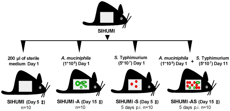
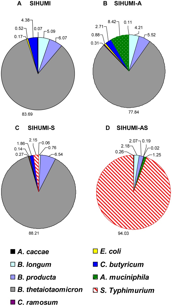
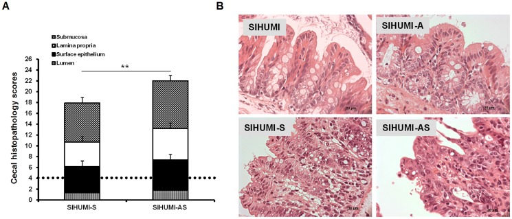
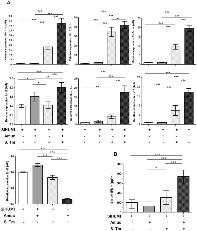
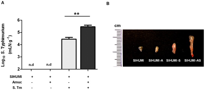
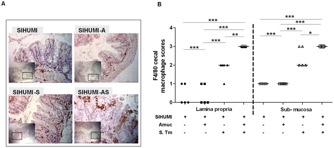
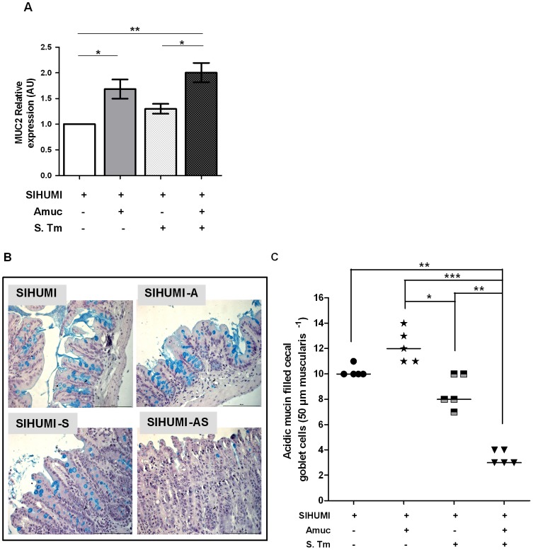
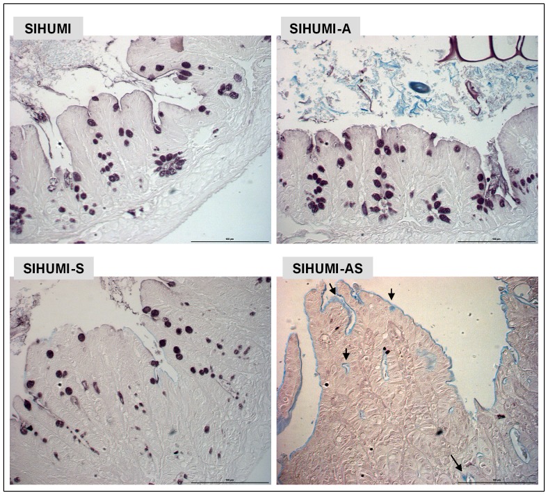
Verification
Claims the paper makes
The claims and statistics of this paper haven’t been checked yet.
Run the check — free, no account needed. It reads the open-access text and checks the sources it cites.
Claims other papers make about this one
Each is checked against this paper’s own text: does it say what the citing paper says it does?
The cited source doesn’t support the claim as written.
In the citing paper
“It has been reported that the abundance of g_Akkermansia has a negative correlation with the value of body mass index (BMI) [59] and that g_Akkermansia has a protective effect for the mucus layer in pro-inflammation [60].”
From Preventive Effects and Mechanisms of Garlic on Dyslipidemia and Gut Microbiome Dysbiosis (2019) · cites this paper as [60]
Checked by a reasoning model — may be wrong.
Experimental — accuracy not yet measuredIn the citing paper
“The Gram-negative bacteria, Akkermansia, can degrade mucin, cause intestinal inflammation and permeability, and increase endotoxemia and systemic inflammation, eventually promoting neuropathology [53,54,55].”
From Neuroprotective Effects of Sodium Butyrate by Restoring Gut Microbiota and Inhibiting TLR4 Signaling in Mice with MPTP-Induced Parkinson’s Disease (2023) · cites this paper as [55]
In the citing paper
“its mucin degrading activity might also damage the gut-barrier triggering inflammation and promoting gut permeability [83,84]”
From The Interplay between Gut Microbiota and Parkinson’s Disease: Implications on Diagnosis and Treatment (2022) · cites this paper as [84]
Self-check
Percentages, sums and reported differences re-computed from the paper’s own numbers.
The self-check needs the paper’s full text.
Citations
Cites
Gut microbiota in health and disease
Sekirov, I., Russell, S. L., Antunes, L. C. M., & Finlay, B. B. (2010) · Physiological Reviews · doi:10.1152/physrev.00045.2009 · Cited by 4,182
Akkermansia muciniphila gen. nov., sp. nov., a human intestinal mucin-degrading bacterium
Derrien, M., Vaughan, E. E., Plugge, C. M., & de Vos, W. M. (2004) · International Journal of Systematic and Evolutionary Microbiology · doi:10.1099/ijs.0.02873-0 · Cited by 2,094 · cited 1×
“muciniphila is known for its ability to degrade mucins [16,17].”
BackgroundMechanisms of Disease: pathogenesis of Crohn's disease and ulcerative colitis
Sartor, R. B. (2006) · Nature Clinical Practice Gastroenterology & Hepatology · doi:10.1038/ncpgasthep0528 · Cited by 1,806
Intestinal Goblet Cells and Mucins in Health and Disease: Recent Insights and Progress
Kim, Y. S., & Ho, S. B. (2010) · Current Gastroenterology Reports · doi:10.1007/s11894-010-0131-2 · Cited by 1,531 · cited 1×
“muciniphila is the main intestinal representative of this phylum [2], suggesting that A.”
BackgroundMucin dynamics and enteric pathogens
McGuckin, M. A., Lindén, S. K., Sutton, P., & Florin, T. H. (2011) · Nature Reviews Microbiology · doi:10.1038/nrmicro2538 · Cited by 1,428 · cited 1×
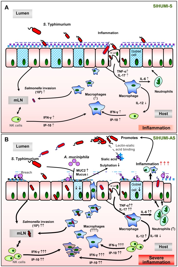
“(q increased; Q decreased; grey dotted line: assumed processes including lectin-sialic acid binding [56], M-cells for pathogen transit [43,58,59]; black line: supported by data of the present study).”
ResultGut inflammation provides a respiratory electron acceptor for Salmonella
Winter, S., Thiennimitr, P., Winter, M. G., Butler, B. P., Huseby, D. L., Crawford, R. W., Russell, J. M., Bevins, C. L., Adams, L. G., Tsolis, R. M., et al. (2010) · Nature · doi:10.1038/nature09415 · Cited by 1,271 · cited 1×
“[21,22] and therefore represents a highly suitable model for”
BackgroundStecher, B., Robbiani, R., Walker, A. W., Westendorf, A. M., Barthel, M., Kremer, M., Chaffron, S., Macpherson, A. J., Buer, J., Parkhill, J., et al. (2007) · PLoS Biology · doi:10.1371/journal.pbio.0050244 · Cited by 1,048 · cited 4×
“Typhimurium-induced gut inflammation [14,50] or in inflamed IL-10 mice [51].”
BackgroundInfluentialHost-Derived Nitrate Boosts Growth of E. coli in the Inflamed Gut
Winter, S. E., Winter, M. G., Xavier, M. N., Thiennimitr, P., Poon, V., Keestra, A. M., Laughlin, R. C., Gomez, G., Wu, J., Lawhon, S. D., et al. (2013) · Science · doi:10.1126/science.1232467 · Cited by 1,019 · cited 2×
“Typhimurium-induced gut inflammation [14,50] or in inflamed IL-10 mice [51].”
BackgroundHelicobacter pylori SabA Adhesin in Persistent Infection and Chronic Inflammation
Mahdavi, J., Sondén, B., Hurtig, M., Olfat, F. O., Forsberg, L., Roche, N., Ångström, J., Larsson, T. P., Teneberg, S., Karlsson, K., et al. (2002) · Science · doi:10.1126/science.1069076 · Cited by 935 · cited 1×
“Interestingly, changes in intestinal mucin composition characterized by a lower degree of sulphation [7,46] and a higher degree of sialylation have previously been reported to occur in UC, Crohn’s Disease (CD) and gastric ulcer caused by Helicobacter pylori-induced inflammation [7,46,47].”
BackgroundCollado, M. C., Derrien, M., Isolauri, E., de Vos, W. M., & Salminen, S. (2007) · Applied and Environmental Microbiology · doi:10.1128/aem.01477-07 · Cited by 706
Cited by
Sorted by citations
Dao, M. C., Everard, A., Aron-Wisnewsky, J., Sokolovska, N., Prifti, E., Verger, E. O., Kayser, B. D., Levenez, F., Chilloux, J., Hoyles, L., et al. (2016) · Gut · doi:10.1136/gutjnl-2014-308778 · Cited by 1,931
Intestinal permeability – a new target for disease prevention and therapy
Bischoff, S. C., Barbara, G., Buurman, W., Ockhuizen, T., Schulzke, J.-D., Serino, M., Tilg, H., Watson, A., & Wells, J. M. (2014) · BMC Gastroenterology · doi:10.1186/s12876-014-0189-7 · Cited by 1,755
“The normal intestinal microbiota triggers the epithelium for synthesizing mucus sugars, while selected commensal bacteria such as Akkermansia muciniphila regulate mucus layers by utilizing mucins as an energy source [67,68].”
BackgroundAlterations of the human gut microbiome in multiple sclerosis
Jangi, S., Gandhi, R., Cox, L. M., Li, N., von Glehn, F., Yan, R., Patel, B., Mazzola, M. A., Liu, S., Glanz, B. L., et al. (2016) · Nature Communications · doi:10.1038/ncomms12015 · Cited by 1,307
Akkermansia muciniphila and its role in regulating host functions
Derrien, M., Belzer, C., & de Vos, W. M. (2017) · Microbial Pathogenesis · doi:10.1016/j.micpath.2016.02.005 · Cited by 1,044
Cekanaviciute, E., Yoo, B. B., Runia, T. F., Debelius, J. W., Singh, S., Nelson, C., Kanner, R., Bencosme, Y., Lee, Y. K., Hauser, S. L., et al. (2017) · Proceedings of the National Academy of Sciences · doi:10.1073/pnas.1711235114 · Cited by 1,040
“muciniphila has been reported to exacerbate inflammation during infection (23).”
Microbiota and diabetes: an evolving relationship
Tilg, H., & Moschen, A. R. (2014) · Gut · doi:10.1136/gutjnl-2014-306928 · Cited by 853
Mucin glycan foraging in the human gut microbiome
Tailford, L. E., Crost, E. H., Kavanaugh, D. W., & Juge, N. (2015) · Frontiers in Genetics · doi:10.3389/fgene.2015.00081 · Cited by 881
Impact of Omega-3 Fatty Acids on the Gut Microbiota
Costantini, L., Molinari, R., Farinon, B., & Merendino, N. (2017) · International Journal of Molecular Sciences · doi:10.3390/ijms18122645 · Cited by 741
“Furthermore, long-term EPA/DHA administration in separated mice improved the inflammatory condition typically associated with stress by increasing the abundance of butyrate-producing bacteria and decreasing the levels of pro-inflammatory bacterial genera, such as Akkermansia and Flexibacter, which have been reported t…”
BackgroundBedarf, J. R., Hildebrand, F., Coelho, L. P., Sunagawa, S., Bahram, M., Goeser, F., Bork, P., & Wüllner, U. (2017) · Genome Medicine · doi:10.1186/s13073-017-0428-y · Cited by 642
“On the other hand, inflammatory and regulatory properties have been reported for Akkermansia, probably mediated due to an increased exposure of immune cells to microbial antigens upon breaking down the mucosal mucin layer [47].”
BackgroundCarbohydrate-active enzymes (CAZymes) in the gut microbiome
Wardman, J. F., Bains, R. K., Rahfeld, P., & Withers, S. G. (2022) · Nature Reviews Microbiology · doi:10.1038/s41579-022-00712-1 · Cited by 597
Shono, Y., Docampo, M. D., Peled, J. U., Perobelli, S. M., Velardi, E., Tsai, J. J., Slingerland, A. E., Smith, O. M., Young, L. F., Gupta, J., et al. (2016) · Science Translational Medicine · doi:10.1126/scitranslmed.aaf2311 · Cited by 592
Gut microbiota disturbance during antibiotic therapy: a multi-omic approach
Pérez‐Cobas, A. E., Gosalbes, M. J., Friedrichs, A. K., Knecht, H., Artacho, A., Eismann, K., Otto, W., Rojo, D., Bargiela, R., von Bergen�, M., et al. (2012) · Gut · doi:10.1136/gutjnl-2012-303184 · Cited by 631
Gut Microbiota and Colonization Resistance against Bacterial Enteric Infection
Ducarmon, Q. R., Zwittink, R. D., Hornung, B. V. H., van Schaik, W., Young, V. B., & Kuijper, E. J. (2019) · Microbiology and Molecular Biology Reviews · doi:10.1128/mmbr.00007-19 · Cited by 566
“Typhimurium infection by thinning the mucus layer, thereby promoting translocation of the pathogen to the epithelial layer (157).”
BackgroundGut Microbiota–Derived Short-Chain Fatty Acids Promote Poststroke Recovery in Aged Mice
Lee, J., d’Aigle, J., Atadja, L. A., Quaicoe, V. A., Honarpisheh, P., Ganesh, B. P., Hassan, A. F., Graf, J., Petrosino, J. F., Putluri, N., et al. (2020) · Circulation Research · doi:10.1161/circresaha.119.316448 · Cited by 567
Self-citationGut microbiota, obesity and diabetes
Patterson, E., Ryan, P. M., Cryan, J. F., Dinan, T. G., Ross, R. P., Fitzgerald, G. F., & Stanton, C. (2016) · Postgraduate Medical Journal · doi:10.1136/postgradmedj-2015-133285 · Cited by 552
Bian, X., Wu, W., Yang, L., Lv, L., Wang, Q., Li, Y., Ye, J., Fang, D., Wu, J., Jiang, X., et al. (2019) · Frontiers in Microbiology · doi:10.3389/fmicb.2019.02259 · Cited by 535
The gut microbiota influences anticancer immunosurveillance and general health
Routy, B., Gopalakrishnan, V., Daillère, R., Zitvogel, L., Wargo, J. A., & Kroemer, G. (2018) · Nature Reviews Clinical Oncology · doi:10.1038/s41571-018-0006-2 · Cited by 523
Ottman, N., Reunanen, J., Meijerink, M., Pietilä, T. E., Kainulainen, V., Klievink, J., Huuskonen, L., Aalvink, S., Skurnik, M., Boeren, S. A., et al. (2017) · PLoS ONE · doi:10.1371/journal.pone.0173004 · Cited by 536
“muciniphila administration in a minimal community appeared to aggravate Salmonella enterica Typhimurium-induced gut inflammation in a gnotobiotic mouse model [22].”
BackgroundMicrobial shifts in the aging mouse gut
Langille, M. G., Meehan, C. J., Koenig, J. E., Dhanani, A. S., Rose, R. A., Howlett, S. E., & Beiko, R. G. (2014) · Microbiome · doi:10.1186/s40168-014-0050-9 · Cited by 437
“Akkermansia has been linked to healthy microbiomes in some studies [39,40], although members of this genus may also exacerbate certain types of infection via mucin degradation [41].”
BackgroundA next generation probiotic, Akkermansia muciniphila
Zhai, Q., Feng, S., Narbad, A., & Chen, W. (2018) · Critical Reviews in Food Science and Nutrition · doi:10.1080/10408398.2018.1517725 · Cited by 447
“To date, very limited reports indicated the potential deleterious impacts of this strain, while a study showed an exacerbation of gut inflammation in Salmonella Typhimurium-infected gnotobiotic mice by A. muciniphila (Ganesh et al. 2013).”
BackgroundXu, Y., Wang, N., Tan, H.-Y., Li, S., Zhang, C., & Feng, Y. (2020) · Frontiers in Microbiology · doi:10.3389/fmicb.2020.00219 · Cited by 420
“muciniphila in the ecosystem can be changed in response to the presence of inflammation (Ganesh et al., 2013).”
BackgroundRodrigues, V. F., Elias‐Oliveira, J., Pereira, Í. S., Pereira, J. A., Barbosa, S. C., Machado, M. S. G., & Carlos, D. (2022) · Frontiers in Immunology · doi:10.3389/fimmu.2022.934695 · Cited by 434
NLRP6 Protects Il10 Mice from Colitis by Limiting Colonization of Akkermansia muciniphila
Seregin, S. S., Golovchenko, N., Schaf, B., Chen, J., Pudlo, N. A., Mitchell, J., Baxter, N. T., Zhao, L., Schloss, P. D., Martens, E. C., et al. (2017) · Cell Reports · doi:10.1016/j.celrep.2017.03.080 · Cited by 386
“muciniphila also exacerbated Salmonella-induced colitis and is positively correlatedwith ulcerative colitis patientswith activepouchitis and IBDpatients that are treatment resistant (Ganesh et al., 2013;White et al., 2009; Zella et al., 2011).”
BackgroundZhai, R., Xue, X., Zhang, L., Yang, X., Zhao, L., & Zhang, C. (2019) · Frontiers in Cellular and Infection Microbiology · doi:10.3389/fcimb.2019.00239 · Cited by 368
“However, the same strain was demonstrated to aggravate gut inflammation in gnotobiotic mice infected with Salmonella typhimurium (Ganesh et al., 2013; Kang et al., 2013).”
BackgroundGenetic and environmental control of host-gut microbiota interactions
Org, E., Parks, B. W., Joo, J. W. J., Emert, B., Schwartzman, W., Kang, E. Y., Mehrabian, M., Pan, C., Knight, R., Gunsalus, R., et al. (2015) · Genome Research · doi:10.1101/gr.194118.115 · Cited by 381
“A. muciniphila (ATCC BAA-835) was grown in a Columbia broth medium supplemented with 0.05 % hog gastric Mucin type III (Sigma) under anaerobic conditions (Ganesh et al. 2013).”
BackgroundMethodInfluentialShang, Q., Shan, X., Cai, C., Hao, J., Li, G., & Yu, G. (2016) · Food & Function · doi:10.1039/c6fo00309e · Cited by 363
Understanding the Molecular Mechanisms of the Interplay Between Herbal Medicines and Gut Microbiota
Xu, J., Chen, H., & Li, S. (2017) · Medicinal Research Reviews · doi:10.1002/med.21431 · Cited by 346
Dietary simple sugars alter microbial ecology in the gut and promote colitis in mice
Khan, S., Waliullah, S., Godfrey, V., Khan, M. A. W., Ramachandran, R. A., Cantarel, B. L., Behrendt, C. L., Peng, L., Hooper, L. V., & Zaki, H. (2020) · Science Translational Medicine · doi:10.1126/scitranslmed.aay6218 · Cited by 322
Zhou, X., Baumann, R., Gao, X., Mendoza, M., Singh, S., Sand, I. K., Xia, Z., Cox, L. M., Chitnis, T., Yoon, H., et al. (2022) · Cell · doi:10.1016/j.cell.2022.08.021 · Cited by 326
Intestinal dysbiosis and probiotic applications in autoimmune diseases
de Oliveira, G. L. V., Leite, A. Z., Higuchi, B. S., Gonzaga, M. I., & Mariano, V. S. (2017) · Immunology · doi:10.1111/imm.12765 · Cited by 324
The Role of Gut Microbiota in Intestinal Inflammation with Respect to Diet and Extrinsic Stressors
Lobionda, S., Sittipo, P., Kwon, H. Y., & Lee, Y. K. (2019) · Microorganisms · doi:10.3390/microorganisms7080271 · Cited by 326
Mining the microbiota for microbial and metabolite-based immunotherapies
Skelly, A. N., Sato, Y., Kearney, S., & Honda, K. (2019) · Nature Reviews Immunology · doi:10.1038/s41577-019-0144-5 · Cited by 306
Zhou, K. (2017) · Journal of Functional Foods · doi:10.1016/j.jff.2017.03.045 · Cited by 309
“…A. muciniphila, despite its utilization of mucin as a source of nutrients, actually were positively associated with the mucus thickness and intestinal barrier integrity in humans and animals (Collado, Derrien, Isolauri, de Vos, & Salminen, 2007; Everard et al., 2013; Ganesh, 2013; Zhong, 2015).”
BackgroundBaxter, N. T., Zackular, J. P., Chen, G. Y., & Schloss, P. D. (2014) · Microbiome · doi:10.1186/2049-2618-2-20 · Cited by 299
Jakobsdottir, G., Xu, J., Molin, G., Ahrné, S., & NYMAN, M. E. (2013) · PLoS ONE · doi:10.1371/journal.pone.0080476 · Cited by 295
Kim, S., Shin, Y.-C., Kim, T., Kim, Y., Lee, Y., Lee, S.-H., Kim, M., Eunju, O., Kim, K. S., & Kweon, M. (2021) · Gut Microbes · doi:10.1080/19490976.2021.1892441 · Cited by 303
“muciniphila diminished microbiome diversity, leading to a deleterious modification of the gut environment.(44) Furthermore, an accumulation of antimicrobial peptide produced by Paneth cells may contribute to a change in gut microbiota.”
BackgroundDirect impact of commonly used dietary emulsifiers on human gut microbiota
Naimi, S., Viennois, É., Gewirtz, A. T., & Chassaing, B. (2021) · Microbiome · doi:10.1186/s40168-020-00996-6 · Cited by 329
Shaw, K. A., Bertha, M., Hofmekler, T., Chopra, P., Vatanen, T., Srivatsa, A., Prince, J., Kumar, A., Sauer, C. G., Zwick, M. E., et al. (2016) · Genome Medicine · doi:10.1186/s13073-016-0331-y · Cited by 278
Ketogenic diet modifies the gut microbiota in a murine model of autism spectrum disorder
Newell, C., Bomhof, M. R., Reimer, R. A., Hittel, D. S., Rho, J. M., & Shearer, J. (2016) · Molecular Autism · doi:10.1186/s13229-016-0099-3 · Cited by 272
“muciniphila—the predominant representative of the Verrucomicrobia phylum within the GI tract [27].”
Liu, Y., Yang, M., Tang, L., Wang, F., Huang, S., Liu, S., Lei, Y., Wang, S., Xie, Z., Wang, W., et al. (2022) · Microbiome · doi:10.1186/s40168-022-01296-x · Cited by 273
“A prior study demonstrated that the presence of A. muciniphila exacerbated the severity of colon inflammation caused by Salmonella typhimurium (S. typhimurium) infection in mice colonized with a simplified human gut microbiota (SIHUMI) [51].”
BackgroundInfluentialTomas, J., Mulet, C., Saffarian, A., Cavin, J., Ducroc, R. A., Regnault, B., Tan, C. K., Duszka, K., Burcelin, R., Wahli, W., et al. (2016) · Proceedings of the National Academy of Sciences · doi:10.1073/pnas.1612559113 · Cited by 260
“In our model collapse of the mucus barrier could favor the emergence of such bacteria able to use it as substrate, or diet-mediated increase in this species may further participate in the alteration of the mucus barrier (67, 73).”
BackgroundQu, S., Fan, L., Qi, Y., Xu, C., Hu, Y., Chen, S., Liu, W., Liu, W., & Si, J. (2021) · Microbiology Spectrum · doi:10.1128/spectrum.00730-21 · Cited by 251
“muciniphila exacerbates gut inflammation in Salmonella enterica serovar Typhimuriuminfected mice (35).”
BackgroundZhang, Q., Wu, Y., Wang, J., Wu, G. G., Long, W., Xue, Z., Wang, L., Zhang, X., Pang, X., Zhao, Y., et al. (2016) · Scientific Reports · doi:10.1038/srep27572 · Cited by 229
Pusceddu, M. M., El Aidy, S., Crispie, F., O’Sullivan, O., Cotter, P., Stanton, C., Kelly, P., Cryan, J. F., & Dinan, T. G. (2015) · PLOS ONE · doi:10.1371/journal.pone.0139721 · Cited by 214
“In an inflammatory milieu, Akkermansia, the mucus degrader, facilitates the microbial translocation to come in direct contact with the intestinal epithelium, therefore exacerbating gut inflammation [34, 35].”
BackgroundIntroduction to host microbiome symbiosis in health and disease
Malard, F., Dore, J. M., Gaugler, B., & Mohty, M. (2020) · Mucosal Immunology · doi:10.1038/s41385-020-00365-4 · Cited by 219
Stoll, M. L., Kumar, R., Morrow, C. D., Lefkowitz, E. J., Cui, X., Genin, A., Cron, R. Q., & Elson, C. O. (2014) · Arthritis Research & Therapy · doi:10.1186/s13075-014-0486-0 · Cited by 208
Morgan, X. C., Kabakchiev, B., Waldron, L., Tyler, A. D., Tickle, T. L., Milgrom, R., Stempak, J. M., Gevers, D., Xavier, R. J., Silverberg, M. S., et al. (2015) · Genome biology · doi:10.1186/s13059-015-0637-x · Cited by 198
“Ganesh BP, Klopfleisch R, Loh G, Blaut M. Commensal Akkermansia muciniphila exacerbates gut inflammation in Salmonella Typhimurium-infected gnotobiotic mice.”
BackgroundInfluentialEarley, H., Lennon, G., Balfe, Á., Coffey, J. C., Winter, D. C., & O’Connell, P. R. (2019) · Scientific Reports · doi:10.1038/s41598-019-51878-3 · Cited by 210
Mucosal adherent bacterial dysbiosis in patients with colorectal adenomas
Lü, Y., Chen, J., Zheng, J.-Y., Hu, G., Wang, J., Huang, C., Lou, L., Wang, X., & Zeng, Y. (2016) · Scientific Reports · doi:10.1038/srep26337 · Cited by 203
“As we know, Akkermansia is known to break down mucin, while mucin degradation has been linked to intestinal inflammation and can facilitate colonization of intestinal pathogens 23 .”
The Gut Microbiota in Multiple Sclerosis: An Overview of Clinical Trials
Schepici, G., Silvestro, S., Bramanti, P., & Mazzon, E. (2019) · Cell Transplantation · doi:10.1177/0963689719873890 · Cited by 196
Gorecki, A. M., Preskey, L., Bakeberg, M. C., Kenna, J. E., Gildenhuys, C., MacDougall, G., Dunlop, S. A., Mastaglia, F. L., Akkari, P. A., Koengten, F., et al. (2019) · Frontiers in Neuroscience · doi:10.3389/fnins.2019.00839 · Cited by 184
Akkermansia muciniphila and Faecalibacterium prausnitzii in Immune-Related Diseases
Effendi, R. M. R. A., Anshory, M., Kalim, H., Dwiyana, R. F., Suwarsa, O., Pardo, L. M., Nijsten, T. E. C., & Thio, H. B. (2022) · Microorganisms · doi:10.3390/microorganisms10122382 · Cited by 193
The potential of Akkermansia muciniphila in inflammatory bowel disease
Zhang, T., Ji, X., Lu, G., & Zhang, F. (2021) · Applied Microbiology and Biotechnology · doi:10.1007/s00253-021-11453-1 · Cited by 179
Intestinal Microbiota as a Host Defense Mechanism to Infectious Threats
Iacob, S. A., Iacob, D. G., & Luminos, L. M. (2019) · Frontiers in Microbiology · doi:10.3389/fmicb.2018.03328 · Cited by 191
Jian, H., Liu, Y., Wang, X., Dong, X., & Zou, X. (2023) · International Journal of Molecular Sciences · doi:10.3390/ijms24043900 · Cited by 186
“TLR4 knockout exacerbated DSS-induced colitis of TLR4−/− mice mediated by gut microbiota and the abundance of Akk was significantly decreased in TLR4−/− mice, whereas it was enriched in wild-type mice [108].”
BackgroundInfluentialThe "Gut Feeling": Breaking Down the Role of Gut Microbiome in Multiple Sclerosis
Freedman, S. N., Shahi, S. K., & Mangalam, A. K. (2018) · Neurotherapeutics · doi:10.1007/s13311-017-0588-x · Cited by 168
“muciniphilia exacerbates colonic pathology in Salmonella typhimurium-induced intestinal inflammation [205], it has also been shown to exert anti-inflammatory prop-”
Recent findings in Akkermansia muciniphila-regulated metabolism and its role in intestinal diseases
Liu, M., Yang, J.-Y., Yan, Z.-H., Hu, S., Li, J.-Q., Xu, Z., & Jian, Y. (2022) · Clinical Nutrition · doi:10.1016/j.clnu.2022.08.029 · Cited by 176
Chemical and pathogen-induced inflammation disrupt the murine intestinal microbiome
Borton, M. A., Sabag-Daigle, A., Wu, J., Solden, L. M., O’Banion, B. S., Daly, R. A., Wolfe, R. A., Gonzalez, J. F., Wysocki, V. H., Ahmer, B. M. M., et al. (2017) · Microbiome · doi:10.1186/s40168-017-0264-8 · Cited by 149
When pathogenic bacteria meet the intestinal microbiota
Rolhion, N., & Chassaing, B. (2016) · Philosophical Transactions of the Royal Society B Biological Sciences · doi:10.1098/rstb.2015.0504 · Cited by 169
Childhood adversity impact on gut microbiota and inflammatory response to stress during pregnancy
Hantsoo, L., Jašarević, E., Criniti, S., McGeehan, B., Tanes, C., Sammel, M. D., Elovitz, M. A., Compher, C., Wu, G., & Epperson, C. N. (2019) · Brain, Behavior, and Immunity · doi:10.1016/j.bbi.2018.11.005 · Cited by 156
“Relative abundance of specific gut bacteria including Akkermansia, Flexibacter and Prevotella have been associated with inflammation in rodents (Ganesh et al., 2013; Pusceddu et al., 2015), and Megasphaera and Proteobacteria were associated with elevated inflammatory markers in humans (Mukhopadhya et al.”
BackgroundChu, F., Shi, M., Lang, Y., Shen, D., Jin, T., Zhu, J., & Cui, L. (2018) · Mediators of Inflammation · doi:10.1155/2018/8168717 · Cited by 164
The role of Akkermansia muciniphila in obesity, diabetes and atherosclerosis
Hasani, A., Ebrahimzadeh, S., Hemmati, F., Khabbaz, A., Hasani, A., & Gholizadeh, P. (2021) · Journal of Medical Microbiology · doi:10.1099/jmm.0.001435 · Cited by 152
Lv, L., Li, S., Li, S.-C., Zhong, Z.-C., Duan, H., Cheng, T., Li, H., He, W., Chen, M.-C., He, T., et al. (2019) · Frontiers in Cellular and Infection Microbiology · doi:10.3389/fcimb.2019.00224 · Cited by 156
“Preeclampsia (PE), the second leading cause of maternal mortality worldwide (Huppertz, 2008; Ghulmiyyah and Sibai, 2012; Mol et al., 2016), is characterized by severe hypertension and multiple organ damage (Brown et al., 2018), and it can result in fetal intrauterine growth retardation, premature…”
BackgroundTriggering Akkermansia with dietary polyphenols: A new weapon to combat the metabolic syndrome?
Anhê, F. F., Pilon, G., Roy, D., Desjardins, Y., Levy, E. H., & Marette, A. (2016) · Gut Microbes · doi:10.1080/19490976.2016.1142036 · Cited by 155
Akkermansia muciniphila : A promising probiotic against inflammation and metabolic disorders
Zhao, Y., Yang, H., Wu, P., Yang, S., Xue, W., Xu, B., Zhang, S., Tang, B., & Xu, D. (2024) · Virulence · doi:10.1080/21505594.2024.2375555 · Cited by 155
“Ganesh et al. reported that the symptoms of colitis were aggravated by oral administration A. mucini-phila in mice infected with Salmonella typhimurium [80].”
BackgroundResultPetriello, M. C., Hoffman, J. B., Vsevolozhskaya, O. A., Morris, A. J., & Hennig, B. (2018) · Environmental Pollution · doi:10.1016/j.envpol.2018.07.039 · Cited by 141
“muciniphila, and there are reports that an abnormally high abundance of Akkermansia may actually be detrimental due to the excessive degradation of the mucus layer lining the intestine in such a way that the mucin producing goblet cells cannot compensate (Ganesh et al. 2013; Hamilton et al. 2015).”
BackgroundLi, F., Han, Y., Cai, X., Gu, M., Sun, J., Qi, C., Goulette, T., Song, M., Li, Z., & Xiao, H. (2019) · Food & Function · doi:10.1039/c9fo01519a · Cited by 141
Exploiting host immunity: the Salmonella paradigm
Behnsen, J., Pérez-López, A., Nuccio, S., & Raffatellu, M. (2015) · Trends in Immunology · doi:10.1016/j.it.2014.12.003 · Cited by 138
Mechanistic links between gut microbial community dynamics, microbial functions and metabolic health
Ha, C. W. (2014) · World Journal of Gastroenterology · doi:10.3748/wjg.v20.i44.16498 · Cited by 137
Qu, S., Zheng, Y., Huang, Y., Feng, Y., Xu, K., Zhang, W., Wang, Y., Nie, K., & Qin, M. (2023) · Frontiers in Microbiology · doi:10.3389/fmicb.2023.1111911 · Cited by 146
“muciniphila interferes with mucosal reconstitution and exacerbates inflammation in Salmonella enterica typhimurium-induced gut inflammation (Ganesh et al., 2013).”
BackgroundRuiz, V. E., Battaglia, T., Kurtz, Z. D., Bijnens, L., Ou, A., Engstrand, I., Zheng, X., Iizumi, T., Mullins, B. J., Müller, C. L., et al. (2017) · Nature Communications · doi:10.1038/s41467-017-00531-6 · Cited by 137
The Interaction of the Gut Microbiota with the Mucus Barrier in Health and Disease in Human
Corfield, A. P. (2018) · Microorganisms · doi:10.3390/microorganisms6030078 · Cited by 140
“Such models have been widely used [334] [335] [336] [337] .”
MethodHan, Y., Song, M., Gu, M., Ren, D., Zhu, X., Cao, X., Li, F., Wang, W., Cai, X., Yuan, B., et al. (2019) · Journal of Agricultural and Food Chemistry · doi:10.1021/acs.jafc.8b05581 · Cited by 133
Interplay between bile acids and the gut microbiota promotes intestinal carcinogenesis
Wang, S., Dong, W., Liu, L., Xu, M., Wang, Y., Liu, T., Zhang, Y., Wang, B., & Cao, H. (2019) · Molecular Carcinogenesis · doi:10.1002/mc.22999 · Cited by 135
“These bacteria are associated with increased intestinal inflammation and impaired gut barrier function.(33,34) In particular, our findings demonstrated decreased ZO‐1, claudin 3, claudin”
Intestinal colonization resistance in the context of environmental, host, and microbial determinants
Woelfel, S., Silva, M. S., & Stecher, B. (2024) · Cell Host & Microbe · doi:10.1016/j.chom.2024.05.002 · Cited by 151
Akkermansia muciniphila: a novel functional microbe with probiotic properties
Gómez-Gallego, C., Pohl, S., Salminen, S., de Vos, W. M., & Kneifel, W. (2016) · Beneficial Microbes · doi:10.3920/bm2016.0009 · Cited by 133
Bladder cancer-related microbiota: examining differences in urine and tissue samples
Mansour, B., Monyók, Á., Makra, N., Gajdács, M., Vadnay, I., Ligeti, B., Juhász, J., Szabó, D., & Ostorházi, E. (2020) · Scientific Reports · doi:10.1038/s41598-020-67443-2 · Cited by 128
Hamouda, N., Sano, T., Oikawa, Y., Ozaki, T., Shimakawa, M., Matsumoto, K., Amagase, K., Higuchi, K., & Kato, S. (2017) · Basic & Clinical Pharmacology & Toxicology · doi:10.1111/bcpt.12793 · Cited by 132
Honarpisheh, P., Reynolds, C. R., Conesa, M. P. B., Moruno-Manchon, J. F., Putluri, N., Bhattacharjee, M. B., Urayama, A., McCullough, L. D., & Ganesh, B. P. (2020) · International Journal of Molecular Sciences · doi:10.3390/ijms21051711 · Cited by 129
“Mucus is the primary protective barrier separating luminal antigens and the intestinal epithelium [37,38].”
BackgroundSelf-citationSynthetic microbial communities (SynComs) of the human gut: design, assembly, and applications
van Leeuwen, P. T., Brul, S., Zhang, J., & Wortel, M. T. (2023) · FEMS Microbiology Reviews · doi:10.1093/femsre/fuad012 · Cited by 132
Akkermansia muciniphilaandHelicobacter typhloniusmodulate intestinal tumor development in mice
der Spek, C. D.-V., Belzer, C., van Hijum, S. A. F. T., Günthel, M., Salvatori, D. C. F., den Dunnen, J. T., Kuijper, E. J., Devilee, P., de Vos, W. M., van Ommen, G. B., et al. (2015) · Carcinogenesis · doi:10.1093/carcin/bgv120 · Cited by 125
“muciniphila increased the number of goblet cells, but upon co-colonization exacerbated the decrease of goblet cell number by Salmonella thyphimurium (39).”
BackgroundInfluentialClavel, T., Neto, J. C. G., Lagkouvardos, I., & Ramer‐Tait, A. E. (2017) · Immunological Reviews · doi:10.1111/imr.12578 · Cited by 132
Chiang, H., Li, J., Liu, C.-C., Liu, P., Chen, H., Chen, Y., Lan, J., & Chen, D. (2019) · Journal of Clinical Medicine · doi:10.3390/jcm8111770 · Cited by 152
“These proinflammatory effects may be related to its ability to degrade mucus and thus increase the exposure of resident immune cells to gut microbial antigens [45].”
BackgroundRevisiting the role of Akkermansia muciniphila as a therapeutic bacterium
Si, J., Kang, H., You, H. J., & Ko, G. (2022) · Gut Microbes · doi:10.1080/19490976.2022.2078619 · Cited by 130
“Typhimurium only (SIHUMI-S).(67) Reduced numbers of mucin-filled goblet cells and thinner mucus layers were found in SIHUMI-AS mice indicating that mucin degradation by A.”
BackgroundImpact of air pollution on intestinal redox lipidome and microbiome
Feng, J., Cavallero, S., Hsiai, T. K., & Li, R. (2020) · Free Radical Biology and Medicine · doi:10.1016/j.freeradbiomed.2019.12.044 · Cited by 120
Luo, Y., Lan, C., Li, H., Ouyang, Q., Kong, F., Wu, A., Ren, Z., Tian, G., Cai, J., Yu, B., et al. (2022) · npj Biofilms and Microbiomes · doi:10.1038/s41522-022-00338-4 · Cited by 130
Wu, R., Xiong, R., Li, Y., Chen, J., & Yan, R. (2023) · Journal of Autoimmunity · doi:10.1016/j.jaut.2023.103062 · Cited by 124
Wang, J., Bose, S., Kim, G.-C., Hong, S.-U., Kim, J.-H., Kim, J.-E., & Kim, H. (2014) · PLoS ONE · doi:10.1371/journal.pone.0086117 · Cited by 123
“reported that Akkermansia muciniphila exacerbates Salmonella typhimurium induced gut inflammation in animal model [69], recently two studies have reported that Akkermansia muciniphila ameliorates high fat diet induced obesity, metabolic endotoxemia and type 2 diabetes via modulation of Foxp3 regulatory T cells [10,70].”
ResultSilveira‐Nunes, G., Durso, D. F., Júnior, L. R. A. D. O., Cunha, E. H. M., Maioli, T. U., Vieira, A. T., Speziali, E., Corrêa‐Oliveira, R., Martins‐Filho, O. A., Teixeira‐Carvalho, A., et al. (2020) · Frontiers in Pharmacology · doi:10.3389/fphar.2020.00258 · Cited by 121
Shah, R., Cope, J. L., Nagy‐Szakal, D., Dowd, S. E., Versalovic, J. A., Hollister, E. B., & Kellermayer, R. (2016) · Gut Microbes · doi:10.1080/19490976.2016.1190073 · Cited by 115
“was demonstrated by experiments co-colonizing Akkermansia and Salmonella in gnotobiotic mice.(34)”
MethodGill, T., Asquith, M. J., Brooks, S. R., Rosenbaum, J. T., & Colbert, R. A. (2017) · Arthritis & Rheumatology · doi:10.1002/art.40405 · Cited by 116
A review of a potential and promising probiotic candidate— Akkermansia muciniphila
Cheng, D., & Xie, M. Z. (2020) · Journal of Applied Microbiology · doi:10.1111/jam.14911 · Cited by 117
“municiphila comes from its capacity to restore the mucus thickness and intestinal barrier integrity (Ganesh et al. 2013; Zhong 2015) because it provides preferable energy sources (e.”
BackgroundSafety of pasteurised Akkermansia muciniphila as a novel food pursuant to Regulation (EU) 2015/2283
EFSA Panel on Nutrition, N. F. A. F. A. (., Turck, D., Bohn, T., Castenmiller, J. J. M., Henauw, S. D., Hirsch‐Ernst, K. I., Maciuk, A., Mangelsdorf, I., McArdle, H. J., Naska, A., et al. (2021) · EFSA Journal · doi:10.2903/j.efsa.2021.6780 · Cited by 117
Gut microbiota disturbance during antibiotic therapy
Ferrer, M., dos Santos, V. A. P. M., Ott, S. J., & Moyá, A. (2013) · Gut Microbes · doi:10.4161/gmic.27128 · Cited by 116
Pröbstel, A.-K., & Baranzini, S. E. (2018) · Neurotherapeutics · doi:10.1007/s13311-017-0587-y · Cited by 110
“The proinflammatory feature most likely results from its mucus-degrading ability leading to an exposure of immune cells to microbial antigens as has been shown in an intestinal inflammation model of Salmonella typhimurium where Akkermansia lead to disease exacerbation [63].”
Wang, F., Cai, K., Xiao, Q., He, L., Xie, L., & Liu, Z. (2021) · Journal of Cancer · doi:10.7150/jca.63578 · Cited by 111
“typhimurium-induced intestinal inflammation with reduced goblet cell numbers [17].”
BackgroundZhang, Z., Cao, H., Song, N., Zhang, L., Cao, Y., & Tai, J. (2020) · Food and Chemical Toxicology · doi:10.1016/j.fct.2020.111237 · Cited by 107
Aging and serum MCP-1 are associated with gut microbiome composition in a murine model
Conley, M. N., Wong, C. P., Duyck, K. M., Hord, N. G., Ho, E., & Sharpton, T. J. (2016) · PeerJ · doi:10.7717/peerj.1854 · Cited by 111
Rolhion, N., Chassaing, B., Nahori, M., Bodt, J. D., Moura, A., Lecuit, M., Dussurget, O., Berard, M., Marzorati, M., Fehlner-Peach, H., et al. (2019) · Cell Host & Microbe · doi:10.1016/j.chom.2019.10.016 · Cited by 113
“Typhimurium-induced intestinal inflammation by disturbing host mucus homeostasis (Ganesh et al., 2013).”
BackgroundWade, H., Pan, K., Duan, Q., Kaluzny, S., Pandey, E., Fatumoju, L., Saraswathi, V., Wu, R., Harris, E. N., & Su, Q. (2023) · Journal of Biomedical Science · doi:10.1186/s12929-023-00935-1 · Cited by 112
“Typhimurium)-induced intestinal inflammation in a gnotobiotic mouse model [70].”
Background
Metadata
- Authors
- Ganesh, B. P., Klopfleisch, R., Loh, G., & Blaut, M.
- Affiliations
- German Institute of Human Nutrition (DE)
- Freie Universität Berlin (DE)
- Corresponding author
- Loh, Gunnar
- Venue
- PLoS ONE
- Publisher
- Public Library of Science (PLoS)
- Published
- 2013-09-10
- Volume
- 8
- Issue
- 9
- Pages
- e74963
- ISSN
- 1932-6203 (ISSN-L)
- PMC
- PMC3769299
- OpenAlex
- W2085991415
- Cited by
- 522
- References
- 60
- Citations by year
- 2026: 27 · 2025: 47 · 2024: 69 · 2023: 56 · 2022: 59 · 2021: 50 · 2020: 46 · 2019: 41 · 2018: 37 · 2017: 27 · 2016: 36 · 2015: 13 · 2014: 11 · 2013: 2
- FWCI
- 9.26
- Type
- journal article
- Language
- en
- Open access
- gold
- doi.org
- Locations
- PLoS ONE (publishedVersion, cc-by)
- Europe PMC (PubMed Central) (publishedVersion, other-oa)
- PubMed Central (publishedVersion, cc-by)
- PubMed (publishedVersion)
- DOAJ (DOAJ: Directory of Open Access Journals) (submittedVersion, cc-by-sa)
- Figshare (submittedVersion, cc-by)
- Refubium (Universitätsbibliothek der Freien Universität Berlin) (submittedVersion, cc-by)
- Universitätsbibliothek der FU Berlin Hochschulschriftenstelle u. Dokumentenserver (cc-by)
- Funders
- Deutsche Forschungsgemeinschaft
- Topics
- Life Sciences › Biochemistry, Genetics and Molecular Biology › Molecular Biology › Gut microbiota and health
- Health Sciences › Medicine › Gastroenterology › Inflammatory Bowel Disease
- Health Sciences › Medicine › Gastroenterology › Gastrointestinal motility and disorders
- Keywords
- Akkermansia muciniphila, Salmonella typhimurium, intestinal inflammation, mucin degradation, commensal bacteria, gnotobiotic mice, goblet cells, Bacteroides thetaiotaomicron, inflammatory cytokines, interferon-gamma, gut microbiota, Salmonella enteritis
- MeSH terms
- Animals, Germ-Free Life, Inflammation, Interferon-gamma, Interleukin-1, Intestines, Lymph Nodes, Macrophages, Mice, Inbred C3H, RNA, Messenger, Salmonella Infections, Salmonella typhimurium, Symbiosis, Tumor Necrosis Factor-alpha, Interleukin-6, Interleukin-12, Mice, Chemokine CXCL10, Mucin-2, Verrucomicrobia
About these checks
Registry, retraction, Cites and statistics results are computed, not judged, with one exception: a printed reference that no index lists is read once by a model before it counts as a citation error. Claims are extracted by a model and kept only when their quote appears verbatim in the text; each cited source is then checked, with a model’s help, for whether it says what the claim relies on. Those verdicts can be wrong — read the claim’s quote and the cited source before relying on one.
Statistics and arithmetic are recomputed, not judged.
Confidence bands are shown only for fields calibrated on our annotated test set. None is calibrated yet, so no band is shown.
What counts as a claim ›How we measure confidence ›How we check this ›
Are you an author, or do you see a problem with this page? Request a review
