

The DOI matches its Crossref record; 62 of the 62 references it cites were found in a registry or an index.
Oxidative Stress Induces Mitochondrial Dysfunction in a Subset of Autism Lymphoblastoid Cell Lines in a Well-Matched Case Control Cohort
Open accessAbstract
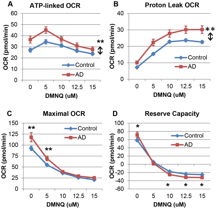
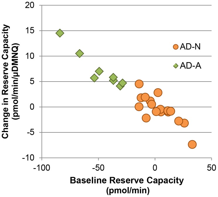
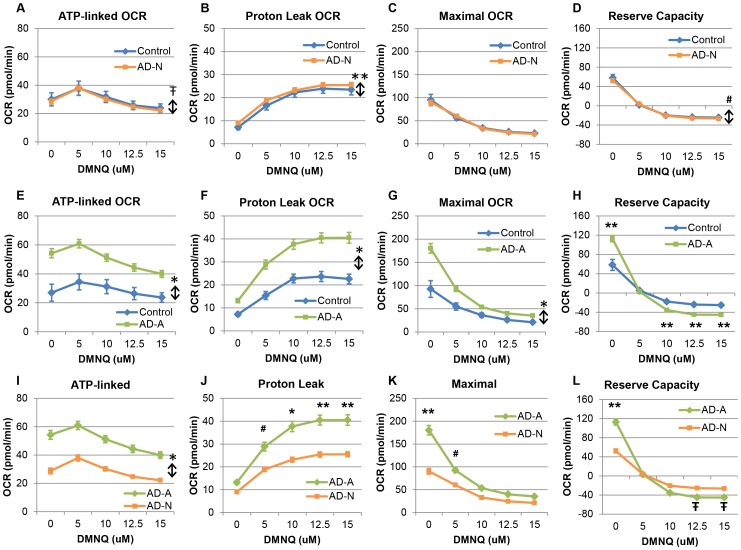
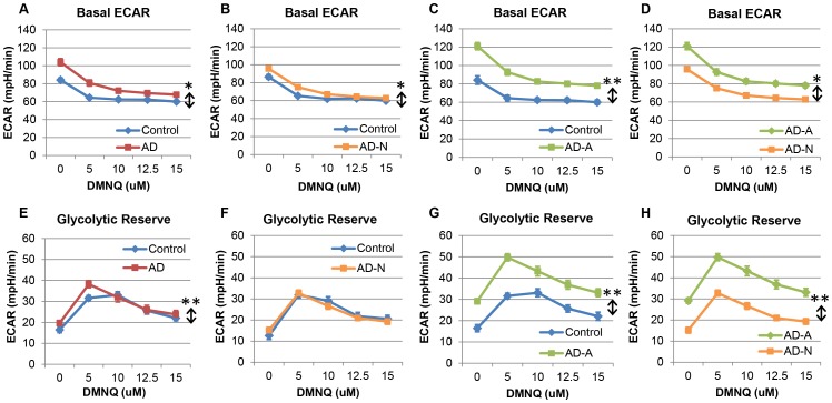
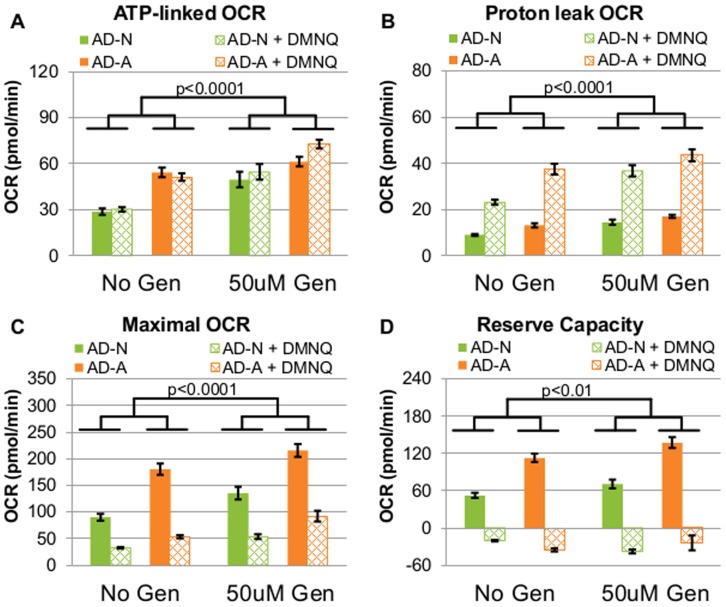
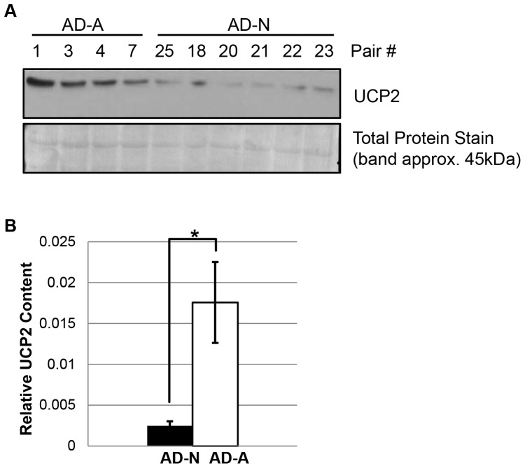
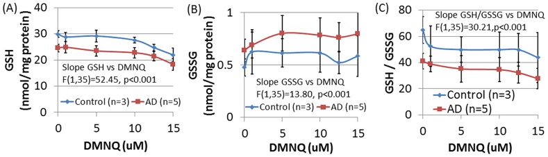
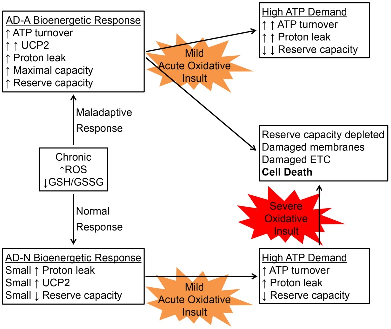
There is increasing recognition that mitochondrial dysfunction is associated with the autism spectrum disorders. However, little attention has been given to the etiology of mitochondrial dysfunction or how mitochondrial abnormalities might interact with other physiological disturbances associated with autism, such as oxidative stress. In the current study we used respirometry to examine reserve capacity, a measure of the mitochondrial ability to respond to physiological stress, in lymphoblastoid cell lines (LCLs) derived from children with autistic disorder (AD) as well as age and gender-matched control LCLs. We demonstrate, for the first time, that LCLs derived from children with AD have an abnormal mitochondrial reserve capacity before and after exposure to increasingly higher concentrations of 2,3-dimethoxy-1,4-napthoquinone (DMNQ), an agent that increases intracellular reactive oxygen species (ROS). Specifically, the AD LCLs exhibit a higher reserve capacity at baseline and a sharper depletion of reserve capacity when ROS exposure is increased, as compared to control LCLs. Detailed investigation indicated that reserve capacity abnormalities seen in AD LCLs were the result of higher ATP-linked respiration and maximal respiratory capacity at baseline combined with a marked increase in proton leak respiration as ROS was increased. We further demonstrate that these reserve capacity abnormalities are driven by a subgroup of eight (32%) of 25 AD LCLs. Additional investigation of this subgroup of AD LCLs with reserve capacity abnormalities revealed that it demonstrated a greater reliance on glycolysis and on uncoupling protein 2 to regulate oxidative stress at the inner mitochondria membrane. This study suggests that a significant subgroup of AD children may have alterations in mitochondrial function which could render them more vulnerable to a pro-oxidant microenvironment derived from intrinsic and extrinsic sources of ROS such as immune activation and pro-oxidant environmental toxicants. These findings are consistent with the notion that AD is caused by a combination of genetic and environmental factors.
Overview
The claims and statistics of this paper haven’t been checked yet.
Run the check — free, no account needed. It reads the open-access text and checks the sources it cites.
Integrity signals
Registry
Cites
Statistics
Figures
Verification
Claims the paper makes
The claims and statistics of this paper haven’t been checked yet.
Run the check — free, no account needed. It reads the open-access text and checks the sources it cites.
Claims other papers make about this one
Each is checked against this paper’s own text: does it say what the citing paper says it does?
In the citing paper
“There are presently several lines of evidence indicating oxidative stress and mitochondrial dysfunction are prevalent in individuals with ASD [3,10,11,12,13,14,15,16].”
From Vitamin D and Autism Spectrum Disorder: A Literature Review (2016) · cites this paper as [13]
Self-check
Percentages, sums and reported differences re-computed from the paper’s own numbers.
The self-check needs the paper’s full text.
Citations
Cites
Diagnostic and Statistical Manual of Mental Disorders
Williams, J. B. W., & First, M. B. (2013) · Oxford University Press eBooks · doi:10.1093/acrefore/9780199975839.013.104 · Cited by 109,845
Hierarchical Grouping to Optimize an Objective Function
Ward, J. H. (1963) · Journal of the American Statistical Association · doi:10.1080/01621459.1963.10500845 · Cited by 21,341
Random-Effects Models for Longitudinal Data
Laird, N. M., & Ware, J. H. (1982) · Biometrics · doi:10.2307/2529876 · Cited by 8,923
Hierarchical Grouping to Optimize an Objective Function
Ward, J. H. (1963) · Journal of the American Statistical Association · doi:10.2307/2282967 · Cited by 3,294
Genetic Heritability and Shared Environmental Factors Among Twin Pairs With Autism
Hallmayer, J. (2011) · Archives of General Psychiatry · doi:10.1001/archgenpsychiatry.2011.76 · Cited by 1,970
Mitochondrial dysfunction in autism spectrum disorders: a systematic review and meta-analysis
Rossignol, D. A., & Frye, R. E. (2011) · Molecular Psychiatry · doi:10.1038/mp.2010.136 · Cited by 940
Self-citationTOXIC AND ESSENTIAL METAL INTERACTIONS
Goyer, R. A. (1997) · Annual Review of Nutrition · doi:10.1146/annurev.nutr.17.1.37 · Cited by 791
Rice, C. E. (2009) · PsycEXTRA Dataset · doi:10.1037/e562722010-001 · Cited by 762
The role of immune dysfunction in the pathophysiology of autism
Onore, C., Careaga, M., & Ashwood, P. (2012) · Brain, Behavior, and Immunity · doi:10.1016/j.bbi.2011.08.007 · Cited by 635
James, S. J., Melnyk, S., Jernigan, S., Cleves, M. A., Halsted, C. H., Wong, D. H., Cutler, P., Bock, K., Boris, M., Bradstreet, J., et al. (2006) · American Journal of Medical Genetics Part B Neuropsychiatric Genetics · doi:10.1002/ajmg.b.30366 · Cited by 618
Self-citation
Research works printed but not linked: 1
- Anderberg M (1973) Cluster analyses for applications. New York: Academic Press.
Cited by
Sorted by citations
Mitochondria and Mood: Mitochondrial Dysfunction as a Key Player in the Manifestation of Depression
Allen, J., Romay-Tallón, R., Brymer, K. J., Caruncho, H. J., & Kalynchuk, L. E. (2018) · Frontiers in Neuroscience · doi:10.3389/fnins.2018.00386 · Cited by 362
“, 2017), autism (Rose et al., 2014), anxiety (Kumar and Chanana, 2017), Alzheimer’s disease (recently reviewed in Tramutola et al.”
BackgroundNaviaux, J. C., Wang, L., Li, K., Bright, A., Alaynick, W. A., Williams, K. R., Powell, S. B., & Naviaux, R. K. (2015) · Molecular Autism · doi:10.1186/2040-2392-6-1 · Cited by 285
The many roads to mitochondrial dysfunction in neuroimmune and neuropsychiatric disorders
Morris, G., & Berk, M. (2015) · BMC Medicine · doi:10.1186/s12916-015-0310-y · Cited by 245
“In fact, the causative role of chronic oxidative stress in the development of mitochondrial damage and localized or systemic bioenergetic failure has now been established beyond reasonable doubt [4,14-16].”
BackgroundNiyazov, D. M., Kahler, S. G., & Frye, R. E. (2016) · Molecular Syndromology · doi:10.1159/000446586 · Cited by 245
“Autism spectrum disorder (ASD) is a multifactorial neurodevelopmental disorder in which genetic and environmental factors appear to contribute equally [Hallmayer et al., 2011; Sandin et al., 2014].”
BackgroundInfluentialSelf-citationButyrate enhances mitochondrial function during oxidative stress in cell lines from boys with autism
Rose, S., Bennuri, S. C., Davis, J. E., Wynne, R., Slattery, J. C., Tippett, M., Delhey, L., Melnyk, S., Kahler, S. G., MacFabe, D. F., et al. (2018) · Translational Psychiatry · doi:10.1038/s41398-017-0089-z · Cited by 226
Self-citationClinical and Molecular Characteristics of Mitochondrial Dysfunction in Autism Spectrum Disorder
Rose, S., Niyazov, D. M., Rossignol, D. A., Goldenthal, M. J., Kahler, S. G., & Frye, R. E. (2018) · Molecular Diagnosis & Therapy · doi:10.1007/s40291-018-0352-x · Cited by 237
“The LCLs derived from children with autistic disorder (AD) are classified into two groups: those with normal mitochondrial function (AD-N) and those with atypical mitochondrial function (AD-A) [170, 171, 173].”
BackgroundInfluentialSelf-citationZawadzka, A., Cieślik, M., & Adamczyk, A. (2021) · International Journal of Molecular Sciences · doi:10.3390/ijms222111516 · Cited by 197
“About one-third of ASD-LCLs showed elevated respiratory rates while, at the same time, being more sensitive to ROS exposure [139].”
BackgroundMitochondrial Dysfunction in Autism Spectrum Disorder: Unique Abnormalities and Targeted Treatments
Frye, R. E. (2020) · Seminars in Pediatric Neurology · doi:10.1016/j.spen.2020.100829 · Cited by 161
Self-citationMorris, G., Berk, M., Gałecki, P., Walder, K. R., & Maes, M. H. J. (2015) · Molecular Neurobiology · doi:10.1007/s12035-015-9090-9 · Cited by 163
Metabolic Dysfunction Underlying Autism Spectrum Disorder and Potential Treatment Approaches
Cheng, N., Rho, J. M., & Masino, S. A. (2017) · Frontiers in Molecular Neuroscience · doi:10.3389/fnmol.2017.00034 · Cited by 162
“Rose et al., 2014 25 Mitochondrial dysfunction observed in a subset of autism lymphoblastoid cell lines.”
BackgroundMazahery, H., Stonehouse, W., Delshad, M., Kruger, M. C., Conlon, C. A., Beck, K. L., & von Hurst, P. R. (2017) · Nutrients · doi:10.3390/nu9020155 · Cited by 146
Frye, R. E., Rose, S., Slattery, J. M., & MacFabe, D. F. (2015) · Microbial Ecology in Health and Disease · doi:10.3402/mehd.v26.27458 · Cited by 145
“Related to this, studies from our laboratory have demonstrated that a subset of approximately one-third of the lymphoblastoid cell lines derived from ASD patients has increased mitochondrial respiratory activity (48, 49) as compared to control and other ASD lymphoblastoid cell lines.”
ResultSelf-citationFrye, R. E., Rose, S., Chacko, J., Wynne, R., Bennuri, S. C., Slattery, J. C., Tippett, M., Delhey, L., Melnyk, S., Kahler, S. G., et al. (2016) · Translational Psychiatry · doi:10.1038/tp.2016.189 · Cited by 130
Self-citationGolestaneh, N., Chu, Y., Cheng, S. K., Cao, H., Poliakov, E., & Berinstein, D. M. (2016) · Journal of Translational Medicine · doi:10.1186/s12967-016-1101-8 · Cited by 128
“Mitochondria is known to be the major source of ROS production and an excess of ROS can induce mitochondrial damage and lead to diseases [58, 59].”
Frye, R. E., Rincon, N., McCarty, P. J., Brister, D., Scheck, A. C., & Rossignol, D. A. (2024) · Neurobiology of Disease · doi:10.1016/j.nbd.2024.106520 · Cited by 132
Self-citationYui, K., Imataka, G., Nakamura, H., Ohara, N., & Naito, Y. (2015) · Current Neuropharmacology · doi:10.2174/1570159x13666151102103305 · Cited by 122
“Lipid peroxidation has been shown to contribute to pathophysiology of many psychiatric disease including neurodegenerative disorders [17] and autism spectrum disorder (ASD) [18, 19].”
BackgroundFrye, R. E. (2015) · Epilepsy & Behavior · doi:10.1016/j.yebeh.2014.08.134 · Cited by 117
“Novel types ofmitochondrial dysfunction have been described in children with ASD [69,96,133] and in cell lines derived from children with ASD [134,135].”
Self-citationDave, K. M., Stolz, D. B., Venna, V. R., Quaicoe, V. A., Maniskas, M. E., Reynolds, M. J., Babidhan, R., Dobbins, D. X., Farinelli, M. N., Sullivan, A., et al. (2021) · Journal of Controlled Release · doi:10.1016/j.jconrel.2023.01.025 · Cited by 119
Vitamin D and Autism Spectrum Disorder: A Literature Review
Mazahery, H., Camargo, C., Conlon, C., Beck, K., Kruger, M., & Von Hurst, P. (2016) · Nutrients · doi:10.3390/nu8040236 · Cited by 110
“There are presently several lines of evidence indicating oxidative stress and mitochondrial dysfunction are prevalent in individuals with ASD [3,10–16].”
BackgroundMetabolic Pathology of Autism in Relation to Redox Metabolism
Frye, R. E., & James, S. J. (2014) · Biomarkers in Medicine · doi:10.2217/bmm.13.158 · Cited by 107
Self-citationMitochondrial function and abnormalities implicated in the pathogenesis of ASD
Castora, F. J. (2018) · Progress in Neuro-Psychopharmacology and Biological Psychiatry · doi:10.1016/j.pnpbp.2018.12.015 · Cited by 107
“It was postulated that a subgroup of ASD patients with altered mitochondrial functions are vulnerable to elevations of ROS resulting from immune activation and/ or exposure to environmental toxins (Rose et al., 2014).”
Frye, R. E., & Rossignol, D. A. (2016) · Clinical Medicine Insights Pediatrics · doi:10.4137/cmped.s38337 · Cited by 96
Self-citationNeuropathological Mechanisms of Seizures in Autism Spectrum Disorder
Frye, R. E., Casanova, M. F., Fatemi, S. H., Folsom, T. D., Reutiman, T. J., Brown, G. L., Edelson, S. M., Slattery, J. C., & Adams, J. B. (2016) · Frontiers in Neuroscience · doi:10.3389/fnins.2016.00192 · Cited by 100
Self-citationBankapalli, K., Saladi, S., Awadia, S. S., Goswami, A. V., Samaddar, M., & D’Silva, P. (2015) · Journal of Biological Chemistry · doi:10.1074/jbc.m115.673624 · Cited by 90
“Additionally, acute oxidative stress also causes organellar damage leading to organellar dysfunction (25, 26).”
BackgroundModern Biomarkers for Autism Spectrum Disorder: Future Directions
Jensen, A., Lane, A. L., Werner, B. A., McLees, S. E., Fletcher, T. S., & Frye, R. E. (2022) · Molecular Diagnosis & Therapy · doi:10.1007/s40291-022-00600-7 · Cited by 98
“manifestations is a decrease in reduced glutathione, the body’s major antioxidant, along with an increase in oxidized glutathione in plasma [81–83], brain [84], and cell lines [85].”
BackgroundSelf-citationTreatments for Biomedical Abnormalities Associated with Autism Spectrum Disorder
Frye, R. E., & Rossignol, D. A. (2014) · Frontiers in Pediatrics · doi:10.3389/fped.2014.00066 · Cited by 90
“Novel types of mitochondrial dysfunction have been described in children with ASD (28, 51, 52) and in cell lines derived from children with ASD (53, 54).”
BackgroundSelf-citationKhemakhem, A. M., Frye, R. E., El-Ansary, A. K., AL-Ayadhi, L. Y., & Bacha, A. G. B. (2017) · Metabolic Brain Disease · doi:10.1007/s11011-017-0085-2 · Cited by 85
Self-citationRose, S., Bennuri, S. C., Murray, K. F., Buie, T., Winter, H., & Frye, R. E. (2017) · PLOS ONE · doi:10.1371/journal.pone.0186377 · Cited by 82
Self-citationChuang, K.-C., Chang, C., Chang, S.-H., Huang, S., Chuang, S., Li, Z., Wang, S.-T., Kao, J., Chen, Y., & Shieh, J. (2020) · Journal of Dermatological Science · doi:10.1016/j.jdermsci.2020.03.009 · Cited by 83
Rose, S., Frye, R. E., Slattery, J., Wynne, R. A., Tippett, M., Melnyk, S., & James, S. J. (2015) · Translational Psychiatry · doi:10.1038/tp.2015.29 · Cited by 78
Self-citationMitochondrial and redox abnormalities in autism lymphoblastoid cells: a sibling control study
Rose, S., Bennuri, S. C., Wynne, R. A., Melnyk, S., James, S. J., & Frye, R. E. (2016) · The FASEB Journal · doi:10.1096/fj.201601004r · Cited by 77
“Previously, we demonstrated that these concentrations of DMNQ increase oxidative stress in LCLs (9).”
BackgroundMethodResultInfluentialSelf-citationThe Bioenergetic Health Index is a sensitive measure of oxidative stress in human monocytes
Chacko, B. K., Zhi, D., Darley-Usmar, V. M., & Mitchell, T. (2015) · Redox Biology · doi:10.1016/j.redox.2015.12.008 · Cited by 74
“The concept that cells isolated from patients may exhibit increased susceptibility to the oxidants generated by DMNQ has recently been tested in subjects with autistic disorder [31].”
BackgroundMatsuhashi, T., Sato, T., Kanno, S., Suzuki, T., Matsuo, A., Oba, Y., Kikusato, M., Ogasawara, E., Kudo, T., Suzuki, K., et al. (2017) · EBioMedicine · doi:10.1016/j.ebiom.2017.05.016 · Cited by 71
“In such cells, MA-5 treatment did not affect the FCCP-stimulated OCR value and ECAR values (Fig.”
MethodInfluentialFrye, R. E., Cakir, J., Rose, S., Delhey, L. M., Bennuri, S. C., Tippett, M., Melnyk, S., James, S. J., Palmer, R. F., Austin, C. E., et al. (2020) · Molecular Psychiatry · doi:10.1038/s41380-020-00885-2 · Cited by 71
Self-citationCytokines profile and peripheral blood mononuclear cells morphology in Rett and autistic patients
Pecorelli, A., Cervellati, F., Belmonte, G., Montagner, G., Waldon, P., Hayek, J., Gambari, R., & Valacchi, G. (2015) · Cytokine · doi:10.1016/j.cyto.2015.10.002 · Cited by 70
Developmental regression and mitochondrial function in children with autism
Singh, K., Singh, I. N., Diggins, E., Connors, S. L., Karim, M. A., Lee, D. J., Zimmerman, A. W., & Frye, R. E. (2020) · Annals of Clinical and Translational Neurology · doi:10.1002/acn3.51034 · Cited by 63
“Rose et al.16 hypothesized that higher than normal Reserve Capacity may be a protective adaptation in response to environmental stressors while Bennuri et al47 showed that prolonged exposure to low-levels of oxidative stress could induce this increase in baseline Reserve Capacity.”
BackgroundInfluentialSelf-citationPKM2 regulates endothelial cell junction dynamics and angiogenesis via ATP production
Gómez-Escudero, J., Clemente, C., García-Weber, D., Acín‐Pérez, R., Millán, J., Enríquez, J. A., Bentley, K., Carmeliet, P., & Arroyo, A. G. (2019) · Scientific Reports · doi:10.1038/s41598-019-50866-x · Cited by 62
Bridging the Gap Between Physical Health and Autism Spectrum Disorder
Sala, R., Amet, L., Blagojevic-Stokic, N., Shattock, P., & Whiteley, P. (2020) · Neuropsychiatric Disease and Treatment · doi:10.2147/ndt.s251394 · Cited by 68
Dave, K. M., Venna, V. R., Rao, K., Stolz, D. B., Brady, B. T., Quaicoe, V. A., Maniskas, M. E., Hildebrand, E. E., Green, D., Chen, M., et al. (2024) · Journal of Controlled Release · doi:10.1016/j.jconrel.2024.07.065 · Cited by 59
Frye, R. E., Cakir, J., Rose, S., Delhey, L. M., Bennuri, S. C., Tippett, M., Palmer, R. F., Austin, C. E., Curtin, P. C. P., & Arora, M. (2020) · Translational Psychiatry · doi:10.1038/s41398-020-00905-3 · Cited by 58
Self-citationÁbrigo, J., Olguín, H. C., Tacchi, F., Orozco-Aguilar, J., Valero‐Breton, M., Soto, J. A., Castro‐Sepúlveda, M., Elorza, Á. A., Simón, F., & Cabello‐Verrugio, C. (2023) · Biological Research · doi:10.1186/s40659-023-00436-3 · Cited by 59
Matsuo, K., Yabuki, Y., & Fukunaga, K. (2020) · Neuropharmacology · doi:10.1016/j.neuropharm.2020.107975 · Cited by 53
Targeting mitochondrial metabolism for metastatic cancer therapy
Passaniti, A., Kim, M. S., Polster, B. M., & Shapiro, P. S. (2022) · Molecular Carcinogenesis · doi:10.1002/mc.23436 · Cited by 54
“mechanisms essential for these activities was the demonstration that the CADD522 compound inhibits mitochondrial respiration OCR(35) and maximum respiratory capacity.(83) Mitochondrial respiratory”
BackgroundEmberley, E. D., Pan, A., Chen, J., Dang, R., Gross, M. I., Huang, T. C., Li, W., MacKinnon, A. L., Singh, D., Sotirovska, N., et al. (2021) · PLoS ONE · doi:10.1371/journal.pone.0259241 · Cited by 54
Frye, R. E., Nankova, B., Bhattacharyya, S., Rose, S., Bennuri, S. C., & MacFabe, D. F. (2017) · Frontiers in Immunology · doi:10.3389/fimmu.2017.01670 · Cited by 51
Self-citationMitochondrial Dysfunction may explain symptom variation in Phelan-McDermid Syndrome
Frye, R. E., Cox, D. M., Slattery, J. M., Tippett, M., Kahler, S. G., Granpeesheh, D., Damle, S., Legido, A., & Goldenthal, M. J. (2016) · Scientific Reports · doi:10.1038/srep19544 · Cited by 48
Self-citationDelhey, L. M., Kilinc, E. N., Yin, L., Slattery, J. M., Tippett, M., Rose, S., Bennuri, S. C., Kahler, S. G., Damle, S., Legido, A., et al. (2017) · Journal of Clinical Medicine · doi:10.3390/jcm6020018 · Cited by 47
“Perhaps more unique is the fact that ETC activity in muscle [21,22], skin [23], buccal cells [11–13] and the brain [20] has been documented to be significantly increased, rather than decreased, in individuals with ASD, consistent with in vitro data showing elevated mitochondrial respiration in cell lines derived from…”
ResultSelf-citationPecorelli, A., Ferrara, F., Messano, N., Cordone, V., Schiavone, M. L., Cervellati, F., Woodby, B. L., Cervellati, C., Hayek, J., & Valacchi, G. (2020) · The FASEB Journal · doi:10.1096/fj.201902677r · Cited by 50
Rose, S., Wynne, R. A., Frye, R. E., Melnyk, S., & James, S. J. (2015) · Journal of Toxicology · doi:10.1155/2015/573701 · Cited by 46
Self-citationMitochondrial morphology is associated with respiratory chain uncoupling in autism spectrum disorder
Frye, R. E., Lionnard, L., Singh, I. N., Karim, M. A., Chajra, H., Fréchet, M., Kissa, K., Racine, V., Ammanamanchi, A., McCarty, P. J., et al. (2021) · Translational Psychiatry · doi:10.1038/s41398-021-01647-6 · Cited by 49
Self-citationYui, K., Sato, A., & Imataka, G. (2015) · Mini-Reviews in Medicinal Chemistry · doi:10.2174/1389557515666150324122930 · Cited by 41
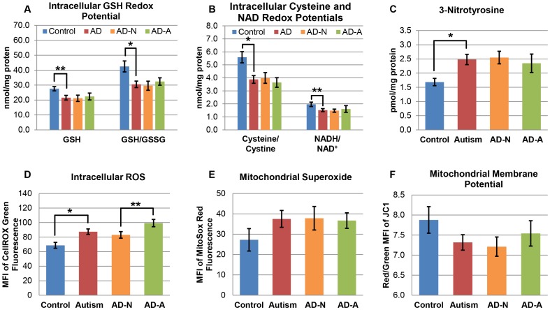
“LCLs derived from children with ASD show decreased glutathione concentrations relative to oxidized disulfide glutathione as well as a decreased ratio of reduced nicotinamide adenine dinucleotide (NADH) to oxidized NAD and higher levels of 3-nitrotyrosine, a marker of protein oxidation that is indicative of chronic oxi…”
BackgroundAntipurinergic therapy for autism—An in-depth review
Naviaux, R. K. (2017) · Mitochondrion · doi:10.1016/j.mito.2017.12.007 · Cited by 42
Burger, B. J., Rose, S., Bennuri, S. C., Gill, P. S., Tippett, M., Delhey, L. M., Melnyk, S., & Frye, R. E. (2017) · Frontiers in Pediatrics · doi:10.3389/fped.2017.00219 · Cited by 39
Self-citationMitochondria May Mediate Prenatal Environmental Influences in Autism Spectrum Disorder
Frye, R. E., Cakir, J., Rose, S., Palmer, R. F., Austin, C. E., Curtin, P. C. P., & Arora, M. (2021) · Journal of Personalized Medicine · doi:10.3390/jpm11030218 · Cited by 38
“To determine if these bioenergetics changes were specific to a subset of ASD LCLs, a cluster analysis was used to separated ASD LCLs into those with normal bioenergetics (AD-N) and those with atypical bioenergetics (AD-A) [109].”
BackgroundMethodSelf-citationOikawa, Y., Izumi, R., Koide, M., Hagiwara, Y., Kanzaki, M., Suzuki, N., Kikuchi, K., Matsuhashi, T., Akiyama, Y., Ichijo, M., et al. (2020) · PLoS ONE · doi:10.1371/journal.pone.0231064 · Cited by 36
“To clarify the mechanism of mitochondrial dysfunction in sIBM, we next examined the mitochondrial bioenergetic function governing the oxygen consumption rate (OCR) and extracellular acidification rate (ECAR) using isolated myoblasts [11, 24, 25].”
BackgroundOyarzábal, A., Bravo‐Alonso, I., Sánchez‐Aragó, M., Rejas, M. T., Merinero, B., García‐Cazorla, À., Artuch, R., Ugarte, M., & Rodríguez‐Pombo, P. (2016) · Biochimica et Biophysica Acta (BBA) - Molecular Basis of Disease · doi:10.1016/j.bbadis.2016.01.016 · Cited by 36
Gut–brain axis in the executive function of austism spectrum disorder
Román, P., Rueda‐Ruzafa, L., Cardona, D., & Cortés‐Rodríguez, A. E. (2018) · Behavioural Pharmacology · doi:10.1097/fbp.0000000000000428 · Cited by 36
Bennuri, S. C., Rose, S., & Frye, R. E. (2019) · Frontiers in Psychiatry · doi:10.3389/fpsyt.2019.00269 · Cited by 33
“Since many environmental stressors may have their biological effect through increases in ROS (21, 44–46) and since ROS is elevated in many ASDderived tissues (45), including LCLs (37) and brain (47, 48), we hypothesized that prolonged environmental exposures, potentially through prolonged exposure to ROS, would alter…”
BackgroundResultInfluentialSelf-citationKoufaris, C., & Sismani, C. (2015) · International Journal of Molecular Sciences · doi:10.3390/ijms16048699 · Cited by 35
“Recently published studies have reported that oxidative stress induces more prominent mitochondrial dysfunction in lymphoblastoid cells derived from subsets of ASD patients [47,48].”
BackgroundMorimoto, M., Hashimoto, T., Tsuda, Y., Nakatsu, T., Kitaoka, T., & Kyotani, S. (2020) · PLoS ONE · doi:10.1371/journal.pone.0233550 · Cited by 31
Yadav, S., Tiwari, V., Singh, M., Yadav, R. K., Roy, S., Devi, U., Gautam, S., Rawat, J. K., Ansari, M. N., Saeedan, A. S., et al. (2016) · Journal of Physiology and Biochemistry · doi:10.1007/s13105-016-0532-2 · Cited by 34
“Pronounced oxidative stress with change in membrane permeability and associated mitochondrial dysfunction are few of the players toward the pathogenesis of autism disorders [14, 44].”
BackgroundBioenergetic variation is related to autism symptomatology
Delhey, L. M., Kilinc, E. N., Yin, L., Slattery, J. M., Tippett, M., Wynne, R. A., Rose, S., Kahler, S. G., Damle, S., Legido, A., et al. (2017) · Metabolic Brain Disease · doi:10.1007/s11011-017-0087-0 · Cited by 32
“Indeed, we have repeatedly demonstrated in the laboratory that a subset of lymphoblastoid cell lines derived from children with ASD have respiratory rates approximately twice that of control cell lines (Rose et al. 2014a, b, 2015a, b; Frye et al. 2016c, 2017a; Rose et al. 2017).”
BackgroundResultInfluentialSelf-citationJyonouchi, H., Geng, L., Rose, S., Bennuri, S. C., & Frye, R. E. (2019) · Frontiers in Psychiatry · doi:10.3389/fpsyt.2019.00071 · Cited by 34
“, North Billerica, MA, United States) was used for measurement of oxygen consumption ratio (OCR), which is an indicator of mitochondrial respiration, in real time in live PBMCs (14, 27).”
MethodSelf-citationCaveolin3 Stabilizes McT1-Mediated Lactate/Proton Transport in Cardiomyocytes
Peper, J., Kownatzki-Danger, D., Weninger, G., Seibertz, F., Pronto, J. R. D., Sutanto, H., Pacheu‐Grau, D., Hindmarsh, R., Brandenburg, S., Kohl, T., et al. (2021) · Circulation Research · doi:10.1161/circresaha.119.316547 · Cited by 30
Garcia, S. M., Marinho, L. S. R., Lunardelli, P. A., Seneda, M. M., & Meirelles, F. V. (2015) · PLoS ONE · doi:10.1371/journal.pone.0119463 · Cited by 29
“In addition to differing numbers of antral follicles, zebu and taurine cows diverge in several aspects, such as estrus manifestation [33], progesterone concentration, follicle size [34] and IGF-I and insulin concentrations [35].”
BackgroundNEK10 interactome and depletion reveal new roles in mitochondria
de Oliveira, A. P., Basei, F. L., Slepicka, P. F., Ferezin, C. D. C., Melo‐Hanchuk, T. D., de Souza, E. E., de Lima, T. I., dos Santos, V. T., Mendes, D., Silveira, L. R., et al. (2020) · Proteome Science · doi:10.1186/s12953-020-00160-w · Cited by 30
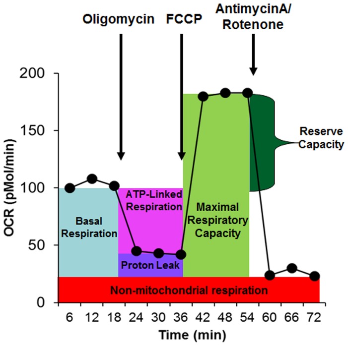
“of experimental oxygen consumption rate (OCR) data type nomenclature, adapted from Rose and coworkers [56].”
Induction of a Senescence-Like Phenotype in Cultured Human Fetal Microglia During HIV-1 Infection
Chen, N. C., Partridge, A. T., Tüzer, F., Cohen, J., Nacarelli, T., Navas, S., Sell, C., Torres, C. A., & Martín‐García, J. (2018) · The Journals of Gerontology Series A · doi:10.1093/gerona/gly022 · Cited by 32
Shaw, G. A., Hyer, M. M., Targett, I. L., Dyer, S. K., Turkson, S., Burns, C. M., & Neigh, G. N. (2020) · Brain Behavior and Immunity · doi:10.1016/j.bbi.2020.05.021 · Cited by 28
Parekh, M., Peh, G. S. L., Mehta, J. S., Ramos, T., Ponzin, D., Ahmad, S., & Ferrari, S. (2019) · Experimental Eye Research · doi:10.1016/j.exer.2019.107814 · Cited by 28
Mertens, R. T., Misra, A., Xiao, P., Baek, S., Rone, J. M., Mangani, D., Sivanathan, K. N., Arojojoye, A. S., Awuah, S. G., Lee, I., et al. (2024) · Immunity · doi:10.1016/j.immuni.2024.06.001 · Cited by 27
Socała, K., Żmudzka, E., Lustyk, K., Zagaja, M. P., Brighenti, V., Costa, A., Andres‐Mach, M., Pytka, K., Martinelli, I., Mandrioli, J., et al. (2024) · Phytotherapy Research · doi:10.1002/ptr.8101 · Cited by 29
Hua, A., Justiniano, R., Perer, J., Park, S., Li, H., Cabello, C. M., & Wondrak, G. T. (2019) · Cancers · doi:10.3390/cancers11050590 · Cited by 26
“A Seahorse Extracellular Flux (XF) 96 Analyzer (Seahorse Bioscience, Inc, North Billerica, MA, USA) using a specialized oxygen consumption rate (OCR) kit (Seahorse XFe96 FluxPaksTM, Agilent, Santa Clara, CA, USA) was employed in 96 well format according to the manufacturer’s specifications measuring OCR as a quantitat…”
MethodAschner, M., Martins, A. C., Oliveira‐Paula, G. H., Skalny, A. V., Zaitseva, I. P., Bowman, A. B., Kirichuk, A. A., Santamarı́a, A., Tizabi, Y., & Tinkov, A. A. (2024) · Current Research in Toxicology · doi:10.1016/j.crtox.2024.100170 · Cited by 26
“In this regard, lymphoblastoid cell lines derived from patients with ASD were more susceptible to oxidative stress and had significant physiological abnormalities in mitochondrial function (Rose et al., 2014).”
BackgroundFrye, R. E., Rose, S., Wynne, R. A., Bennuri, S. C., Blossom, S. J., Gilbert, K. M., Heilbrun, L. P., & Palmer, R. F. (2017) · Scientific Reports · doi:10.1038/s41598-017-04821-3 · Cited by 25
Self-citationThe Effect of Freeze-Thaw Cycles on Gene Expression Levels in Lymphoblastoid Cell Lines
Çalışkan, M., Pritchard, J. K., Ober, C., & Gilad, Y. (2014) · PLoS ONE · doi:10.1371/journal.pone.0107166 · Cited by 24
“Thousands of published studies have used LCLs to address a wide range of questions in human biology, including studies of gene regulatory mechanisms [3,4,6–14], drug toxicity [15,16], response to stress [17,18], and case control studies of human diseases [19,20].”
BackgroundSerum microRNAs in ASD: Association With Monocyte Cytokine Profiles and Mitochondrial Respiration
Jyonouchi, H., Geng, L., Törüner, G., Rose, S., Bennuri, S. C., & Frye, R. E. (2019) · Frontiers in Psychiatry · doi:10.3389/fpsyt.2019.00614 · Cited by 27
“Samples were sent to Dr. R. Frye’s laboratory on dry ice where the Seahorse Extracellular Flux (XF) 96 Analyzer (Seahorse Bioscience, Inc., North Billerica, MA, USA) was used to measure oxygen consumption ratio (OCR; pMol/min), which is an indicator of mitochondrial respiration, in real time in live PBMCs. ATP-linked…”
MethodInfluentialSelf-citationTargeting breast cancer metabolism with a novel inhibitor of mitochondrial ATP synthesis
Kim, M. S., Gernapudi, R., Cedeno, Y. C., Polster, B. M., Martinez, R., Shapiro, P., Kesari, S., Nurmemmedov, E., & Passaniti, A. (2020) · Oncotarget · doi:10.18632/oncotarget.27743 · Cited by 22
“Baseline cellular OCR (BL) was calculated from basal respiration after subtracting NM [111].”
MethodPhysiological mediators of prenatal environmental influences in autism spectrum disorder
Frye, R. E., Cakir, J., Rose, S., Palmer, R. F., Austin, C. E., & Curtin, P. C. P. (2021) · BioEssays · doi:10.1002/bies.202000307 · Cited by 24
“dysregulation of oxidative stress at the inner mitochondria membrane in neurodevelopmental disorders,[15] including ASD[78] and Fragile X,[79] as manifested by proton leak across the inner mitochondrial membrane.”
BackgroundSelf-citationIKCa channels control breast cancer metabolism including AMPK-driven autophagy
Gross, D., Bischof, H., Maier, S., Sporbeck, K., Birkenfeld, A. L., Malli, R., Ruth, P., Proikas‐Cezanne, T., & Łukowski, R. (2022) · Cell Death and Disease · doi:10.1038/s41419-022-05329-z · Cited by 23
Serum starvation affects mitochondrial metabolism of adipose-derived stem/stromal cells
Giannasi, C., Niada, S., Morte, E. D., Casati, S. R., Palma, C. D., & Brini, A. T. (2023) · Cytotherapy · doi:10.1016/j.jcyt.2023.03.004 · Cited by 21
Slattery, J., MacFabe, D., Kahler, S., & Frye, R. (2016) · Current Pharmaceutical Design · doi:10.2174/1381612822666160905123953 · Cited by 20
Self-citationInheritance of HLA-Cw7 Associated With Autism Spectrum Disorder (ASD)
Harville, T., Rhodes-Clark, B. S., Bennuri, S. C., Delhey, L. M., Slattery, J. M., Tippett, M., Wynne, R. A., Rose, S., Kahler, S. G., & Frye, R. E. (2019) · Frontiers in Psychiatry · doi:10.3389/fpsyt.2019.00612 · Cited by 20
Self-citationSaxena, R., Saribas, S., Jadiya, P., Tomar, D., Kaminski, R., Elrod, J. W., & Safak, M. (2020) · Virology · doi:10.1016/j.virol.2020.11.004 · Cited by 18
“That is, this inhibitor provides information about the levels of O2 consumption, directly coupled to ATP production (Supplement 1) (Rose et al., 2014).”
BackgroundEnhancing enterocyte fatty acid oxidation in mice affects glycemic control depending on dietary fat
Ramachandran, D., Clara, R., Fedele, S., Michel, L., Burkard, J., Kaufman, S., Alvarado-Diaz, A., Weissfeld, N., Bock, K. D., Prip‐Buus, C., et al. (2018) · Scientific Reports · doi:10.1038/s41598-018-29139-6 · Cited by 18
Webb, L. M. C., Pascall, J. C., Hepburn, L., Carter, C., Turner, M., & Butcher, G. W. (2014) · PLoS ONE · doi:10.1371/journal.pone.0110294 · Cited by 17
“We used the Seahorse Extracellular Flux (XF) 96 Analyzer (Seahorse Bioscience, Inc, North Billerica, MA, USA), to measure the oxygen consumption rate (OCR), an indicator of mitochondrial respiration, in real-time in live intact B lymphocytes as previously described [51].”
MethodNitrosative Stress in Autism: Supportive Evidence and Implications for Mitochondrial Dysfunction
Frye, R. E., Rose, S., Voinsky, I., & Gurwitz, D. (2024) · Advanced Science · doi:10.1002/advs.202304439 · Cited by 16
Self-citationSon, Y., Jeong, Y. I., Hwang, K. C., Jeong, Y. I., & Hwang, W. S. (2021) · Theriogenology · doi:10.1016/j.theriogenology.2021.01.010 · Cited by 15
Sasagawa, S., Nishimura, Y., Koiwa, J., Nomoto, T., Shintou, T., Murakami, S., Yuge, M., Kawaguchi, K., Kawase, R., Miyazaki, T., et al. (2015) · ACS Chemical Biology · doi:10.1021/acschembio.5b00751 · Cited by 15
Wu, Z., Törő, G., Xu, G., Dang, D., Prater, C., & Yang, Q. (2022) · Cells · doi:10.3390/cells11244067 · Cited by 14
Oxidative stress and neuroimmune proteins in a mouse model of autism
Uddin, M. N., Mondal, T. K., Yao, Y., MANLEY, K. M., & Lawrence, D. A. (2023) · Cell Stress and Chaperones · doi:10.1007/s12192-023-01331-2 · Cited by 17
Bioenergetic signatures of neurodevelopmental regression
Frye, R. E., McCarty, P. J., Werner, B. A., Rose, S., & Scheck, A. C. (2024) · Frontiers in Physiology · doi:10.3389/fphys.2024.1306038 · Cited by 15
Self-citationAlsharabasy, A. M., Lagarias, P. I., Papavasileiou, K. D., Afantitis, A., Farràs, P., Glynn, S. A., & Pandit, A. S. (2024) · Journal of Medicinal Chemistry · doi:10.1021/acs.jmedchem.4c00989 · Cited by 14
Strong, M. D., Hart, M. D., Tang, T. Z., Ojo, B. A., Wu, L., Nacke, M. R., Agidew, W. T., Hwang, H. J., Hoyt, P. R., Bettaieb, A., et al. (2020) · The FASEB Journal · doi:10.1096/fj.202000772r · Cited by 14
Kashyap, B., Barge, S., Bharadwaj, S., Deka, B., Rahman, S., Ghosh, A., Manna, P., Dutta, P. P., Sheikh, Y., Kandimalla, R., et al. (2021) · Phytomedicine · doi:10.1016/j.phymed.2021.153761 · Cited by 14
Association of NGF and Mitochondrial Respiration with Autism Spectrum Disorder
Gevezova, M., Minchev, D. S., Pacheva, I., Todorova, T., Yordanova, R. V., Timova, E., Ivanov, I. S., & Sarafian, V. S. (2022) · International Journal of Molecular Sciences · doi:10.3390/ijms231911917 · Cited by 15
“Furthermore, we found that reserve respiratory capacity is almost 40% higher in ASD children with developmental regression compared to those without, which is regarded by some authors as a protective adaptation in response to environmental stressors [83].”
BackgroundAhmed, S. A., Saikia, K., Basumatary, D., Gurumayum, S., Swargiary, D., & Borah, J. C. (2025) · International Immunopharmacology · doi:10.1016/j.intimp.2025.114825 · Cited by 13
Xiong, J., Bonney, S., Gonçalves, R. V., & Esposito, D. (2022) · Life Sciences · doi:10.1016/j.lfs.2022.120887 · Cited by 12
Kingsley, J., Costello, J. R., Raghunand, N., & Rejniak, K. A. (2021) · PLoS Computational Biology · doi:10.1371/journal.pcbi.1009206 · Cited by 12
“, 1–350 amol/s per cell[29], and 1–120 amol/s per cell[30]), changes reported in the same cells varied no more than 10-fold when the culture conditions were modified[14,31].”
BackgroundRedox System Dysfunction as a Key Mechanism in Autism Spectrum Disorder Pathogenesis
de Oliveira, C. A., Iorio, E. L., & Espíndola, F. S. (2025) · International Journal of Molecular Sciences · doi:10.3390/ijms26209850 · Cited by 12
Monsour, M., Gorsky, A., Nguyen, H., Castelli, V., Lee, J.-Y., & Borlongan, C. V. (2022) · Neuroreport · doi:10.1097/wnr.0000000000001828 · Cited by 12
Metadata
- Authors
- Rose, S., Frye, R. E., Slattery, J., Wynne, R., Tippett, M., Pavliv, O., Melnyk, S., & James, S. J.
- Affiliations
- Arkansas Children's Hospital (US)
- Corresponding author
- Frye, Richard E.
- Venue
- PLoS ONE
- Publisher
- Public Library of Science (PLoS)
- Published
- 2014-01-08
- Volume
- 9
- Issue
- 1
- Pages
- e85436
- ISSN
- 1932-6203 (ISSN-L)
- PMC
- PMC3885720
- OpenAlex
- W2163485668
- Cited by
- 168
- References
- 61
- Citations by year
- 2026: 9 · 2025: 9 · 2024: 20 · 2023: 7 · 2022: 22 · 2021: 17 · 2020: 18 · 2019: 9 · 2018: 10 · 2017: 12 · 2016: 13 · 2015: 16 · 2014: 6
- FWCI
- 6.42
- Type
- journal article
- Language
- en
- Open access
- gold
- doi.org
- Licence
- Locations
- PLoS ONE (publishedVersion, cc-by)
- Europe PMC (PubMed Central) (publishedVersion, cc-by)
- PubMed Central (publishedVersion, cc-by)
- PubMed (publishedVersion)
- DOAJ (DOAJ: Directory of Open Access Journals) (submittedVersion, cc-by-sa)
- Funders
- Arkansas Biosciences Institute
- Topics
- Life Sciences › Biochemistry, Genetics and Molecular Biology › Molecular Biology › Mitochondrial Function and Pathology
- Life Sciences › Neuroscience › Cognitive Neuroscience › Autism Spectrum Disorder Research
- Life Sciences › Biochemistry, Genetics and Molecular Biology › Cell Biology › Autophagy in Disease and Therapy
- Keywords
- autism spectrum disorder, mitochondrial dysfunction, oxidative stress, reserve capacity, lymphoblastoid cell lines, reactive oxygen species, UCP2, glycolysis, ATP-linked respiration, mitochondrial heterogeneity
- MeSH terms
- Uncoupling Protein 2, Autistic Disorder, Cell Line, Child, Child, Preschool, Female, Glycolysis, Humans, Ion Channels, Lymphocytes, Male, Mitochondria, Naphthoquinones, Oxidation-Reduction, Protons, Case-Control Studies, Reactive Oxygen Species, Oxidative Stress, Cell Respiration, Mitochondrial Proteins, Mitochondrial Membranes
About these checks
Registry, retraction, Cites and statistics results are computed, not judged, with one exception: a printed reference that no index lists is read once by a model before it counts as a citation error. Claims are extracted by a model and kept only when their quote appears verbatim in the text; each cited source is then checked, with a model’s help, for whether it says what the claim relies on. Those verdicts can be wrong — read the claim’s quote and the cited source before relying on one.
Statistics and arithmetic are recomputed, not judged.
Confidence bands are shown only for fields calibrated on our annotated test set. None is calibrated yet, so no band is shown.
What counts as a claim ›How we measure confidence ›How we check this ›
Are you an author, or do you see a problem with this page? Request a review
