

The DOI matches its Crossref record; 59 of the 59 references it cites were found in a registry or an index.
Structural and Functional Rich Club Organization of the Brain in Children and Adults
Open accessAbstract
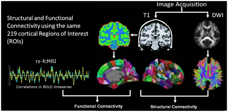
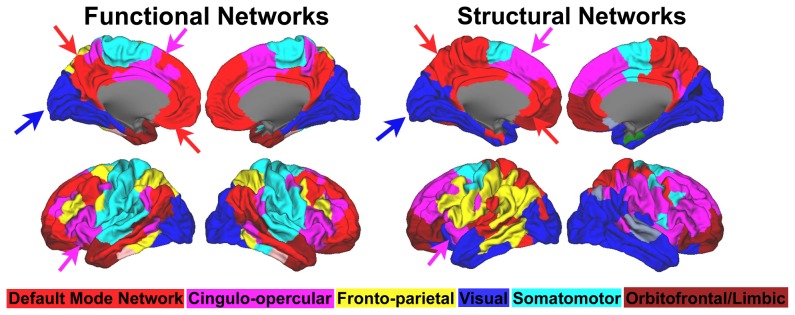
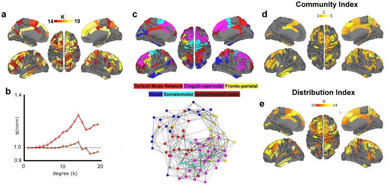
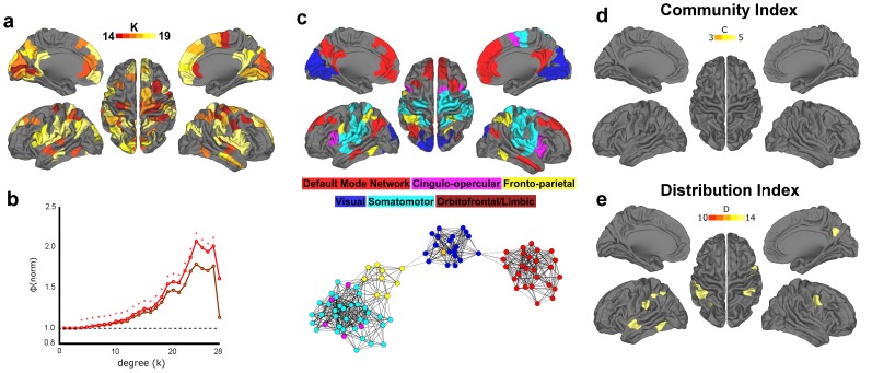
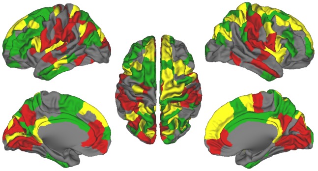
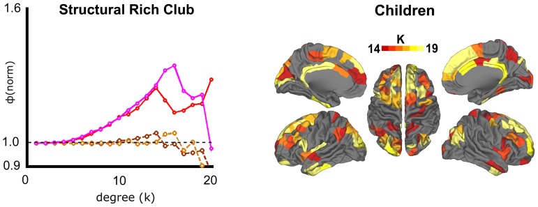
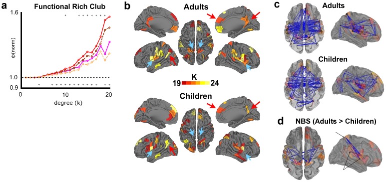
Recent studies using Magnetic Resonance Imaging (MRI) have proposed that the brain's white matter is organized as a rich club, whereby the most highly connected regions of the brain are also highly connected to each other. Here we use both functional and diffusion-weighted MRI in the human brain to investigate whether the rich club phenomena is present with functional connectivity, and how this organization relates to the structural phenomena. We also examine whether rich club regions serve to integrate information between distinct brain systems, and conclude with a brief investigation of the developmental trajectory of rich-club phenomena. In agreement with prior work, both adults and children showed robust structural rich club organization, comprising regions of the superior medial frontal/dACC, medial parietal/PCC, insula, and inferior temporal cortex. We also show that these regions were highly integrated across the brain's major networks. Functional brain networks were found to have rich club phenomena in a similar spatial layout, but a high level of segregation between systems. While no significant differences between adults and children were found structurally, adults showed significantly greater functional rich club organization. This difference appeared to be driven by a specific set of connections between superior parietal, insula, and supramarginal cortex. In sum, this work highlights the existence of both a structural and functional rich club in adult and child populations with some functional changes over development. It also offers a potential target in examining atypical network organization in common developmental brain disorders, such as ADHD and Autism.
Overview
The claims and statistics of this paper haven’t been checked yet.
Run the check — free, no account needed. It reads the open-access text and checks the sources it cites.
Integrity signals
Registry
Cites
Statistics
Figures
Verification
Claims the paper makes
The claims and statistics of this paper haven’t been checked yet.
Run the check — free, no account needed. It reads the open-access text and checks the sources it cites.
Claims other papers make about this one
Each is checked against this paper’s own text: does it say what the citing paper says it does?
In the citing paper
“high strength and betweenness hubs in adulthood are largely located within within the default mode, attentional, sensorimotor, and visual areas (Cao et al., 2014; Fransson et al., 2011; Grayson et al., 2014; Hwang et al., 2013)”
From Development of large-scale functional networks from birth to adulthood: A guide to the neuroimaging literature (2017) · cites this paper as Grayson et al., 2014
In the citing paper
“Among cortical areas, some have reported increasing hubness of frontal nodes (Cao et al., 2014; Wu et al., 2013) while in other lobes there have been mixed reports of increasing (Cao et al., 2014; Grayson et al., 2014) and decreasing (Wu et al., 2013; Zuo et al., 2012), or stable (Hwang et al., 2013) hubness.”
From Development of large-scale functional networks from birth to adulthood: A guide to the neuroimaging literature (2017) · cites this paper as Grayson et al., 2014
In the citing paper
“More detailed network analyses have revealed that specific connections to cortical hub regions increase during childhood and adolescence (Grayson et al., 2014; Hwang et al., 2013) resulting in increased rich club coefficients in adulthood (Cao et al., 2014; Grayson et al., 2014).”
From Development of large-scale functional networks from birth to adulthood: A guide to the neuroimaging literature (2017) · cites this paper as Grayson et al., 2014
In the citing paper
“Returning to the MRI literature, there is convergent evidence that the locations of high-strength and high-betweenness hub regions appear to be stable after 5 years of age (Cao et al., 2014; Grayson et al., 2014; Hwang et al., 2013; Wang et al., 2012; Wu et al., 2013; Zuo et al., 2012).”
From Development of large-scale functional networks from birth to adulthood: A guide to the neuroimaging literature (2017) · cites this paper as Grayson et al., 2014
Self-check
Percentages, sums and reported differences re-computed from the paper’s own numbers.
The self-check needs the paper’s full text.
Citations
Cites
Modularity and community structure in networks
Newman, M. E. J. (2006) · Proceedings of the National Academy of Sciences · doi:10.1073/pnas.0601602103 · Cited by 12,256
Complex brain networks: graph theoretical analysis of structural and functional systems
Bullmore, E., & Sporns, O. (2009) · Nature Reviews Neuroscience · doi:10.1038/nrn2575 · Cited by 11,820
Complex network measures of brain connectivity: Uses and interpretations
Rubinov, M., & Sporns, O. (2010) · NeuroImage · doi:10.1016/j.neuroimage.2009.10.003 · Cited by 11,522
Functional connectivity in the motor cortex of resting human brain using echo‐planar mri
Biswal, B., Zerrin Yetkin, F., Haughton, V. M., & Hyde, J. S. (1995) · Magnetic Resonance in Medicine · doi:10.1002/mrm.1910340409 · Cited by 9,812
The organization of the human cerebral cortex estimated by intrinsic functional connectivity
Thomas Yeo, B. T., Krienen, F. M., Sepulcre, J., Sabuncu, M. R., Lashkari, D., Hollinshead, M., Roffman, J. L., Smoller, J. W., Zöllei, L., Polimeni, J. R., et al. (2011) · Journal of Neurophysiology · doi:10.1152/jn.00338.2011 · Cited by 9,182
The human brain is intrinsically organized into dynamic, anticorrelated functional networks
Fox, M. D., Snyder, A. Z., Vincent, J. L., Corbetta, M., Van Essen, D. C., & Raichle, M. E. (2005) · Proceedings of the National Academy of Sciences · doi:10.1073/pnas.0504136102 · Cited by 8,553
Power, J. D., Barnes, K. A., Snyder, A. Z., Schlaggar, B. L., & Petersen, S. E. (2012) · NeuroImage · doi:10.1016/j.neuroimage.2011.10.018 · Cited by 7,429
The organization of the human cerebellum estimated by intrinsic functional connectivity
Buckner, R. L., Krienen, F. M., Castellanos, A., Diaz, J. C., & Yeo, B. T. T. (2011) · Journal of Neurophysiology · doi:10.1152/jn.00339.2011 · Cited by 7,304
Spontaneous fluctuations in brain activity observed with functional magnetic resonance imaging
Fox, M. D., & Raichle, M. E. (2007) · Nature Reviews Neuroscience · doi:10.1038/nrn2201 · Cited by 6,700
The brainweb: phase synchronization and large-scale integration
Varela, F., Lachaux, J.-P., Rodriguez, E., & Martinerie, J. (2001) · Nature Reviews Neuroscience · doi:10.1038/35067550 · Cited by 4,761
Cited by
Sorted by citations
An Integrative Model of the Maturation of Cognitive Control
Luna, B., Marek, S., Larsen, B., Tervo-Clemmens, B., & Chahal, R. (2015) · Annual Review of Neuroscience · doi:10.1146/annurev-neuro-071714-034054 · Cited by 550
Grayson, D. S., & Fair, D. A. (2017) · NeuroImage · doi:10.1016/j.neuroimage.2017.01.079 · Cited by 411
“Returning to the MRI literature, there is convergent evidence that the locations of high-strength and high-betweenness hub regions appear to be stable after 5 years of age (Cao et al., 2014; Grayson et al., 2014; Hwang et al., 2013; Wang et al., 2012; Wu et al., 2013; Zuo et al., 2012).”
BackgroundInfluentialSelf-citationUnraveling the Miswired Connectome: A Developmental Perspective
Martino, A. D., Fair, D. A., Kelly, C. A. M., Satterthwaite, T. D., Castellanos, F. X., Thomason, M. E., Craddock, R. C., Luna, B., LEVENTHAL, B. L., Zuo, X., et al. (2014) · Neuron · doi:10.1016/j.neuron.2014.08.050 · Cited by 357
Self-citationvan den Heuvel, M. P., Bullmore, E. T., & Sporns, O. (2016) · Trends in Cognitive Sciences · doi:10.1016/j.tics.2016.03.001 · Cited by 328
An update on the comorbidity of ADHD and ASD: a focus on clinical management
Antshel, K. M., Zhang‐James, Y., Wagner, K. E., Ledesma, A., & Faraone, S. V. (2016) · Expert Review of Neurotherapeutics · doi:10.1586/14737175.2016.1146591 · Cited by 276
Metadata
- Authors
- Grayson, D. S., Ray, S., Carpenter, S., Iyer, S., Dias, T. G. C., Stevens, C., Nigg, J. T., & Fair, D. A.
- Affiliations
- Oregon Health & Science University (US)
- Corresponding author
- Grayson, David S.
- Fair, Damien A.
- Venue
- PLoS ONE
- Publisher
- Public Library of Science (PLoS)
- Published
- 2014-02-05
- Volume
- 9
- Issue
- 2
- Pages
- e88297
- ISSN
- 1932-6203 (ISSN-L)
- PMC
- PMC3915050
- OpenAlex
- W2063751901
- Cited by
- 187
- References
- 59
- Citations by year
- 2026: 4 · 2025: 9 · 2024: 12 · 2023: 14 · 2022: 14 · 2021: 14 · 2020: 13 · 2019: 26 · 2018: 19 · 2017: 14 · 2016: 19 · 2015: 18 · 2014: 11
- FWCI
- 11.52
- Type
- journal article
- Language
- en
- Open access
- gold
- doi.org
- Locations
- PLoS ONE (publishedVersion, cc-by)
- Europe PMC (PubMed Central) (publishedVersion, cc-by)
- PubMed Central (publishedVersion, cc-by)
- PubMed (publishedVersion)
- DOAJ (DOAJ: Directory of Open Access Journals) (submittedVersion, cc-by-sa)
- Figshare (submittedVersion, cc-by)
- Funders
- James S. McDonnell Foundation (R01 MH096773)
- National Institutes of Health (MH091238, MH086654)
- Oregon Clinical and Translational Research Institute
- National Institute of Mental Health (MH086654, MH091238, R01MH115357, R00MH091238, R01MH096773, MH096773, R01MH086654)
- Topics
- Life Sciences › Neuroscience › Cognitive Neuroscience › Functional Brain Connectivity Studies
- Health Sciences › Medicine › Radiology, Nuclear Medicine and Imaging › Advanced Neuroimaging Techniques and Applications
- Physical Sciences › Physics and Astronomy › Statistical and Nonlinear Physics › Complex Network Analysis Techniques
- Keywords
- rich club, brain network organization, functional connectivity, structural connectivity, white matter, brain development, children, adults, default mode network, dorsal anterior cingulate cortex, posterior cingulate cortex, insula, inferior temporal cortex, supramarginal cortex, functional segregation, attention deficit hyperactivity disorder, autism spectrum disorder
- MeSH terms
- Adult, Brain, Brain Mapping, Child, Female, Humans, Magnetic Resonance Imaging, Male, Models, Anatomic, Models, Neurological, Nerve Net, Neural Pathways, Young Adult, Diffusion Tensor Imaging
About these checks
Registry, retraction, Cites and statistics results are computed, not judged, with one exception: a printed reference that no index lists is read once by a model before it counts as a citation error. Claims are extracted by a model and kept only when their quote appears verbatim in the text; each cited source is then checked, with a model’s help, for whether it says what the claim relies on. Those verdicts can be wrong — read the claim’s quote and the cited source before relying on one.
Statistics and arithmetic are recomputed, not judged.
Confidence bands are shown only for fields calibrated on our annotated test set. None is calibrated yet, so no band is shown.
What counts as a claim ›How we measure confidence ›How we check this ›
Are you an author, or do you see a problem with this page? Request a review
