

IHC Profiler: An Open Source Plugin for the Quantitative Evaluation and Automated Scoring of Immunohistochemistry Images of Human Tissue Samples
Open accessThe DOI matches its Crossref record; 30 of the 30 references it cites were found in a registry or an index.
Abstract
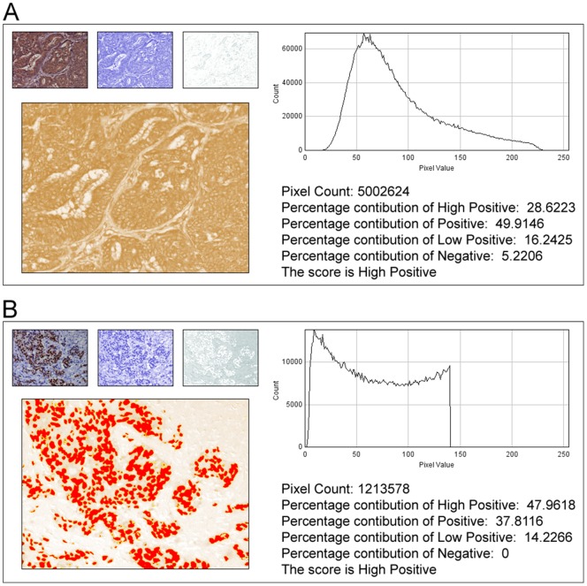
In anatomic pathology, immunohistochemistry (IHC) serves as a diagnostic and prognostic method for identification of disease markers in tissue samples that directly influences classification and grading the disease, influencing patient management. However, till today over most of the world, pathological analysis of tissue samples remained a time-consuming and subjective procedure, wherein the intensity of antibody staining is manually judged and thus scoring decision is directly influenced by visual bias. This instigated us to design a simple method of automated digital IHC image analysis algorithm for an unbiased, quantitative assessment of antibody staining intensity in tissue sections. As a first step, we adopted the spectral deconvolution method of DAB/hematoxylin color spectra by using optimized optical density vectors of the color deconvolution plugin for proper separation of the DAB color spectra. Then the DAB stained image is displayed in a new window wherein it undergoes pixel-by-pixel analysis, and displays the full profile along with its scoring decision. Based on the mathematical formula conceptualized, the algorithm is thoroughly tested by analyzing scores assigned to thousands (n = 1703) of DAB stained IHC images including sample images taken from human protein atlas web resource. The IHC Profiler plugin developed is compatible with the open resource digital image analysis software, ImageJ, which creates a pixel-by-pixel analysis profile of a digital IHC image and further assigns a score in a four tier system. A comparison study between manual pathological analysis and IHC Profiler resolved in a match of 88.6% (P<0.0001, CI = 95%). This new tool developed for clinical histopathological sample analysis can be adopted globally for scoring most protein targets where the marker protein expression is of cytoplasmic and/or nuclear type. We foresee that this method will minimize the problem of inter-observer variations across labs and further help in worldwide patient stratification potentially benefitting various multinational clinical trial initiatives.
Overview
The claims and statistics of this paper haven’t been checked yet.
Free: it reads the open-access text and checks the first 5 claims that cite a source we can check. Create a free account to check every claim.
Integrity signals
Registry
Cites
Statistics
Figures



Verification
Claims the paper makes
The claims and statistics of this paper haven’t been checked yet.
Free: it reads the open-access text and checks the first 5 claims that cite a source we can check. Create a free account to check every claim.
Claims other papers make about this one
Each is checked against this paper’s own text: does it say what the citing paper says it does?
In the citing paper
“For analysis of c-MYC expression in liver sections, random 20× non-tumor fields of the c-MYC IHC scans from liver sections of individual mice were output using NDP.view2 and blindly analyzed using ImageJ (https://imagej.nih.gov/ij/) with the plugin ‘IHC Profiler’62.”
From Tumor-specific cholinergic CD4+ T lymphocytes guide immunosurveillance of hepatocellular carcinoma (2023) · cites this paper as [62]
Self-check
Percentages, sums and reported differences re-computed from the paper’s own numbers.
The self-check needs the paper’s full text.
Citations
Cites
NIH Image to ImageJ: 25 years of image analysis
Schneider, C. A., Rasband, W. S., & Eliceiri, K. W. (2012) · Nature Methods · doi:10.1038/nmeth.2089 · Cited by 64,638 · cited 1×
“The zones were visually identified by using the threshold feature of Image menu of the ImageJ program [22].”
MethodHsu, S. M., Raine, L., & Fanger, H. (1981) · Journal of Histochemistry & Cytochemistry · doi:10.1177/29.4.6166661 · Cited by 14,140
Harvey, J. M., Clark, G. M., Osborne, C. K., & Allred, D. C. (1999) · Journal of Clinical Oncology · doi:10.1200/jco.1999.17.5.1474 · Cited by 2,056
Quantification of histochemical staining by color deconvolution.
Ruifrok, A. C. C., & Johnston, D. A. (2001) · PubMed · Cited by 2,029
Tuominen, V. J., Ruotoistenmäki, S., Viitanen, A., Jumppanen, M., & Isola, J. (2010) · Breast Cancer Research · doi:10.1186/bcr2615 · Cited by 472
McCarty, K. S., Szabo, E. M., Flowers, J. L., Cox, E. B., Leight, G. S., Miller, L. A., Konrath, J. G., Jt, S., Budwit, D. A., & Wt, C. (1986) · PubMed · Cited by 417
A global view of protein expression in human cells, tissues, and organs
Pontén, F., Gry, M., Fagerberg, L., Lundberg, E., Asplund, A., Berglund, L., Oksvold, P., Björling, E., Hober, S., Kampf, C., et al. (2009) · Molecular Systems Biology · doi:10.1038/msb.2009.93 · Cited by 222
Automated Selection of DAB-labeled Tissue for Immunohistochemical Quantification
Brey, E. M., Lalani, Z., Johnston, C., Wong, M., McIntire, L. V., Duke, P. J., & Patrick, C. W. (2003) · Journal of Histochemistry & Cytochemistry · doi:10.1177/002215540305100503 · Cited by 200
Quantitative image analysis of immunohistochemical stains using a CMYK color model
Pham, N.-A., Morrison, A., Schwock, J., Aviel-Ronen, S., Iakovlev, V., Tsao, M.-S., Ho, J., & Hedley, D. W. (2007) · Diagnostic Pathology · doi:10.1186/1746-1596-2-8 · Cited by 179
Quantification of renal pathology by image analysis (Methods in Renal Research)
RANGAN, G. K., & TESCH, G. H. (2007) · Nephrology · doi:10.1111/j.1440-1797.2007.00855.x · Cited by 176
Cited by
Items 901–1,000 of 1,000
Zhang, B., Yu, L., & Tang, L. (2026) · ACS Biomaterials Science & Engineering · doi:10.1021/acsbiomaterials.5c01606 · Cited by 5
NAT10-mediated ac4C modifications regulate glioblastoma progression
Lin, L., Yp, X., Guo, Y., Tu, Z., Luo, P., Fang, Z., Zhang, L., Huang, K., & Wu, L. (2026) · Cell Death and Disease · doi:10.1038/s41419-025-08315-3 · Cited by 5
3-D Textural Analysis of 2-[¹⁸F]FDG PET and Ki67 Expression in Nonsmall Cell Lung Cancer
Hu, X., Liang, X., Antonecchia, E., Chiaravallotti, A., Chu, Q., Han, S., Li, Z., Wan, L., D’Ascenzo, N., Schillaci, O., et al. (2021) · IEEE Transactions on Radiation and Plasma Medical Sciences · doi:10.1109/trpms.2021.3051376 · Cited by 5
Jarosz, P. M., Jasielski, P. P., Zarobkiewicz, M. K., Sławiński, M., Wawryk‐Gawda, E., & Jodłowska‐Jędrych, B. (2022) · Foods · doi:10.3390/foods11081131 · Cited by 4
The prognostic role of p53 and its correlation with CDK9 in urothelial carcinoma
Borowczak, J., Szczerbowski, K., Maniewski, M., Zdrenka, M., Słupski, P., Andrusewicz, H., Łysik-Miśkurka, J., Rutkiewicz, P., Bodnar, M., & Szylberg, Ł. (2022) · Clinical & Translational Oncology · doi:10.1007/s12094-022-02994-6 · Cited by 4
Kim, J. Y., Nam, G., Shin, Y. K., Vilaplana‐Lopera, N., Jeung, H., Moon, E. J., & Lee, I. J. (2023) · Cancers · doi:10.3390/cancers15061882 · Cited by 4
Lazăr, A. D., Dinescu, S., Sleiman, L., Dumitru, A. V., Costache, M., & Costache, M. (2023) · International Journal of Molecular Sciences · doi:10.3390/ijms24076565 · Cited by 7
Osiak-Wicha, C., Muszyński, S., Tomaszewska, E., Kras, K., Ropka‐Molik, K., Zhyla, M. I., & Arciszewski, M. B. (2024) · Annals of Animal Science · doi:10.2478/aoas-2024-0016 · Cited by 5
Szczepanik, K., Dobrowolski, P., & Świątkiewicz, M. (2024) · Journal of Animal Physiology and Animal Nutrition · doi:10.1111/jpn.14024 · Cited by 5
Pei, Q., Li, Z., Zhao, J., Zhang, H., Qin, T., & Zhao, J. (2024) · BMC Biotechnology · doi:10.1186/s12896-024-00879-w · Cited by 4
Wang, X., Xia, H., Li, T., Zuo, Q., Wang, Z., Yan, K., Xu, Z., Xue, W., Sun, G., Liu, Z., et al. (2024) · Advanced Healthcare Materials · doi:10.1002/adhm.202401625 · Cited by 6
Liu, J.-R., Xiangni, W., Cui, Y., Xu, G., Lu, J., He, Z., Xu, Y., Rongrong, L., Lingge, G., Wang, H., et al. (2025) · Journal of Physics D Applied Physics · doi:10.1088/1361-6463/adb04a · Cited by 4
Deng, X., Liu, H., Zhao, W., Wu, R., Chen, K., Li, Q., Yazlık, M. O., Özkan, H., Ren, J., Zhang, J., et al. (2025) · BMC Veterinary Research · doi:10.1186/s12917-025-04622-1 · Cited by 6
Subdermal nitrous oxide delivery increases skin microcirculation and random flap survival in rats
Serin, M., Altınel, D., Leblebıcı, C., Biltekin, B., Hüseyinbaş, Ö., Yazar, S. K., Irmak, F., Sönmez, A., & Bayrami̇çli̇, M. (2018) · Journal of Plastic Surgery and Hand Surgery · doi:10.1080/2000656x.2018.1531013 · Cited by 4
Circulating levels of hydroxylated bradykinin function as an indicator of tissue HIF-1α expression
Liu, Y., Gu, Y., Ng, S., Deng, Z., Lyon, C. J., Koay, E. J., Ning, B., Katz, M. H. G., Chiao, P. J., Fan, J., et al. (2020) · 中国科学通报:英文版 · doi:10.1016/j.scib.2020.04.023 · Cited by 6
Ye, X., Zhang, F., Li, Z., Wei, Y., Zhang, L., Wang, L., Tang, H., Chen, Z., Kerr, W. G., Zheng, T., et al. (2021) · Scientific Reports · doi:10.1038/s41598-021-88677-8 · Cited by 5
Moawad, R. S., Fattah, E. R. A. E., & Alsemeh, A. E. (2021) · The Egyptian Journal of Histology · doi:10.21608/ejh.2021.71825.1459 · Cited by 4
Taidi, Z., Zhou, T., Moore, K. H., Mansfield, K. J., & Liu, L. (2021) · Frontiers in Pharmacology · doi:10.3389/fphar.2021.682520 · Cited by 4
Lu, Y., Ma, J., Lin, J., Tian, Y., Ma, Y., Wang, W., Li, J., Zhang, H., & Jiao, P. (2021) · Bioengineered · doi:10.1080/21655979.2021.2010314 · Cited by 4
Loss of MAGEC3 Expression Is Associated with Prognosis in Advanced Ovarian Cancers
Ellegate, J., Mastri, M., Isenhart, E., Krolewski, J. J., Chatta, G. S., KAUFFMAN, E. C., Moffitt, M. N., & Eng, K. H. (2022) · Cancers · doi:10.3390/cancers14030731 · Cited by 5
Eid, A. A., Aly, R. G. M., Elkholy, S. A. E., & Sorror, O. A. (2022) · Photodermatology Photoimmunology & Photomedicine · doi:10.1111/phpp.12790 · Cited by 4
A Method for Unsupervised Semi-Quantification of Inmunohistochemical Staining with Beta Divergences
Sarmiento, A., Durán-Díaz, I., Fondón, I., Tomé, M., Bodineau, C., & Durán, R. V. (2022) · Entropy · doi:10.3390/e24040546 · Cited by 4
In utero exposure to selective serotonin re‐uptake inhibitor affects murine mandibular development
Boyce, M., Durham, E. L., Kuo, S., Taylor, J. M., Kishinchand, R., LaRue, A. C., & Cray, J. (2022) · Orthodontics and Craniofacial Research · doi:10.1111/ocr.12624 · Cited by 4
Depletion of vascular adaptive mechanisms in hypertension-induced injury of the heart and kidney
Stanchev, S., Kotov, G. N., Landzhov, B. V., Kirkov, V., Gaydarski, L., & Iliev, A. A. (2023) · Bratislavské lekárske listy/Bratislava medical journal · doi:10.4149/bll_2023_021 · Cited by 4
Chen, Y., Gao, L.-L., Kong, L., Guan, X., Yang, H., Li, Y., Lv, Z.-Y., Zhang, X., Liang, H., Chen, H., et al. (2023) · Cancer Treatment and Research Communications · doi:10.1016/j.ctarc.2023.100684 · Cited by 6
Liu, Y., Liu, S., Song, Z., Chen, D., Album, Z. M., Green, S., Deng, X., & Rodeo, S. R. (2023) · Journal of Clinical Medicine · doi:10.3390/jcm12030999 · Cited by 4
Jin, B., Yang, J., Zhen, J., Xu, Y. Y., Wang, C., Jing, Q., & Shang, Y. (2023) · Journal of Computer Assisted Tomography · doi:10.1097/rct.0000000000001515 · Cited by 4
Bastos‐Silva, V. J., Spineli, H., Guimarães, J. C., Borbely, K. S. C., Ursulino, J. S., de Aquino, T. M., Bento, E. S., Scariot, P. P. M., Sousa, F. A. D. B., & de Araújo, G. G. (2024) · Brazilian Journal of Medical and Biological Research · doi:10.1590/1414-431x2024e13276 · Cited by 4
Luminance Adaptive Biomarker Detection in Digital Pathology Images
Liu, J., Qiu, G., & Shen, L. (2016) · Procedia Computer Science · doi:10.1016/j.procs.2016.07.032 · Cited by 4
Kusmardi, K., Shavera, D., Estuningtyas, A., Tedjo, A., & Priosoeryanto, B. P. (2017) · Asian Journal of Pharmaceutical and Clinical Research · doi:10.22159/ajpcr.2017.v10i12.20560 · Cited by 4
Prognostic value and functional role of ROCK2 in pediatric Ewing sarcoma
Vieira, G. M., Roberto, G. M., Lira, R. ⁄. G., Engel, E. E., Tone, L. G., & Brassesco, M. S. (2017) · Oncology Letters · doi:10.3892/ol.2017.7571 · Cited by 5
Yin, H.-L., Fan, Y., Mu, D., Song, F., Tian, F., & Yin, Q. (2021) · Evidence-based Complementary and Alternative Medicine · doi:10.1155/2021/5584099 · Cited by 4
TESTICULAR INJURIES DUE TO SUBACUTE COMBINED EXPOSURE TO CADMIUM AND LEAD IN RATS
Solcan, C. (2022) · FARMACIA · doi:10.31925/farmacia.2022.4.15 · Cited by 4
Analyzing Immunohistochemically Stained Whole-Slide Images of Ovarian Carcinoma
Bug, D., Grote, A., Schüler, J., Feuerhake, F., & Merhof, D. (2017) · Informatik aktuell · doi:10.1007/978-3-662-54345-0_41 · Cited by 4
San‐Marina, S., Prummer, C. M., Voss, S. G., Hunter, D. E., Madden, B. J., Charlesworth, M. C., Ekbom, D. C., & Janus, J. R. (2021) · Tissue Engineering Part A · doi:10.1089/ten.tea.2020.0117 · Cited by 4
Li, R., Xiong, Z., Ma, Y., Li, Y., Yang, Y., Ma, S., & Ha, C. (2024) · World Journal of Surgical Oncology · doi:10.1186/s12957-024-03359-9 · Cited by 3
Fernandes, D. C., Eto, S. F., Baldassi, A. C., Balbuena, T. S., Charlie‐Silva, I., Belo, M. A. D. A., & Pizauro, J. M. (2024) · Fish & Shellfish Immunology · doi:10.1016/j.fsi.2024.109687 · Cited by 4
Refai, A. A., Jumaa, M. I., Yousef, E. M., Aljehani, A. M., Alduraywish, R. A., Elkabary, M. R., Hanafy, S. M., Seleem, H. S., & El-Roghy, E. S. (2025) · Medicina · doi:10.3390/medicina61040751 · Cited by 4
From stem cells to skin: ADSCP6 peptide’s role in transforming scar therapy
Li, J., Zeng, S., Zhou, B., Yan, Q., Sun, Y., Chen, L., Zhang, E., & Li, J. (2025) · Biochemical Pharmacology · doi:10.1016/j.bcp.2025.116980 · Cited by 3
Yang, B., Fu, S., Sun, N., Wang, H., Qiu, Y., Xu, Y., Zhang, J., & Miao, J. (2025) · International Immunopharmacology · doi:10.1016/j.intimp.2025.115724 · Cited by 3
Čížková, K. (2025) · International Journal of Molecular Sciences · doi:10.3390/ijms262110799 · Cited by 3
Huang, J., Zhang, Y., Nie, P., Luo, Q., Hu, R., Zhang, R., Zhao, Y. M., Yu, M., & Xu, H. (2026) · Particle and Fibre Toxicology · doi:10.1186/s12989-026-00667-8 · Cited by 5
Romano, L. E. L., Tsukagoshi, S., Davey, E. E., Ajredini, R., Manasi, K., Ortiz, T. V. R., Rijos, E., Bourgon, N. J., Ames, E. S., Garrett, T. J., et al. (2026) · Life Science Alliance · doi:10.26508/lsa.202503555 · Cited by 3
Differential roles of SALL transcription factors in breast cancer: Potential biomarkers
Sisodiya, S., Singh, P., Joshi, T., Khan, A., Mishra, N., Kumar, S., Tanwar, P., Agrawal, U., & Hussain, S. (2026) · Computers in Biology and Medicine · doi:10.1016/j.compbiomed.2026.111604 · Cited by 4
Gomes, J., Rodrigues, E. S., Carvalhinho-Lopes, P. S., Comis-Neto, A. A., Arena, R. V. P., Birmann, P. T., Fidélis, E. M., Meus, S. S., Rodrigues, B. G., Jesus, G. F. A., et al. (2026) · Neuropharmacology · doi:10.1016/j.neuropharm.2026.111011 · Cited by 4
Eldomiaty, M. A., A.Hassan, Z., Halawa, A. M., Elnajar, A. M., & Almohammadi, N. H. (2022) · Anatomical Science International · doi:10.1007/s12565-022-00686-3 · Cited by 4
Dajnowska, A., Osiak-Wicha, C., Piech, M., Muszyński, S., Tomaszewska, E., Ropka‐Molik, K., Krzysiak, M. K., & Arciszewski, M. B. (2023) · Animals · doi:10.3390/ani13243789 · Cited by 3
The Reparative Function of MMP13 in Tertiary Reactionary Dentinogenesis after Tooth Injury
Duncan, H. F., Kobayashi, Y., Yamauchi, Y., & Shimizu, E. (2024) · International Journal of Molecular Sciences · doi:10.3390/ijms25020875 · Cited by 4
Hassan, N. H., Saleh, D. M., El‐khair, S. M. A., Almasry, S. M., & Ibrahim, A. (2024) · Tissue and Cell · doi:10.1016/j.tice.2024.102395 · Cited by 3
Jia, M., Lin, L., Yu, H., Dong, Z., Pan, X., & Song, X. (2025) · Scientific Reports · doi:10.1038/s41598-025-85900-8 · Cited by 4
Genetic diversity leads to differential inflammatory responses to cigarette smoke in mice
Faizan, M. I., Kaur, G., Shaikh, S. B., Effah, F., Unwalla, H. J., & Rahman, I. (2025) · Physiological Reports · doi:10.14814/phy2.70199 · Cited by 3
Gu, W., Zhang, W., Wu, Z. B., & Cai, Y. (2025) · Journal of Neuro-Oncology · doi:10.1007/s11060-025-04949-7 · Cited by 3
Wicaksono, A. W., Anggorowati, N., Kusumanto, A., Kurniadi, A., & Zucha, M. A. (2025) · Indian Journal of Gynecologic Oncology · doi:10.1007/s40944-025-00978-2 · Cited by 3
Sandwich-Layered Structure Nanofiber Conduits Facilitating the Repair of Long-Gap Nerve Defects
Ye, L., Chen, P., Liu, Y., Shen, J., Saijilafu, Zhang, P., Liu, Y., & Xu, R. (2025) · ACS Omega · doi:10.1021/acsomega.5c02439 · Cited by 3
Liu, J., Ning, J., Wang, Y., He, X., Wang, D., Zhou, J., & Wang, J. (2025) · Oncogene · doi:10.1038/s41388-025-03564-0 · Cited by 5
Wang, J. H., & Lee, B. H. (2018) · BioMed Research International · doi:10.1155/2018/3762580 · Cited by 3
Ristić, N., Ajdžanović, V. Z., Manojlović‐Stojanoski, M., Maliković, J., Ušćebrka, G., Marković, Z., & Milošević, V. (2017) · PubMed Central · doi:10.22074/cellj.2017.4334 · Cited by 3
Expression of iron regulatory proteins in full‐term swine placenta
Rajamanickam, K., Leela, V., Suganya, G., Basha, S. H., Parthiban, M., Pazhanivel, N., & Gowri, A. M. (2020) · Reproduction in Domestic Animals · doi:10.1111/rda.13730 · Cited by 3
Cárdenas-Cárdenas, E., Tenorio‐Torres, A., Méndez, J. P., Orozco-Arguelles, L., Leal-García, M., Coral‐Vázquez, R. M., Vega-García, C. C., Bautista‐Piña, V., & Canto, P. (2021) · Annals of Diagnostic Pathology · doi:10.1016/j.anndiagpath.2021.151705 · Cited by 7
Madi, N. M., Gheit, R. E. A. E., Barhoma, R. A., Saadany, A. A. E., Alghazaly, G. M., Marea, K. E., & El‐Saka, M. H. (2021) · Physiology International · doi:10.1556/2060.2021.00176 · Cited by 4
Mahran, H. A., Khedr, Y. I., Gawaan, Y. M., & El-Gerbed, M. S. A. (2022) · The Journal of Basic and Applied Zoology · doi:10.1186/s41936-022-00302-w · Cited by 3
Wuest, M., Bailey, J. J., Dufour, J. H., Glubrecht, D. D., Omana, V. N., Johnston, T. H., Brotchie, J. M., & Schirrmacher, R. (2022) · EJNMMI Research · doi:10.1186/s13550-022-00915-w · Cited by 3
Deng, J., Zhao, N., Lv, L.-P., Ma, P., Zhang, Y., Xu, J.-B., Zhou, X., Chen, Z.-A., & Zhang, Y. (2022) · Frontiers in Immunology · doi:10.3389/fimmu.2022.1022850 · Cited by 3
Rapid progression of subcutaneous glioblastoma: A case report and literature review
Wang, F., Dong, J., Zhang, J., Zhao, H., Wang, N., Jin, J., Yan, X., Gao, X., Liu, H., & Hu, S. (2023) · Frontiers in Oncology · doi:10.3389/fonc.2023.935944 · Cited by 3
Role of galectin-3 in cardiac dysfunction induced by subarachnoid hemorrhage
Huang, X., Zhang, Q., Pang, X.-X., Lin, H., He, W., Yuan, D., Guo, W., Zhang, H., & Li, F. (2023) · Experimental Neurology · doi:10.1016/j.expneurol.2023.114418 · Cited by 3
Luan, Y., Chen, Y., Wang, Y., & Shu, M. (2023) · International Journal of Molecular Sciences · doi:10.3390/ijms241612880 · Cited by 4
Israr, F., Hasan, S. M. U., Hussain, M., Qazi, F. U. R., & Hasan, A. (2023) · Journal of Endodontics · doi:10.1016/j.joen.2023.08.017 · Cited by 3
A platform-independent framework for phenotyping of multiplex tissue imaging data
Ahmadian, M., Rickert, C., Minic, A. D., Wrobel, J., Bitler, B. G., Xing, F., Angelo, M., Hsieh, E. W. Y., Ghosh, D., & Jordan, K. R. (2023) · PLoS Computational Biology · doi:10.1371/journal.pcbi.1011432 · Cited by 3
Pagliarani, S., Johnston, S. D., Beagley, K. W., & Palmieri, C. (2024) · Veterinary Pathology · doi:10.1177/03009858231225499 · Cited by 3
Siegfried, L. G., Bilik, S. M., Burgess, J. L., Catanuto, P., Jozic, I., Pastar, I., Stone, R. C., & Tomic‐Canic, M. (2024) · JID Innovations · doi:10.1016/j.xjidi.2024.100270 · Cited by 4
Kobayashi, G., Ito, R., Taga, M., Koyama, K., Yano, S., Endo, T., Kai, T., Yamamoto, T., Hiratsuka, T., & Tsuruyama, T. (2024) · Journal of Proteomics · doi:10.1016/j.jprot.2024.105196 · Cited by 4
Soltanipur, M., Bastami, M. R., Shahshenas, S., Ghorbanniadelavar, Z., Barouh, A., Shadmand, A., Nemch, M. K., Yarmohammadi, H., Siadat, S. D., & Nadoushan, M. J. (2024) · Indian Journal of Otolaryngology and Head & Neck Surgery · doi:10.1007/s12070-024-05172-y · Cited by 5
Computationally Designed Nanobinders as Affinity Ligands in Diagnostic and Therapeutic Applications
Jeon, J., Liu, Q. J., Woo, H., Barth, I., Choi, Y., Sang, L. J., & Lee, H. (2025) · Journal of the American Chemical Society · doi:10.1021/jacs.5c11289 · Cited by 3
Proteomics in India: the clinical aspect
Mukherjee, S., & Bandyopadhyay, A. (2016) · Clinical Proteomics · doi:10.1186/s12014-016-9122-0 · Cited by 3
Distinct expression and function of breast cancer metastasis suppressor 1 in mutant P53 glioblastoma
Babu, D., Chintal, R., Panigrahi, M. K., & Babu, P. P. (2022) · Cellular Oncology · doi:10.1007/s13402-022-00729-x · Cited by 3
Heterogeneity of GRIM-19 Expression in the Adult Mouse Brain
Hwang, S.-N., Kim, J.-C., & Kim, S.-Y. (2019) · Cellular and Molecular Neurobiology · doi:10.1007/s10571-019-00689-1 · Cited by 3
Khalil, N., & Nagui, D. (2020) · Egyptian Dental Journal /Egyptian Dental Journal · doi:10.21608/edj.2020.77535 · Cited by 3
Estuningtyas, A., Widiasari, S., Louisa, M., & Kusmardi, K. (2019) · Journal of international dental and medical research · Cited by 3
Li, L., Lao, Y.-H., & Zhang, N. (2021) · Biomedical Reports · doi:10.3892/br.2021.1451 · Cited by 3
Hsia, Y., Huang, H.-C., & Chang, W.-C. (2022) · Applied immunohistochemistry & molecular morphology · doi:10.1097/pai.0000000000001020 · Cited by 3
Tomaszewska, E., Rudyk, H., Dobrowolski, P., Arciszewski, M. B., Donaldson, J., Kras, K., Abramowicz, B., Kuc, D., & Muszyński, S. (2023) · Animals · doi:10.3390/ani13091538 · Cited by 3
Oral contact allergy to carvone : with a focus on oral lichen
Kroona, L. (2018) · doi:10.24834/978-91-7104-966-7 · Cited by 4
Ram, A., Oblak, M. L., Matuszewska, K., Singh, A., & Petrik, J. (2020) · bioRxiv (Cold Spring Harbor Laboratory) · doi:10.1101/2020.03.17.994210 · Cited by 3
Ivelja, I., Vukcevic, J., Stanić, B., Kojić, D. K., Pogrmic‐Majkic, K., Andric, N. L., & Marković, J. (2024) · Toxicology and Applied Pharmacology · doi:10.1016/j.taap.2024.117182 · Cited by 2
Clinicopathologic significance of FUT8, STX4, and calpain2 expression in ovarian cancer
Yang, Y. (2025) · American Journal of Translational Research · doi:10.62347/sqkl1957 · Cited by 2
Hassan, G. S., Abouzaid, B. H., & Saleh, R. G. (2025) · BMC Oral Health · doi:10.1186/s12903-025-05627-w · Cited by 2
Tan, X., Kuang, W., Deng, F., Huang, Z.-P., Ouyang, Q., Huang, X.-T., Ho, W. I., Liang, M., Huo, F., & Chen, H. (2025) · Hepatobiliary & pancreatic diseases international · doi:10.1016/j.hbpd.2025.02.003 · Cited by 2
Hussain, I., Boza, J., Lukande, R. L., Ayanga, R., Semeere, A., Cesarman, E., Martin, J. N., Maurer, T. A., & Erickson, D. (2025) · JCO Global Oncology · doi:10.1200/go-24-00536 · Cited by 6
Ether-lipids accumulation promotes hepatocellular carcinoma progression linked to PPARα deficiency
Liao, P.-Y., Lin, W.-J., Shen, P.-C., Yang, C.-R., Yu, Y., Yeh, C.-C., Jeng, L., Lai, H.-C., Cheng, W., & Ma, W. (2025) · Journal of Biomedical Science · doi:10.1186/s12929-025-01178-y · Cited by 6
Immuno-RAFT Fluorescence Array Sensing for High-Throughput Analysis of CD81 Protein
Ma, N., Zhang, Y., Li, W., Kong, J., & Zhang, X. (2025) · Analytical Chemistry · doi:10.1021/acs.analchem.5c04189 · Cited by 2
Park, J. M., Seo, Y., Kim, J.-H., Jin, W., Yeo, J. Y., Kim, J., Kim, M., & Lee, M. (2025) · Archives of Toxicology · doi:10.1007/s00204-025-04240-8 · Cited by 2
Ou, L.-H., Lee, H.-T., Lin, C.-I., Li, W., Lin, C.-S., Lin, C.-S., & Chien, C. (2025) · World Journal of Surgical Oncology · doi:10.1186/s12957-025-03998-6 · Cited by 2
Zhao, D., Liu, Y., Liu, Z., Guo, X., Zhang, L., Zhang, H., Li, Q., Yang, L., Zheng, C., & Liang, B. (2025) · International Journal of Biological Macromolecules · doi:10.1016/j.ijbiomac.2025.149868 · Cited by 3
Li, Y., Li, C., Wang, Y., Zhou, J. H., Zhou, B., Mei, L., Niu, H. J., Hu, Z., Tian, J., & Li, C. (2026) · Cancer Letters · doi:10.1016/j.canlet.2026.218241 · Cited by 2
Zhang, J., Deng, X., Lv, J., Lu, J., Wang, L., Wang, W., Lei, H., Wang, Y., & Tian, J. (2026) · Frontiers in Pharmacology · doi:10.3389/fphar.2026.1759454 · Cited by 2
Liquid metal cryoprotectants for scalable biosamples cryopreservation
Hou, Y., Wang, X., Zhang, J., Wang, X., Yang, F., Lu, C., Li, X., Liu, J., & Rao, W. (2026) · Matter · doi:10.1016/j.matt.2026.102764 · Cited by 2
de Barros, A., dos Santos, D. C. D., Lourenço, T., Lopes, C. C. C., Madureira, T. V., & Rocha, E. (2026) · Animals · doi:10.3390/ani16071073 · Cited by 2
Xu, Y., Zhang, S., Mi, J., Lu, X., An, Y., Liu, J., Sun, J., Wang, Y., & Xu, G. (2026) · npj Parkinson s Disease · doi:10.1038/s41531-026-01333-8 · Cited by 1
Zhu, H., Cheng, L., Liu, D., Ma, Y., Fan, H., He, S., Liang, W., Mei, D., Ma, X., Li, R. H., et al. (2026) · Journal of Neuroinflammation · doi:10.1186/s12974-026-03930-z · Cited by 3
Computer Assisted Immunohistochemical Score Prediction via Simplified Image Acquisition Technique
Jawad, S. N., & Abdullah, B. H. (2017) · Journal of Baghdad College of Dentistry · doi:10.12816/0038631 · Cited by 2
Metadata
- Authors
- Varghese, F., Bukhari, A. B., Malhotra, R., & De, A.
- Affiliations
- Tata Memorial Hospital (IN)
- Corresponding author
- De, Abhijit
- Venue
- PLoS ONE
- Publisher
- Public Library of Science (PLoS)
- Published
- 2014-05-06
- Volume
- 9
- Issue
- 5
- Pages
- e96801
- ISSN
- 1932-6203 (ISSN-L)
- PMC
- PMC4011881
- OpenAlex
- W1998734250
- Cited by
- 1,318
- References
- 30
- Citations by year
- 2026: 90 · 2025: 129 · 2024: 172 · 2023: 172 · 2022: 171 · 2021: 144 · 2020: 141 · 2019: 101 · 2018: 69 · 2017: 60 · 2016: 44 · 2015: 22 · 2014: 3
- FWCI
- 42.82
- Type
- journal article
- Language
- en
- Open access
- gold
- doi.org
- Locations
- PLoS ONE (publishedVersion, cc-by)
- Europe PMC (PubMed Central) (publishedVersion, cc-by)
- PubMed Central (publishedVersion, cc-by)
- PubMed (publishedVersion)
- RePEc: Research Papers in Economics (submittedVersion)
- DOAJ (DOAJ: Directory of Open Access Journals) (submittedVersion, cc-by-sa)
- Figshare (submittedVersion, cc-by)
- Funders
- Indian Council of Medical Research
- Advanced Centre for Treatment, Research and Education in Cancer
- Topics
- Physical Sciences › Computer Science › Artificial Intelligence › AI in cancer detection
- Life Sciences › Biochemistry, Genetics and Molecular Biology › Biophysics › Cell Image Analysis Techniques
- Physical Sciences › Computer Science › Computer Vision and Pattern Recognition › Digital Imaging for Blood Diseases
- Keywords
- immunohistochemistry image analysis, automated grading, ImageJ plugin, interobserver variability, digital pathology, human tissue samples, protein expression profiling
- MeSH terms
- Automation, Cell Nucleus, Cytoplasm, Humans, Image Processing, Computer-Assisted, Immunohistochemistry, Neoplasms, Software, Biomarkers, Tumor
About these checks
Registry, retraction, Cites and statistics results are computed, not judged, with one exception: a printed reference that no index lists is read once by a model before it counts as a citation error. Claims are extracted by a model and kept only when their quote appears verbatim in the text; each cited source is then checked, with a model’s help, for whether it says what the claim relies on. Those verdicts can be wrong — read the claim’s quote and the cited source before relying on one.
Statistics and arithmetic are recomputed, not judged.
Confidence bands are shown only for fields calibrated on our annotated test set. None is calibrated yet, so no band is shown.
What counts as a claim ›How we measure confidence ›How we check this ›
Are you an author, or do you see a problem with this page? Request a review
Data sources
Bibliographic data: Semantic Scholar, OpenAlex, Crossref.
Contains information from the Semantic Scholar Academic Graph Datasets, made available under the ODC Attribution License (ODC-BY 1.0).
Semantic Scholar’s data is described in: Kinney, R., et al. (2023). The Semantic Scholar Open Data Platform. arXiv:2301.10140.
