

IHC Profiler: An Open Source Plugin for the Quantitative Evaluation and Automated Scoring of Immunohistochemistry Images of Human Tissue Samples
Open accessThe DOI matches its Crossref record; 30 of the 30 references it cites were found in a registry or an index.
Abstract
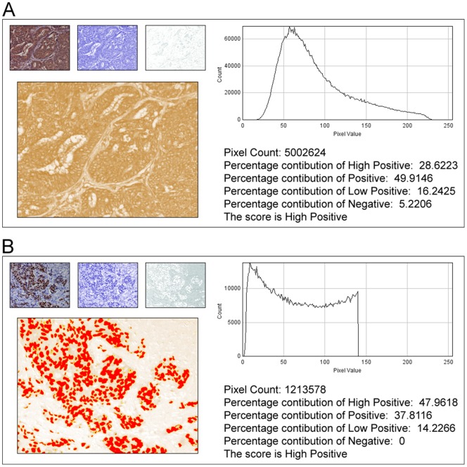
In anatomic pathology, immunohistochemistry (IHC) serves as a diagnostic and prognostic method for identification of disease markers in tissue samples that directly influences classification and grading the disease, influencing patient management. However, till today over most of the world, pathological analysis of tissue samples remained a time-consuming and subjective procedure, wherein the intensity of antibody staining is manually judged and thus scoring decision is directly influenced by visual bias. This instigated us to design a simple method of automated digital IHC image analysis algorithm for an unbiased, quantitative assessment of antibody staining intensity in tissue sections. As a first step, we adopted the spectral deconvolution method of DAB/hematoxylin color spectra by using optimized optical density vectors of the color deconvolution plugin for proper separation of the DAB color spectra. Then the DAB stained image is displayed in a new window wherein it undergoes pixel-by-pixel analysis, and displays the full profile along with its scoring decision. Based on the mathematical formula conceptualized, the algorithm is thoroughly tested by analyzing scores assigned to thousands (n = 1703) of DAB stained IHC images including sample images taken from human protein atlas web resource. The IHC Profiler plugin developed is compatible with the open resource digital image analysis software, ImageJ, which creates a pixel-by-pixel analysis profile of a digital IHC image and further assigns a score in a four tier system. A comparison study between manual pathological analysis and IHC Profiler resolved in a match of 88.6% (P<0.0001, CI = 95%). This new tool developed for clinical histopathological sample analysis can be adopted globally for scoring most protein targets where the marker protein expression is of cytoplasmic and/or nuclear type. We foresee that this method will minimize the problem of inter-observer variations across labs and further help in worldwide patient stratification potentially benefitting various multinational clinical trial initiatives.
Overview
The claims and statistics of this paper haven’t been checked yet.
Free: it reads the open-access text and checks the first 5 claims that cite a source we can check. Create a free account to check every claim.
Integrity signals
Registry
Cites
Statistics
Figures



Verification
Claims the paper makes
The claims and statistics of this paper haven’t been checked yet.
Free: it reads the open-access text and checks the first 5 claims that cite a source we can check. Create a free account to check every claim.
Claims other papers make about this one
Each is checked against this paper’s own text: does it say what the citing paper says it does?
In the citing paper
“For analysis of c-MYC expression in liver sections, random 20× non-tumor fields of the c-MYC IHC scans from liver sections of individual mice were output using NDP.view2 and blindly analyzed using ImageJ (https://imagej.nih.gov/ij/) with the plugin ‘IHC Profiler’62.”
From Tumor-specific cholinergic CD4+ T lymphocytes guide immunosurveillance of hepatocellular carcinoma (2023) · cites this paper as [62]
Self-check
Percentages, sums and reported differences re-computed from the paper’s own numbers.
The self-check needs the paper’s full text.
Citations
Cites
NIH Image to ImageJ: 25 years of image analysis
Schneider, C. A., Rasband, W. S., & Eliceiri, K. W. (2012) · Nature Methods · doi:10.1038/nmeth.2089 · Cited by 64,638 · cited 1×
“The zones were visually identified by using the threshold feature of Image menu of the ImageJ program [22].”
MethodHsu, S. M., Raine, L., & Fanger, H. (1981) · Journal of Histochemistry & Cytochemistry · doi:10.1177/29.4.6166661 · Cited by 14,140
Harvey, J. M., Clark, G. M., Osborne, C. K., & Allred, D. C. (1999) · Journal of Clinical Oncology · doi:10.1200/jco.1999.17.5.1474 · Cited by 2,056
Quantification of histochemical staining by color deconvolution.
Ruifrok, A. C. C., & Johnston, D. A. (2001) · PubMed · Cited by 2,029
Tuominen, V. J., Ruotoistenmäki, S., Viitanen, A., Jumppanen, M., & Isola, J. (2010) · Breast Cancer Research · doi:10.1186/bcr2615 · Cited by 472
McCarty, K. S., Szabo, E. M., Flowers, J. L., Cox, E. B., Leight, G. S., Miller, L. A., Konrath, J. G., Jt, S., Budwit, D. A., & Wt, C. (1986) · PubMed · Cited by 417
A global view of protein expression in human cells, tissues, and organs
Pontén, F., Gry, M., Fagerberg, L., Lundberg, E., Asplund, A., Berglund, L., Oksvold, P., Björling, E., Hober, S., Kampf, C., et al. (2009) · Molecular Systems Biology · doi:10.1038/msb.2009.93 · Cited by 222
Automated Selection of DAB-labeled Tissue for Immunohistochemical Quantification
Brey, E. M., Lalani, Z., Johnston, C., Wong, M., McIntire, L. V., Duke, P. J., & Patrick, C. W. (2003) · Journal of Histochemistry & Cytochemistry · doi:10.1177/002215540305100503 · Cited by 200
Quantitative image analysis of immunohistochemical stains using a CMYK color model
Pham, N.-A., Morrison, A., Schwock, J., Aviel-Ronen, S., Iakovlev, V., Tsao, M.-S., Ho, J., & Hedley, D. W. (2007) · Diagnostic Pathology · doi:10.1186/1746-1596-2-8 · Cited by 179
Quantification of renal pathology by image analysis (Methods in Renal Research)
RANGAN, G. K., & TESCH, G. H. (2007) · Nephrology · doi:10.1111/j.1440-1797.2007.00855.x · Cited by 176
Cited by
Items 201–300 of 1,000
Zhou, H., Boys, A. J., Harrod, J. B., Bonassar, L. J., & Estroff, L. A. (2020) · Acta Biomaterialia · doi:10.1016/j.actbio.2020.05.032 · Cited by 37
Araki, T., Mitsuyama, K., Yamasaki, H., Morita, M., Tsuruta, K., Mori, A., Yoshimura, T., Fukunaga, S., Kuwaki, K., Yoshioka, S., et al. (2021) · Journal of Crohn s and Colitis · doi:10.1093/ecco-jcc/jjab033 · Cited by 34
Xiao, X., Li, Y., Wang, Y., Zhang, Y., Chen, J., Liu, W., Tang, J., Yue, F., & Yang, J. (2021) · International Immunopharmacology · doi:10.1016/j.intimp.2021.108427 · Cited by 38
CREB Inactivation by HDAC1/PP1γ Contributes to Dopaminergic Neurodegeneration in Parkinson's Disease
Xu, X., He, X., Zhang, Z., Chen, Y., Li, J., Ma, S., Huang, Q., & Li, M. (2022) · Journal of Neuroscience · doi:10.1523/jneurosci.1419-21.2022 · Cited by 39
Rosas, E., Roberts, J. T., O’Neill, K. I., Christenson, J. L., Williams, M. M., Hanamura, T., Spoelstra, N. S., Vahrenkamp, J. M., Gertz, J., & Richer, J. K. (2020) · Endocrinology · doi:10.1210/endocr/bqaa226 · Cited by 37
Niu, M., Guo, H.-X., Cai, J.-W., Duan, B., Chen, Y., & Xiang-chen, M. (2023) · Food Bioscience · doi:10.1016/j.fbio.2023.103020 · Cited by 35
Mechanosensitive hormone signaling promotes mammary progenitor expansion and breast cancer risk
Northey, J. J., Hayward, M.-K., Yui, Y., Stashko, C., Kai, F., Mouw, J. K., Thakar, D., Lakins, J. N., Ironside, A. J., Samson, S. M., et al. (2024) · Cell stem cell · doi:10.1016/j.stem.2023.12.002 · Cited by 41
Xiang, B., Zhang, M., Li, K., Zhang, Z., Liu, Y., Gao, M., Wang, X., Xiao, X., Sun, Y., He, C. H., et al. (2025) · Nature Immunology · doi:10.1038/s41590-024-02047-w · Cited by 47
CXXC5 mediates growth plate senescence and is a target for enhancement of longitudinal bone growth
Choi, S., Kim, H.-Y., Cha, P.-H., Seo, S. H., Lee, C., Choi, Y., Shin, W., Heo, Y., Han, G., Lee, W., et al. (2019) · Life Science Alliance · doi:10.26508/lsa.201800254 · Cited by 33
Xu, D., Liang, S., Yang, H., Bruggmann, R., Berezowska, S., Zhang, Y., Marti, T. M., Hall, S. R. R., Gao, Y., Kocher, G. J., et al. (2020) · Molecular Cancer Therapeutics · doi:10.1158/1535-7163.mct-19-0724 · Cited by 35
Zheng, L., Feng, Z., Tao, S., Gao, J., Lin, Y., Wei, X., Zheng, B., Huang, B. H., Zheng, Z., Zhang, X., et al. (2022) · Clinical and Translational Medicine · doi:10.1002/ctm2.652 · Cited by 36
CCN2/CTGF promotes liver fibrosis through crosstalk with the Slit2/Robo signaling
Pi, L., Sun, C., Jn‐Simon, N., Basha, S., Thomas, H., Figueroa, V., Zarrinpar, A., Cao, Q., & Petersen, B. E. (2022) · Journal of Cell Communication and Signaling · doi:10.1007/s12079-022-00713-y · Cited by 38
Blanco, R., Domínguez, E., Morales, O., Blanco, D., Bedoya, D. M., Rengifo, C. E., Viada, C., Cedeño, M., Rengifo, E., & Carr, A. (2015) · Pathology Research International · doi:10.1155/2015/132326 · Cited by 34
N-Butylphthalide Alleviates Blood–Brain Barrier Impairment in Rats Exposed to Carbon Monoxide
Bi, M., Zhang, M., Guo, D., Bi, W., Liu, B., Zou, Y., & Li, Q. (2016) · Frontiers in Pharmacology · doi:10.3389/fphar.2016.00394 · Cited by 39
AKT1 interacts with DHX9 to Mitigate R Loop–Induced Replication Stress in Ovarian Cancer
Huang, T., Chiang, C.-Y., Nair, J. R., Wilson, K. M., Cheng, K. C.-C., & Lee, J. (2024) · Cancer Research · doi:10.1158/0008-5472.can-23-1908 · Cited by 41
Mesothelin promotes brain metastasis of non-small cell lung cancer by activating MET
Xia, S., Duan, W., Xu, M., Li, M., Tang, M., Wei, S., Lin, M., Li, E., Liu, W., & Wang, Q. (2024) · Journal of Experimental & Clinical Cancer Research · doi:10.1186/s13046-024-03015-w · Cited by 37
Interaction between inflammatory mediators and miRNAs in Helicobacter pylori infection
Rossi, A. F. T., Cadamuro, A. C. T., Biselli, J. M., Leite, K. R. M., Severino, F. E., Reis, P. P., Cordeiro, J. A., & Silva, A. E. (2016) · Cellular Microbiology · doi:10.1111/cmi.12587 · Cited by 36
IHC Color Histograms for Unsupervised Ki67 Proliferation Index Calculation
Geread, R. S., Morreale, P., Dony, R. D., Brouwer, E. R., Wood, G. A., Androutsos, D., & Khademi, A. (2019) · Frontiers in Bioengineering and Biotechnology · doi:10.3389/fbioe.2019.00226 · Cited by 33
Raut, P. K., & Park, P. (2020) · Biochemical Pharmacology · doi:10.1016/j.bcp.2020.114186 · Cited by 34
Rahman, M. S., & Rahman, M. S. (2021) · Cell Stress and Chaperones · doi:10.1007/s12192-021-01232-2 · Cited by 36
Lee, P.-J., Ho, C., Ho, H. H., Chen, W.-J., Lin, C.-H., Lai, Y., Juan, Y., Chu, W.-C., Lee, J.-H., Su, S., et al. (2021) · Theranostics · doi:10.7150/thno.62676 · Cited by 39
Li, G., Tang, X., Zhang, S., Deng, Z., Wang, B., Shi, W., Xie, H., Liu, B., & Li, J. (2022) · Journal of Investigative Dermatology · doi:10.1016/j.jid.2022.03.026 · Cited by 37
Ge, F., Li, Z., Hu, J., Pu, Y., Zhao, F., & Kong, L. (2022) · Frontiers in Pharmacology · doi:10.3389/fphar.2022.1002565 · Cited by 35
Transcriptome Analysis Uncovers a Growth-Promoting Activity of Orosomucoid-1 on Hepatocytes
Qin, X., Hara, M., Arner, E., Kawaguchi, Y., Inoue, I., Tatsukawa, H., Furutani, Y., Nagatsuma, K., Matsuura, T., Wei, F., et al. (2017) · EBioMedicine · doi:10.1016/j.ebiom.2017.09.008 · Cited by 34
Kim, S., Lim, E., Yoo, K.-C., Zhao, Y., Kang, J.-H., Lim, E., Shin, I., Kang, S., Lim, H. W., & Lee, S. (2022) · Clinical and Translational Medicine · doi:10.1002/ctm2.997 · Cited by 36
Perelli, L., Carbone, F., Zhang, L., Huang, J. K.-L., Le, C. N., Khan, H., Citron, F., Poggetto, E. D., Gutschner, T., Tomihara, H., et al. (2023) · Nature Cancer · doi:10.1038/s43018-023-00584-1 · Cited by 37
Duran, C. L., Lee, D. W., Jung, J.-U., Ravi, S., Pogue, C. B., Toussaint, L. G., Bayless, K. J., & Sitcheran, R. (2016) · Oncogenesis · doi:10.1038/oncsis.2016.39 · Cited by 37
CB2R agonist prevents nicotine induced lung fibrosis
Wawryk‐Gawda, E., Chłapek, K., Zarobkiewicz, M. K., Lis-Sochocka, M., Chylińska‐Wrzos, P., Boguszewska‐Czubara, A., Sławiński, M., Franczak, A., Jodłowska‐Jędrych, B., & Biała, G. (2018) · Experimental Lung Research · doi:10.1080/01902148.2018.1543368 · Cited by 34
Georgiev, G. P., Kotov, G. N., Iliev, A. A., Slavchev, S. A., Ovtscharoff, W. A., & Landzhov, B. V. (2019) · Annals of Anatomy - Anatomischer Anzeiger · doi:10.1016/j.aanat.2019.04.002 · Cited by 33
Holyoak, D. T., Chlebek, C., Kim, M., Wright, T. M., Otero, M., & van der Meulen, M. C. H. (2019) · Osteoarthritis and Cartilage · doi:10.1016/j.joca.2019.06.005 · Cited by 36
Chlorogenic acid attenuates cyclophosphamide-induced rat interstitial cystitis
Luo, J., Yang, C., Luo, X., Yang, Y., Li, J., Song, B., Zhao, J., & Li, L. (2020) · Life Sciences · doi:10.1016/j.lfs.2020.117590 · Cited by 32
Identification of genes associated with endometrial cell ageing
Kawamura, T., Tomari, H., Onoyama, I., Araki, H., Yasunaga, M., Lin, C., Kawamura, K., Yokota, N., Yoshida, S., Yagi, H., et al. (2020) · Molecular Human Reproduction · doi:10.1093/molehr/gaaa078 · Cited by 40
Chang, T.-Y., Lan, K., Chiu, C., Sheu, M., & Liu, S. (2022) · Clinical Science · doi:10.1042/cs20220043 · Cited by 32
Singh, R. S., Chaudhary, D. K., Mohan, A., Kumar, P., Chaturvedi, C. P., ECELBARGER, C. A., Godbole, M. M., & Tiwari, S. (2016) · Scientific Reports · doi:10.1038/srep38034 · Cited by 37
Tang, H., He, K., Zhao, K., Chen, Z., Wu, W., Jin, W., Yang, L., & Xie, B. (2023) · Journal of Neurotrauma · doi:10.1089/neu.2023.0150 · Cited by 31
FMT rescues mice from DSS-induced colitis in a STING-dependent manner
Pu, D., Yao, Y., Zhou, C., Liu, R., Wang, Z., Liu, Y., Wang, D., Wang, B., Wang, Y., Liu, Z., et al. (2024) · Gut Microbes · doi:10.1080/19490976.2024.2397879 · Cited by 32
Zanotto‐Filho, A., Rajamanickam, S., Loranc, E., Masamsetti, V. P., Gorthi, A., Romero, J. C., Tonapi, S. S., Gonçalves, R. M., Reddick, R. L., Benavides, R., et al. (2018) · Cancer Letters · doi:10.1016/j.canlet.2018.03.037 · Cited by 33
Fang, X., Wu, L., Yang, L., Song, L., Cai, J., Luo, F., Wei, J., Zhou, L., & Wang, D. (2018) · The Journal of Steroid Biochemistry and Molecular Biology · doi:10.1016/j.jsbmb.2018.04.011 · Cited by 31
Vetuschi, A., Pompili, S., Gallone, A., D’Alfonso, A., Carbone, M. G., Carta, G., Festuccia, C., Gaudio, E., Colapietro, A., & Sferra, R. (2018) · Journal of Histochemistry & Cytochemistry · doi:10.1369/0022155418772798 · Cited by 34
PES1 enhances proliferation and tumorigenesis in hepatocellular carcinoma via the PI3K/AKT pathway
Wang, J., Sun, J. L., Zhang, N., Yang, R., Li, H., Zhang, Y., Chen, K., & Kong, D. (2019) · Life Sciences · doi:10.1016/j.lfs.2018.12.054 · Cited by 36
Humanized anti-CD271 monoclonal antibody exerts an anti-tumor effect by depleting cancer stem cells
Morita, S., Mochizuki, M., Wada, K., Shibuya, R., Nakamura, M., Yamaguchi, K., Yamazaki, T., Imai, T., Asada, Y., Matsuura, K., et al. (2019) · Cancer Letters · doi:10.1016/j.canlet.2019.07.011 · Cited by 31
Du, J., Dong, Z., Tan, L., Tan, M., Zhang, F., Zhang, K., Pan, G., Li, C., Shi, S., Zhang, Y., et al. (2020) · Frontiers in Cell and Developmental Biology · doi:10.3389/fcell.2020.607757 · Cited by 31
Yang, W., Wu, A.-C., Hsu, T., Liou, J., Lo, W.-L., Chang, K., Chen, P., Kikkawa, U., Yang, S.-T., Kao, T., et al. (2021) · Cell Death and Disease · doi:10.1038/s41419-021-04182-w · Cited by 32
Hou, M., Tian, B., Bai, B., Ci, Z., Liu, Y., Zhang, Y., Zhou, G., & Cao, Y. (2021) · Bioactive Materials · doi:10.1016/j.bioactmat.2021.11.007 · Cited by 36
He, Y., Dong, Y., Zhang, X., Ding, Z., Song, Y., Huang, X., Chen, S., Wang, Z., Ni, Y., & Ding, L. (2022) · Frontiers in Oncology · doi:10.3389/fonc.2022.824235 · Cited by 36
Mohammed, A. T., Mohamed, A. A.-R., & Ali, H. A. (2017) · Acta Histochemica · doi:10.1016/j.acthis.2017.03.004 · Cited by 31
Selective serotonin re-uptake inhibitor sertraline inhibits bone healing in a calvarial defect model
Howie, R. N., Herberg, S., Durham, E. L., Grey, Z., Bennfors, G., Elsalanty, M. E., LaRue, A. C., Hill, W., & Cray, J. (2018) · International Journal of Oral Science · doi:10.1038/s41368-018-0026-x · Cited by 36
Chen, C., Ye, L., Yi, J., Liu, T., & Li, Z. (2023) · Breast Cancer Research and Treatment · doi:10.1007/s10549-023-07032-9 · Cited by 30
de Carvalho, F. M. C., de Lima, V. C. O., Costa, I. D. S., Luz, A. B. S., Ladd, F. V. L., Serquiz, A. C., Bortolin, R. H., Silbiger, V. N., Maciel, B. L. L., dos Santos, E. A., et al. (2019) · Nutrients · doi:10.3390/nu11030512 · Cited by 31
Cancer‐associated V‐ATPase induces delayed apoptosis of protumorigenic neutrophils
Ibrahim, S. A.-S., Kulshrestha, A., Katara, G. K., Riehl, V. E., Sahoo, M., & Beaman, K. D. (2020) · Molecular Oncology · doi:10.1002/1878-0261.12630 · Cited by 33
Injectable Mesenchymal Stem Cell‐Laden Matrigel Microspheres for Endometrium Repair and Regeneration
Xu, B., Cao, Y., Zheng, Z., Galan, E. A., Hu, Z., Ge, J., Xing, X., & Ma, S. (2021) · Advanced Biology · doi:10.1002/adbi.202000202 · Cited by 33
Rezq, S., Hassan, R. A. R. A., & Mahmoud, M. F. (2021) · International Immunopharmacology · doi:10.1016/j.intimp.2021.108140 · Cited by 31
Ahmed, O. M., Galaly, S. R., Mostafa, M.-A. M. A., Eed, E. M., Ali, T. M., Fahmy, A. M., & Zaky, M. Y. (2022) · Oxidative Medicine and Cellular Longevity · doi:10.1155/2022/6702773 · Cited by 30
RetractedZhao, J., Chen, S., Yang, C., Zhou, M., Yang, T., Sun, B., Zhu, J., Zhang, H., Lu, Q., Li, L., et al. (2022) · Biochemical Pharmacology · doi:10.1016/j.bcp.2022.115047 · Cited by 33
Córdova, C., Muñoz, R., Olivares, R., Minonzio, J.-G., Lozano, C., González, P., Marchant, I., González‐Arriagada, W. A., & Olivero, P. (2022) · Oncology Letters · doi:10.3892/ol.2022.13630 · Cited by 35
Deng, S., Banerjee, S., Chen, H., Pochampally, S., Wang, Y., Yun, M., White, S. W., Parmar, K. R., Meibohm, B., Hartman, K. L., et al. (2022) · Cancer Letters · doi:10.1016/j.canlet.2022.216046 · Cited by 31
Trahearn, N., Epstein, D., Cree, I. A., Snead, D. R. J., & Rajpoot, N. (2017) · Scientific Reports · doi:10.1038/s41598-017-05511-w · Cited by 34
Chatterjee, S. B., Hou, J., Bandaru, V. V. R., Pezhouh, M. K., Mannan, A. A. S. R., & Sharma, R. B. (2018) · Biochemical and Biophysical Research Communications · doi:10.1016/j.bbrc.2018.11.149 · Cited by 30
Mittal, P., Klingler‐Hoffmann, M., Arentz, G., Winderbaum, L., Kaur, G., Anderson, L. A., Scurry, J., Leung, Y. C., Stewart, C. J. R., Carter, J. R., et al. (2016) · Biochimica et Biophysica Acta (BBA) - Proteins and Proteomics · doi:10.1016/j.bbapap.2016.10.010 · Cited by 30
Toeda, Y., Kasamatsu, A., Koike, K., Endo‐Sakamoto, Y., Fushimi, K., Kasama, H., Yamano, Y., Shiiba, M., Tanzawa, H., & Uzawa, K. (2018) · Molecular Carcinogenesis · doi:10.1002/mc.22889 · Cited by 29
Mild Methylenetetrahydrofolate Reductase Deficiency Alters Inflammatory and Lipid Pathways in Liver
Leclerc, D., Christensen, K. E., Cauvi, O., Yang, E., Fournelle, F., Bahous, R. H., Malysheva, O., Deng, L.-Y., Wu, Q. H., Zhou, Z., et al. (2018) · Molecular Nutrition & Food Research · doi:10.1002/mnfr.201801001 · Cited by 30
Lee, Y. S., Yeo, I. J., Kim, K. C., Han, S., & Hong, J. T. (2019) · Frontiers in Immunology · doi:10.3389/fimmu.2019.01379 · Cited by 33
D’Amico, R., Cordaro, M., Fusco, R., Peritore, A. F., Genovese, T., Gugliandolo, E., Crupi, R., Mandalari, G., Caccamo, D., Cuzzocrea, S., et al. (2022) · Nutrients · doi:10.3390/nu14071474 · Cited by 29
Yao, S., Liang, Z., Lee, Y. W., Yung, P. S. H., & Lui, P. P. Y. (2023) · The American Journal of Sports Medicine · doi:10.1177/03635465221135770 · Cited by 29
Sferra, R., Pompili, S., Festuccia, C., Marampon, F., Gravina, G. L., Ventura, L., Cesare, E. D., Cicchinelli, S., Gaudio, E., & Vetuschi, A. (2017) · European Journal of Histochemistry · doi:10.4081/ejh.2017.2732 · Cited by 31
Rab11a drives adhesion molecules to the surface of endometrial epithelial cells
Kakar, R., Brahmbhatt, K., Chauhan, B., Katkam, R. R., Bashir, T., Gawde, H., Mayadeo, N., Chaudhari, U. K., & Sachdeva, G. (2018) · Human Reproduction · doi:10.1093/humrep/dey365 · Cited by 28
Xu, X., Huang, Z., Huang, Z., Lv, X., Jiang, D., Huang, Z., Han, B., Lin, G., Liu, G., Li, S., et al. (2024) · International Immunopharmacology · doi:10.1016/j.intimp.2024.113305 · Cited by 36
Bastola, S., Pavlyukov, M. S., Sharma, N., Ghochani, Y., Nakano, M. A., Muthukrishnan, S. D., Yu, S. Y., Kim, M., Sohrabi, A., Biscola, N. P., et al. (2025) · Nature Communications · doi:10.1038/s41467-024-55487-1 · Cited by 35
The strong propensity of Cadherin‐23 for aggregation inhibits cell migration
Sannigrahi, M. K., Srinivas, C. S., Deokate, N., & Rakshit, S. (2019) · Molecular Oncology · doi:10.1002/1878-0261.12469 · Cited by 29
Kuhla, B., Kaever, V., Tuchscherer, A., & Kuhla, A. (2019) · Neuroendocrinology · doi:10.1159/000501208 · Cited by 32
Li, K., Pan, W., Ma, Y., Xu, X., Gao, Y., He, Y., Wei, L., & Zhang, J. (2019) · Breast Cancer · doi:10.1007/s12282-019-01024-8 · Cited by 28
Sendoya, J. M., Iseas, S., Coraglio, M. F., Golubicki, M., Robbio, J., Salanova, R., Kujaruk, M., Mikolaitis, V., Rizzolo, M., Ruiz, G., et al. (2020) · Cancers · doi:10.3390/cancers12082227 · Cited by 31
Redant, V., Favoreel, H. W., Dallmeier, K., Campe, W. V., & Regge, N. D. (2020) · Journal of Neuroinflammation · doi:10.1186/s12974-020-01974-3 · Cited by 39
Comprehensive Assessment of High-Risk Plaques by Dual-Modal Imaging Catheter in Coronary Artery
Kim, S., Nam, H. S., Lee, M. W., Kim, H. J., Kang, W. J., Song, J. W., Han, J., Kang, D. O., Oh, W., Yoo, H., et al. (2021) · JACC Basic to Translational Science · doi:10.1016/j.jacbts.2021.10.005 · Cited by 30
Price, D. R., Benedetti, E., Hoffman, K. L., Gómez-Escobar, L. G., Alvarez-Mulett, S. L., Capili, A. M., Sarwath, H., Parkhurst, C. N., LaFond, E., Weidman, K., et al. (2022) · American Journal Of Pathology · doi:10.1016/j.ajpath.2022.04.002 · Cited by 29
Guan, Q.-Y., & Zhao, X. (2022) · Food Chemistry · doi:10.1016/j.foodchem.2022.133741 · Cited by 28
Sato, S., Hiruma, T., Koizumi, M., Yoshihara, M., Nakamura, Y., Tadokoro, H., Motomatsu, S., Yamanaka, T., Washimi, K., Okubo, Y., et al. (2023) · Cancer Science · doi:10.1111/cas.15786 · Cited by 31
SIPA1 promotes invasion and migration in human oral squamous cell carcinoma by ITGB1 and MMP7
Takahara, T., Kasamatsu, A., Yamatoji, M., Iyoda, M., Kasama, H., Saito, T., Takeuchi, S., Endo‐Sakamoto, Y., Shiiba, M., Tanzawa, H., et al. (2017) · Experimental Cell Research · doi:10.1016/j.yexcr.2017.02.026 · Cited by 28
Biofilm-Induced Type 2 Innate Immunity in a Cystic Fibrosis Model of Pseudomonas aeruginosa
Bielen, K., Jongers, B. ‘., Boddaert, J., Raju, T. K., Lammens, C., Malhotra‐Kumar, S., Jorens, P. G., Goossens, H., & Kumar‐Singh, S. (2017) · Frontiers in Cellular and Infection Microbiology · doi:10.3389/fcimb.2017.00274 · Cited by 32
Allen, L., Matyas, J. R., Ungrin, M., Hart, D. A., & Sen, A. (2019) · Stem Cells International · doi:10.1155/2019/4607461 · Cited by 32
Collagen XI mutation lowers susceptibility to load‐induced cartilage damage in mice
Holyoak, D. T., Otero, M., Armar, N. S., Ziemian, S. N., Otto, A., Cullinane, D., Wright, T. M., Goldring, S. R., Goldring, M. B., & van der Meulen, M. C. H. (2017) · Journal of Orthopaedic Research® · doi:10.1002/jor.23731 · Cited by 29
Li, K., Xu, X., He, Y., Tian, Y., Pan, W., Liu, X., Ma, Y., Gao, Y., Gao, J., Qi, Y., et al. (2018) · Journal of Cancer · doi:10.7150/jca.24934 · Cited by 27
Mishra, A., Galvankar, M., Vaidya, S., Chaudhari, U. K., & Modi, D. N. (2020) · Journal of Biosciences · doi:10.1007/s12038-020-00073-y · Cited by 28
Nakatani, K., Maehama, T., Nishio, M., Otani, J., Yamaguchi, K., Fukumoto, M., Hikasa, H., Hagiwara, S., Nishina, H., Mak, T. W., et al. (2021) · Cancer Science · doi:10.1111/cas.15079 · Cited by 28
Zhou, J., Sun, C., Yang, L., Wang, J., Jn‐Simon, N., Zhou, C., Bryant, A. J., Cao, Q., Li, C., Petersen, B. E., et al. (2022) · The FASEB Journal · doi:10.1096/fj.202101686r · Cited by 31
Morioka, S., Sakaguchi, H., Yamaguchi, T., Ninoyu, Y., Mohri, H., Nakamura, T., Hisa, Y., Ogita, K., Saito, N., & Ueyama, T. (2018) · Journal of Neurochemistry · doi:10.1111/jnc.14451 · Cited by 28
Alsemeh, A. E., Moawad, R. S., & Abdelfattah, E. R. (2019) · Anatomical Science International · doi:10.1007/s12565-019-00488-0 · Cited by 29
Du, P., Lai, Y., Yao, D., Chen, J., & Ding, N. (2019) · Oncology Letters · doi:10.3892/ol.2019.10571 · Cited by 29
Basha, G., Cottle, A. G., Pretheeban, T., Chan, K. Y. T., Witzigmann, D., Young, R. N., Rossi, F. M. V., & Cullis, P. R. (2022) · Molecular Therapy · doi:10.1016/j.ymthe.2022.06.012 · Cited by 29
Gupta, P., Suman, S., Mishra, M., Mishra, S., Srivastava, N., Kumar, V., Singh, P. K., & Shukla, Y. (2016) · PROTEOMICS - CLINICAL APPLICATIONS · doi:10.1002/prca.201500138 · Cited by 26
Effect of voluntary running on expression of myokines in brains of rats with depression
Algaidi, S. A., Eldomiaty, M. A., Elbastwisy, Y. M., Almasry, S. M., Desouky, M. K., & Elnaggar, A. M. (2019) · International Journal of Immunopathology and Pharmacology · doi:10.1177/2058738419833533 · Cited by 28
Stošić, M., Matavulj, M., & Marković, J. (2018) · Acta Histochemica · doi:10.1016/j.acthis.2018.02.002 · Cited by 26
Bellido-Carreras, N., Argüello, H., Zaldívar‐López, S., Jiménez-Marín, Á., Martins, R. P., Arce, C., Morera, L., Carvajal, A., & Garrido, J. J. (2019) · Veterinary Pathology · doi:10.1177/0300985819843682 · Cited by 32
Li, K., Pan, W., Ma, Y., Xu, X., Gao, Y., He, Y., Wei, L., & Zhang, J. (2019) · Pathology - Research and Practice · doi:10.1016/j.prp.2019.152573 · Cited by 29
The ameliorative effects of methylene blue on testicular damage induced by cisplatin in rats
Jourabi, F. G., Yari, S., Amiri, P., Heidarianpour, A., & Hashemi, H. (2020) · Andrologia · doi:10.1111/and.13850 · Cited by 27
A novel atmospheric‐pressure air plasma jet for wound healing
Guo, P., Liu, Y., Li, J., Zhang, N., Zhou, M., Li, Y., Zhao, G., Wang, N., Wang, A., Wang, Y., et al. (2021) · International Wound Journal · doi:10.1111/iwj.13652 · Cited by 28
Shin, S.-U., Cho, H.-M., Das, R., Gil-Henn, H., Ramakrishnan, S., Bayati, A. A., Carroll, S. F., Zhang, Y., Sankar, A. P., Elledge, C., et al. (2021) · Cells · doi:10.3390/cells10112904 · Cited by 26
Zhao, H., Wu, W., Li, X., & Chen, W. (2022) · Translational Oncology · doi:10.1016/j.tranon.2022.101340 · Cited by 29
Khaled, S. S., Soliman, H. A., Abdel‐Gabbar, M., Ahmed, N. A., El‐Nahass, E., & Ahmed, O. M. (2023) · Environmental Science and Pollution Research · doi:10.1007/s11356-023-28454-4 · Cited by 28
Zhou, S., Parham, D. M., Yung, E. E., Pattengale, P. K., & Wang, L. L. (2015) · Histopathology · doi:10.1111/his.12730 · Cited by 27
Metadata
- Authors
- Varghese, F., Bukhari, A. B., Malhotra, R., & De, A.
- Affiliations
- Tata Memorial Hospital (IN)
- Corresponding author
- De, Abhijit
- Venue
- PLoS ONE
- Publisher
- Public Library of Science (PLoS)
- Published
- 2014-05-06
- Volume
- 9
- Issue
- 5
- Pages
- e96801
- ISSN
- 1932-6203 (ISSN-L)
- PMC
- PMC4011881
- OpenAlex
- W1998734250
- Cited by
- 1,318
- References
- 30
- Citations by year
- 2026: 90 · 2025: 129 · 2024: 172 · 2023: 172 · 2022: 171 · 2021: 144 · 2020: 141 · 2019: 101 · 2018: 69 · 2017: 60 · 2016: 44 · 2015: 22 · 2014: 3
- FWCI
- 42.82
- Type
- journal article
- Language
- en
- Open access
- gold
- doi.org
- Locations
- PLoS ONE (publishedVersion, cc-by)
- Europe PMC (PubMed Central) (publishedVersion, cc-by)
- PubMed Central (publishedVersion, cc-by)
- PubMed (publishedVersion)
- RePEc: Research Papers in Economics (submittedVersion)
- DOAJ (DOAJ: Directory of Open Access Journals) (submittedVersion, cc-by-sa)
- Figshare (submittedVersion, cc-by)
- Funders
- Indian Council of Medical Research
- Advanced Centre for Treatment, Research and Education in Cancer
- Topics
- Physical Sciences › Computer Science › Artificial Intelligence › AI in cancer detection
- Life Sciences › Biochemistry, Genetics and Molecular Biology › Biophysics › Cell Image Analysis Techniques
- Physical Sciences › Computer Science › Computer Vision and Pattern Recognition › Digital Imaging for Blood Diseases
- Keywords
- immunohistochemistry image analysis, automated grading, ImageJ plugin, interobserver variability, digital pathology, human tissue samples, protein expression profiling
- MeSH terms
- Automation, Cell Nucleus, Cytoplasm, Humans, Image Processing, Computer-Assisted, Immunohistochemistry, Neoplasms, Software, Biomarkers, Tumor
About these checks
Registry, retraction, Cites and statistics results are computed, not judged, with one exception: a printed reference that no index lists is read once by a model before it counts as a citation error. Claims are extracted by a model and kept only when their quote appears verbatim in the text; each cited source is then checked, with a model’s help, for whether it says what the claim relies on. Those verdicts can be wrong — read the claim’s quote and the cited source before relying on one.
Statistics and arithmetic are recomputed, not judged.
Confidence bands are shown only for fields calibrated on our annotated test set. None is calibrated yet, so no band is shown.
What counts as a claim ›How we measure confidence ›How we check this ›
Are you an author, or do you see a problem with this page? Request a review
Data sources
Bibliographic data: Semantic Scholar, OpenAlex, Crossref.
Contains information from the Semantic Scholar Academic Graph Datasets, made available under the ODC Attribution License (ODC-BY 1.0).
Semantic Scholar’s data is described in: Kinney, R., et al. (2023). The Semantic Scholar Open Data Platform. arXiv:2301.10140.
