

IHC Profiler: An Open Source Plugin for the Quantitative Evaluation and Automated Scoring of Immunohistochemistry Images of Human Tissue Samples
Open accessThe DOI matches its Crossref record; 30 of the 30 references it cites were found in a registry or an index.
Abstract
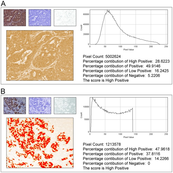
In anatomic pathology, immunohistochemistry (IHC) serves as a diagnostic and prognostic method for identification of disease markers in tissue samples that directly influences classification and grading the disease, influencing patient management. However, till today over most of the world, pathological analysis of tissue samples remained a time-consuming and subjective procedure, wherein the intensity of antibody staining is manually judged and thus scoring decision is directly influenced by visual bias. This instigated us to design a simple method of automated digital IHC image analysis algorithm for an unbiased, quantitative assessment of antibody staining intensity in tissue sections. As a first step, we adopted the spectral deconvolution method of DAB/hematoxylin color spectra by using optimized optical density vectors of the color deconvolution plugin for proper separation of the DAB color spectra. Then the DAB stained image is displayed in a new window wherein it undergoes pixel-by-pixel analysis, and displays the full profile along with its scoring decision. Based on the mathematical formula conceptualized, the algorithm is thoroughly tested by analyzing scores assigned to thousands (n = 1703) of DAB stained IHC images including sample images taken from human protein atlas web resource. The IHC Profiler plugin developed is compatible with the open resource digital image analysis software, ImageJ, which creates a pixel-by-pixel analysis profile of a digital IHC image and further assigns a score in a four tier system. A comparison study between manual pathological analysis and IHC Profiler resolved in a match of 88.6% (P<0.0001, CI = 95%). This new tool developed for clinical histopathological sample analysis can be adopted globally for scoring most protein targets where the marker protein expression is of cytoplasmic and/or nuclear type. We foresee that this method will minimize the problem of inter-observer variations across labs and further help in worldwide patient stratification potentially benefitting various multinational clinical trial initiatives.
Overview
The claims and statistics of this paper haven’t been checked yet.
Free: it reads the open-access text and checks the first 5 claims that cite a source we can check. Create a free account to check every claim.
Integrity signals
Registry
Cites
Statistics
Figures



Verification
Claims the paper makes
The claims and statistics of this paper haven’t been checked yet.
Free: it reads the open-access text and checks the first 5 claims that cite a source we can check. Create a free account to check every claim.
Claims other papers make about this one
Each is checked against this paper’s own text: does it say what the citing paper says it does?
In the citing paper
“For analysis of c-MYC expression in liver sections, random 20× non-tumor fields of the c-MYC IHC scans from liver sections of individual mice were output using NDP.view2 and blindly analyzed using ImageJ (https://imagej.nih.gov/ij/) with the plugin ‘IHC Profiler’62.”
From Tumor-specific cholinergic CD4+ T lymphocytes guide immunosurveillance of hepatocellular carcinoma (2023) · cites this paper as [62]
Self-check
Percentages, sums and reported differences re-computed from the paper’s own numbers.
The self-check needs the paper’s full text.
Citations
Cites
NIH Image to ImageJ: 25 years of image analysis
Schneider, C. A., Rasband, W. S., & Eliceiri, K. W. (2012) · Nature Methods · doi:10.1038/nmeth.2089 · Cited by 64,638 · cited 1×
“The zones were visually identified by using the threshold feature of Image menu of the ImageJ program [22].”
MethodHsu, S. M., Raine, L., & Fanger, H. (1981) · Journal of Histochemistry & Cytochemistry · doi:10.1177/29.4.6166661 · Cited by 14,140
Harvey, J. M., Clark, G. M., Osborne, C. K., & Allred, D. C. (1999) · Journal of Clinical Oncology · doi:10.1200/jco.1999.17.5.1474 · Cited by 2,056
Quantification of histochemical staining by color deconvolution.
Ruifrok, A. C. C., & Johnston, D. A. (2001) · PubMed · Cited by 2,029
Tuominen, V. J., Ruotoistenmäki, S., Viitanen, A., Jumppanen, M., & Isola, J. (2010) · Breast Cancer Research · doi:10.1186/bcr2615 · Cited by 472
McCarty, K. S., Szabo, E. M., Flowers, J. L., Cox, E. B., Leight, G. S., Miller, L. A., Konrath, J. G., Jt, S., Budwit, D. A., & Wt, C. (1986) · PubMed · Cited by 417
A global view of protein expression in human cells, tissues, and organs
Pontén, F., Gry, M., Fagerberg, L., Lundberg, E., Asplund, A., Berglund, L., Oksvold, P., Björling, E., Hober, S., Kampf, C., et al. (2009) · Molecular Systems Biology · doi:10.1038/msb.2009.93 · Cited by 222
Automated Selection of DAB-labeled Tissue for Immunohistochemical Quantification
Brey, E. M., Lalani, Z., Johnston, C., Wong, M., McIntire, L. V., Duke, P. J., & Patrick, C. W. (2003) · Journal of Histochemistry & Cytochemistry · doi:10.1177/002215540305100503 · Cited by 200
Quantitative image analysis of immunohistochemical stains using a CMYK color model
Pham, N.-A., Morrison, A., Schwock, J., Aviel-Ronen, S., Iakovlev, V., Tsao, M.-S., Ho, J., & Hedley, D. W. (2007) · Diagnostic Pathology · doi:10.1186/1746-1596-2-8 · Cited by 179
Quantification of renal pathology by image analysis (Methods in Renal Research)
RANGAN, G. K., & TESCH, G. H. (2007) · Nephrology · doi:10.1111/j.1440-1797.2007.00855.x · Cited by 176
Cited by
Items 501–600 of 1,000
Yuan, R., Zhang, Y., Wang, Y., Chen, H., Zhang, R., Hu, Z., Chai, C., & Chen, T. (2023) · Frontiers in Immunology · doi:10.3389/fimmu.2023.1152678 · Cited by 18
Yuan, Y., Fang, A., Wang, Z., Chen, H., Fu, Z. F., Zhou, M., & Zhao, L. (2024) · Autophagy · doi:10.1080/15548627.2024.2338575 · Cited by 16
Zhu, H., Cheng, L., Liu, D., Ma, X., Chen, Z., Fan, H., Li, R., Zhang, Y., Mi, H., Li, J., et al. (2024) · Neuro-Oncology · doi:10.1093/neuonc/noae224 · Cited by 16
de Oliveira, A. R., da Silva, F. S., Bortolin, R. H., Marques, D. E. D. S., Ramos, G. V., Marqueti, R. D. C., da Silva, N. B., Medeiros, K. C. D. P., Corrêa, M. A., Lima, J. P. M. S., et al. (2019) · PLoS ONE · doi:10.1371/journal.pone.0211643 · Cited by 15
Roberto, G. M., Lira, R. C. P., Delsin, L. E. A., Vieira, G. M., Silva, M. O., Hakime, R. G., Yamashita, M. E. D. A. S., Engel, E. E., Scrideli, C. A., Tone, L. G., et al. (2019) · Pathology & Oncology Research · doi:10.1007/s12253-019-00633-0 · Cited by 15
Modulating autophagy as a therapeutic strategy for the treatment of paediatric high‐grade glioma
Howarth, A., Madureira, P. A., Lockwood, G., Storer, L. C. D., Grundy, R. G., Rahman, R., Pilkington, G. J., & Hill, R. (2019) · Brain Pathology · doi:10.1111/bpa.12729 · Cited by 16
NUDT7 Loss Promotes KrasG12D CRC Development
Song, J., Park, S., Oh, J., Kim, D., Ryu, J. H., Park, W. C., Baek, I., Cheng, X., Lü, X., & Jin, E. (2020) · Cancers · doi:10.3390/cancers12030576 · Cited by 15
Host deficiency in ephrin-A1 inhibits breast cancer metastasis
Shiuan, E. F., Inala, A., Wang, S., Song, W., Youngblood, V., Chen, J., & Brantley‐Sieders, D. M. (2020) · F1000Research · doi:10.12688/f1000research.22689.2 · Cited by 16
TGFβ1 orchestrates renal fibrosis following Escherichia coli pyelonephritis
Hreha, T. N., Collins, C. A., Daugherty, A. L., Twentyman, J., Paluri, S. N., & Hunstad, D. A. (2020) · Physiological Reports · doi:10.14814/phy2.14401 · Cited by 21
Loading Imatinib inside targeted nanoparticles to prevent Bronchiolitis Obliterans Syndrome
Pandolfi, L., Fusco, R., Frangipane, V., D’Amico, R., Giustra, M., Bozzinì, S., Morosini, M., D’Amato, M., Cova, E., Ferrario, G., et al. (2020) · Scientific Reports · doi:10.1038/s41598-020-77828-y · Cited by 16
Ranjan, N., Pandey, V., Panigrahi, M. K., Klumpp, L., Naumann, U., & Babu, P. P. (2021) · Cancers · doi:10.3390/cancers13061245 · Cited by 16
Huang, G.-Y., Choi, S., Jung, H.-D., Kim, H. S., Hwang, C., & Lee, K. (2021) · Clinical Oral Investigations · doi:10.1007/s00784-021-03932-6 · Cited by 15
Long non-coding RNA ELFN1-AS1 in the pathogenesis of pancreatic cancer
Ma, G., Li, G., Gou, A., Xiao, Z., Xu, Y., Song, S., Guo, K., & Liu, Z. (2021) · Annals of Translational Medicine · doi:10.21037/atm-21-2376 · Cited by 15
Host deficiency in ephrin-A1 inhibits breast cancer metastasis
Shiuan, E. F., Inala, A., Wang, S., Song, W., Youngblood, V., Chen, J., & Brantley‐Sieders, D. M. (2020) · F1000Research · doi:10.12688/f1000research.22689.1 · Cited by 15
ANGPTL4 promotes nephrotic syndrome by downregulating podocyte expression of ACTN4 and podocin
Li, Y., Xu, Z., Deng, H., Liu, M. R., Lin, X., Zhang, M., Li, G., Yue, S., & Gao, X. (2022) · Biochemical and Biophysical Research Communications · doi:10.1016/j.bbrc.2022.11.081 · Cited by 15
Zhang, J., Ye, Y., Xu, Z., Luo, M., Wu, C., Zhang, Y., Lv, S., & Wei, Q. (2023) · Biochemical and Biophysical Research Communications · doi:10.1016/j.bbrc.2023.02.083 · Cited by 17
Madka, V., Patlolla, J. M. R., Venkatachalam, K., Zhang, Y., Pathuri, G., Stratton, N. C., Lightfoot, S. A., Janakiram, N. B., Mohammed, A., & Rao, C. V. (2023) · Cancers · doi:10.3390/cancers15154001 · Cited by 15
Biological therapy induces expression changes in Notch pathway in psoriasis
Skarmoutsou, E., Trovato, C., Granata, M., Rossi, G. A., Mosca, A., Longo, V., Gangemi, P., Pettinato, M., D’Amico, F., & Mazzarino, M. C. (2015) · Archives of Dermatological Research · doi:10.1007/s00403-015-1594-7 · Cited by 15
Arecoline-induced pro-fibrotic proteins in LLC-PK1 cells are dependent on c-Jun N-terminal kinase
Lin, S.-H., Chiou, S.-J., Ho, W.-T., Chuang, C., Chuang, L., & Guh, J. (2016) · Toxicology · doi:10.1016/j.tox.2016.02.004 · Cited by 15
Geng, X., Liu, Y., Dreyer, T. F., Bronger, H., Drecoll, E., Magdolen, V., & Dorn, J. (2018) · Europe PMC (PubMed Central) · Cited by 17
Elmaaboud, M. A. A., Khattab, H. A., & Shalaby, S. M. (2020) · Canadian Journal of Physiology and Pharmacology · doi:10.1139/cjpp-2020-0049 · Cited by 15
Differential SATB1 Expression Reveals Heterogeneity of Cutaneous T-Cell Lymphoma
Gao, Y., Liu, F., Sun, J., Wen, Y., Tu, P., Kadin, M. E., & Wang, Y. (2020) · Journal of Investigative Dermatology · doi:10.1016/j.jid.2020.05.120 · Cited by 15
Novel transcripts of EMT driving the malignant transformation of oral submucous fibrosis
Shetty, S. S., Padam, K. S. R., Sharma, M., Kudva, A., Patel, P., & Radhakrishnan, R. A. (2025) · Scientific Reports · doi:10.1038/s41598-025-87790-2 · Cited by 18
Kondo, H., Tazawa, H., Fujiwara, T., Yoshida, A., Kure, M., Demiya, K., Kanaya, N., Hata, T., Uotani, K., Hasei, J., et al. (2025) · Cancer Immunology Immunotherapy · doi:10.1007/s00262-025-04051-x · Cited by 15
Vervaecke, A. J., Carbone, A. D., Abraham, A. C., Bernstein, Z. L., Laudier, D. M., Verborgt, O., Galatz, L. M., & Huang, A. H. (2022) · Journal of Shoulder and Elbow Surgery · doi:10.1016/j.jse.2022.05.004 · Cited by 15
Liang, J., Tsai, M., Hsieh, Y., Liu, H.-S., Chen, S.-W., Huang, Y., Lin, L., Tsai, T., Liang, Y., & Hsu, W. (2023) · Oncology Letters · doi:10.3892/ol.2023.13678 · Cited by 16
Galvão, I. C., Kandratavicius, L., Messias, L. A., Athié, M. C. P., Assis‐Mendonça, G. R., Alvim, M. K. M., Ghizoni, E., Tedeschi, H., Yasuda, C. L., Cendes, F., et al. (2023) · Scientific Reports · doi:10.1038/s41598-023-40240-3 · Cited by 16
Oudin, A., Moreno‐Sanchez, P. M., Baus, V., Niclou, S. P., & Golebiewska, A. (2024) · BMC Cancer · doi:10.1186/s12885-023-11774-6 · Cited by 14
Song, Z., Zhang, Y., Luo, W., Sun, C., Lv, C., Wang, S., He, Q., Xu, R., Bai, Z., Chang, X., et al. (2024) · Current Cancer Drug Targets · doi:10.2174/0115680096279597240219055135 · Cited by 15
Sun, R., Tu, X., Chan, S., Wang, X., Ji, Y., Wang, Z., Yu, Z., Zuo, X., Zhang, Q. H., Chen, J., et al. (2024) · Journal of Cancer · doi:10.7150/jca.92633 · Cited by 14
Citrus flavanones mildly interfere with pituitary-thyroid axis in old-aged male rats
Miler, M., Jarić, I., Živanović, J., Ajdžanović, V. Z., Tanić, N., Milošević, V., & Šošić‐Jurjević, B. (2017) · Acta Histochemica · doi:10.1016/j.acthis.2017.02.005 · Cited by 16
D’Amico, F., Granata, M., Skarmoutsou, E., Trovato, C., Lovero, G., Gangemi, P., Longo, V., Pettinato, M., & Mazzarino, M. C. (2018) · Inflammation Research · doi:10.1007/s00011-018-1147-6 · Cited by 15
Bai, Y., Sha, J., Kanno, T., Miyamoto, K., Hideshima, K., & Matsuzaki, Y. (2019) · Journal of Investigative Surgery · doi:10.1080/08941939.2019.1616859 · Cited by 14
Rezq, S., Nasr, A. M., Shaheen, A. M., & Elshazly, S. M. (2019) · Basic & Clinical Pharmacology & Toxicology · doi:10.1111/bcpt.13371 · Cited by 14
Ma, X., Chi, X., Yuan, L., Wang, Y., Li, Z., Xu, W., Rajput, Z. I., & Hu, S. (2020) · Veterinary Immunology and Immunopathology · doi:10.1016/j.vetimm.2020.110061 · Cited by 14
Tyrosine kinase inhibitor therapy and metabolic remodelling in papillary thyroid cancer
Wagner, M., Wuest, M., Lopez-Campistrous, A., Glubrecht, D. D., Dufour, J. H., Jans, H.-S., Wuest, F. R., & McMullen, T. P. W. (2020) · Endocrine Related Cancer · doi:10.1530/erc-20-0135 · Cited by 15
Kader, G. A., Ibrahim, M. A., Khalifa, A. M., Mirza, U., Rashwan, E. K., & Abdel-Hady, Z. (2020) · Journal of Chemical Neuroanatomy · doi:10.1016/j.jchemneu.2020.101893 · Cited by 14
β-Catenin Regulates Cardiac Energy Metabolism in Sedentary and Trained Mice
Balatskyi, V. V., Palchevska, O., Bortnichuk, L., Gan, A., Myronova, A., Macewicz, L. L., Navrulin, V. O., Tumanovska, L. V., Olichwier, A. J., Dobrzyń, P., et al. (2020) · Life · doi:10.3390/life10120357 · Cited by 17
Vagus Nerve Stimulation Transiently Mitigates Chemotherapy-Induced Peripheral Neuropathy in Rats
Zhang, R., Gan, Y., Li, J., & Feng, Y. (2020) · Journal of Pain Research · doi:10.2147/jpr.s281190 · Cited by 16
DNAJC2 is reversely regulated by miR‑627‑3p, promoting the proliferation of colorectal cancer
Liu, H., Li, J., Zhao, H., Liu, X., & Ye, X. (2021) · Molecular Medicine Reports · doi:10.3892/mmr.2021.12228 · Cited by 14
Kusmardi, K., Wiyarta, E., Rusdi, N. K., Maulana, A. M., Estuningtyas, A., & Sunaryo, H. (2021) · F1000Research · doi:10.12688/f1000research.55385.1 · Cited by 14
Nair, J., Syed, S. B., Mahaddalkar, T., Ketkar, M., Thorat, R. A., Goda, J. S., & Dutt, S. (2021) · Journal of Cell Science · doi:10.1242/jcs.259520 · Cited by 17
Tohamy, H. G., El-Neweshy, M. S., Soliman, M. M., Sayed, S. M. H., Shukry, M., Ghamry, H. I., & Abd-Ellatieff, H. A. (2022) · PLoS ONE · doi:10.1371/journal.pone.0265261 · Cited by 14
Racial differences in transcriptomics and reactive oxygen species burden in myometrium and leiomyoma
Li, Y., McNally, R. P., Yue, F., Kim, J. J., & Wei, J. (2023) · Human Reproduction · doi:10.1093/humrep/dead020 · Cited by 14
Li, J., Yin, Y., Zou, J., Zhang, E., Li, Q., Chen, L., & Li, J. (2023) · Acta Physiologica · doi:10.1111/apha.14010 · Cited by 14
A clinicopathological analysis of KISS 1 and KISS 1R expression in colorectal cancer
Kostakis, I. D., Agrogiannis, G. D., Vaiopoulos, G., Mylona, E. E., Patsouris, E., Kouraklis, G. P., & Koutsilieris, M. (2015) · Apmis · doi:10.1111/apm.12397 · Cited by 18
Zhang, C., Arentz, G., Winderbaum, L., Lokman, N. A., Klingler‐Hoffmann, M., Mittal, P., Carter, C., Oehler, M. K., & Hoffmann, P. (2016) · International Journal of Molecular Sciences · doi:10.3390/ijms17071088 · Cited by 14
Targeting the superoxide/nitric oxide ratio by L -arginine and SOD mimic in diabetic rat skin
Janković, A., Ferreri, C., Filipović, M. R., Ivanović‐Burmazović, I., Stančić, A., Otašević, V., Korać, A., Buzadžić, B., & Korać, B. (2016) · Free Radical Research · doi:10.1080/10715762.2016.1232483 · Cited by 16
Korejo, N. A., Wei, Q., Zheng, K., Mao, D., Korejo, R. A., Shah, A. H., & Shi, F. (2018) · BMC Developmental Biology · doi:10.1186/s12861-018-0174-4 · Cited by 16
Liu, C., Yang, D., Zhang, X., Zhang, W., Li, D., Zhang, C., Qin, C., Du, L., Li, J., Gao, F., et al. (2018) · CNS Neuroscience & Therapeutics · doi:10.1111/cns.13044 · Cited by 13
Stewart, J. L., Canisso, I. F., Ellerbrock, R. E., Mercadante, V. R. G., & de Lima, F. S. (2018) · Animal Reproduction Science · doi:10.1016/j.anireprosci.2018.09.006 · Cited by 14
Nanofibrous Guidance Conduits with Multiple Gradient Cues for Spinal Cord Repair
Zhang, X., Guo, W., Zhang, J., Xiong, F., Yao, Z., Lin, J., Shu-yun, H., Liu, Q., Tian, F., Zhao, N., et al. (2025) · Advanced Materials · doi:10.1002/adma.202503892 · Cited by 15
Gioia, M. D., Poli, V., Tan, P. J., Spreafico, R., Chu, A., Cuenca, A. G., Wu, Z., Benamar, M., Pandolfi, L., Meloni, F., et al. (2025) · Immunity · doi:10.1016/j.immuni.2025.06.017 · Cited by 14
Xu, X., Li, Y., Zhang, R., Chen, Y., Shen, J., Yuan, M., Chen, Y., Chen, M., Liu, S., Wu, J., et al. (2023) · Journal of Ethnopharmacology · doi:10.1016/j.jep.2023.116450 · Cited by 16
Wagdy, R., Abdel-Kader, R. M., El‐Khatib, A. H., Linscheid, M. W., Handoussa, H., & Hamdi, N. (2023) · BMC Complementary Medicine and Therapies · doi:10.1186/s12906-023-03994-x · Cited by 17
Circadian clock disruption in autoimmune thyroiditis
Fu, J., Fan, Z., He, L., Liu, Q., Liu, H., Li, Y., & Guan, H. (2023) · European Thyroid Journal · doi:10.1530/etj-23-0035 · Cited by 14
Tumor-infiltrating platelets promote the growth of lung adenocarcinoma
Li, X., Li, M., Hu, Z., Zhou, L. Q., Zheng, M., Jiao, D., Qin, J., Fu, B., Zheng, X., & Wei, H. (2023) · Translational Oncology · doi:10.1016/j.tranon.2023.101813 · Cited by 14
Liu, Y., Ni, Z., Liao, D., Niu, J., Liu, R., Li, Z., Lei, Z., & Yang, Z. (2024) · Microbiology Spectrum · doi:10.1128/spectrum.02838-23 · Cited by 18
Qiao, L., Dong, C., Jia, W., & Sun, G. (2024) · Endocrine Related Cancer · doi:10.1530/erc-23-0257 · Cited by 13
Zhang, N., Zhou, H., Xu, Y., Zhang, Y., Yu, F., Gui, L., Zhang, Q., & Lu, Y. (2024) · Molecular and Cellular Endocrinology · doi:10.1016/j.mce.2024.112225 · Cited by 16
Unraveling CCL20's role by regulating Th17 cell chemotaxis in experimental autoimmune prostatitis
Zhang, C., Xu, S., Hu, R., Liu, X., Yue, S., Li, X., Dai, B., Liang, C. Y., & Zhan, C. (2024) · Journal of Cellular and Molecular Medicine · doi:10.1111/jcmm.18445 · Cited by 14
Chang, L., Luo, Y., Li, W., Liu, F., Guo, J., Dai, B., Tong, W., Qin, L., Wang, J., & Xu, J. (2024) · Regenerative Biomaterials · doi:10.1093/rb/rbae095 · Cited by 14
Mazewski, C., Luna‐Vital, D. A., Berhow, M. A., & de Mejı́a, E. G. (2020) · Carcinogenesis · doi:10.1093/carcin/bgaa008 · Cited by 14
Germline PTEN mutations are associated with a skewed peripheral immune repertoire in humans and mice
Jaini, R., Loya, M. G., King, A. T., Thacker, S., Sarn, N., Yu, Q., Stark, G. R., & Eng, C. (2020) · Human Molecular Genetics · doi:10.1093/hmg/ddaa118 · Cited by 14
Cha, J., Plá, R., Vignoletti, F., Jung, U., Sanz, J., & Sanz, M. (2021) · Clinical Oral Implants Research · doi:10.1111/clr.13726 · Cited by 13
Zhou, Y., Xiong, H., Chen, R., Wan, L., Kong, Y., Rao, J., Xie, Y., Huang, C., & Zhang, X. (2021) · Frontiers in Cellular and Infection Microbiology · doi:10.3389/fcimb.2021.634915 · Cited by 13
Nie, H., Zhang, R., Yu, X., Zhang, Y., Yan, P., Li, E., Wang, R., & Wu, X. (2021) · Gene · doi:10.1016/j.gene.2021.145672 · Cited by 13
Naproxen inhibits spontaneous lung adenocarcinoma formation in KrasG12V mice
Kumar, G., Madka, V., Singh, A. K., Farooqui, M., Stratton, N. C., Lightfoot, S. A., Mohammed, A., & Rao, C. V. (2021) · Neoplasia · doi:10.1016/j.neo.2021.05.010 · Cited by 18
Chang, T.-Y., Wu, C., Sheu, M., Yang, R., & Liu, S. (2021) · Cancers · doi:10.3390/cancers13235946 · Cited by 13
Chen, Q., Chu, L., Li, X., Li, H., Zhang, Y., Cao, Q., & Zhuang, Q. (2022) · Frontiers in Cell and Developmental Biology · doi:10.3389/fcell.2021.801715 · Cited by 12
Seo, Y., Kang, J., Kim, T. I., & Joo, C. G. (2022) · Scientific Reports · doi:10.1038/s41598-022-09529-7 · Cited by 14
Su, Z., Linnebacher, C. S., & Linnebacher, M. (2023) · Cancers · doi:10.3390/cancers15072065 · Cited by 13
Astrocytic pathology in Alpers’ syndrome
Smith, L. A., Chen, C., Lax, N. Z., Taylor, R. W., Erskine, D., & McFarland, R. (2023) · Acta Neuropathologica Communications · doi:10.1186/s40478-023-01579-w · Cited by 16
MiRNA-494 induces trophoblast senescence by targeting SIRT1
Su, S., Zhong, L., Huang, S., Deng, L., & Pang, L. (2023) · Hypertension in Pregnancy · doi:10.1080/10641955.2023.2219774 · Cited by 15
Effect of argon plasma abutment activation on soft tissue healing: RCT with histological assessment
Canullo, L., Antonacci, D., Savadori, P., Radovanović, S., Iacono, R., & Rakić, M. (2023) · Clinical Implant Dentistry and Related Research · doi:10.1111/cid.13286 · Cited by 15
Li, Y., Hu, J., Zhang, Y., Yan, K., Wang, X., Zhou, S., Xu, S., Yan, X., & Wang, Y. (2024) · International Journal of Biological Macromolecules · doi:10.1016/j.ijbiomac.2024.131863 · Cited by 15
Calabrese, C., Rizzello, F., Gionchetti, P., Calafiore, A., Pagano, N., Fazio, L. D., Valerii, M. C., Cavazza, E., Strillacci, A., Comelli, M. C., et al. (2016) · Carcinogenesis · doi:10.1093/carcin/bgw041 · Cited by 13
Thomas, R. J., Demeter, Z., Kennedy, K. A., Borst, L. B., Singh, K., Valli, V. E., Boedec, K. L., & Breen, M. (2016) · Veterinary and Comparative Oncology · doi:10.1111/vco.12227 · Cited by 13
Floyd, R. V., Mobasheri, A., & Wray, S. (2017) · Physiological Reports · doi:10.14814/phy2.13527 · Cited by 18
Hong, T., Ge, Z., Zhang, B., Meng, R., Zhu, D., & Bi, Y. (2019) · International Journal of Molecular Medicine · doi:10.3892/ijmm.2019.4210 · Cited by 16
Effects of nicotine exposure on murine mandibular development
Durham, E. L., Balog, C., Howie, R. N., Boyce, M., Arand, J. R., Warren, G. W., LaRue, A. C., & Cray, J. (2019) · PLoS ONE · doi:10.1371/journal.pone.0218376 · Cited by 14
Glyoxalase 1 and protein kinase Cλ as potential therapeutic targets for late‑stage breast cancer
Motomura, H., Ozaki, A., Tamori, S., Onaga, C., Nozaki, Y., Waki, Y., Takasawa, R., Yoshizawa, K., Mano, Y., Sato, T., et al. (2021) · Oncology Letters · doi:10.3892/ol.2021.12808 · Cited by 13
Pratiwi, R., Antara, N. Y., Fadliansyah, L. G., Ardiansyah, S. A., Nurhidayat, L., Sholikhah, E. N., Sunarti, S., Widyarini, S., Fadhlurrahman, A. G., Fatmasari, H., et al. (2019) · F1000Research · doi:10.12688/f1000research.20727.1 · Cited by 13
Suliman, S., Mustafa, K., Krueger, A., Steinmüller‐Nethl, D., Finne‐Wistrand, A., Osdal, T., Hamza, A. O., Sun, Y., Parajuli, H., Waag, T., et al. (2016) · Biomaterials · doi:10.1016/j.biomaterials.2016.04.002 · Cited by 13
Application of automated immunohistochemistry in anatomical research: A brief review of the method
Golberg, M., Wysiadecki, G., Kobos, J. W., Brzeziński, P. M., Polguj, M., Clarke, E. M., Barszcz, K., Balawender, K., Radek, M., & Żytkowski, A. (2022) · Translational Research in Anatomy · doi:10.1016/j.tria.2022.100211 · Cited by 13
Qiao, Z., Kong, Y., Zhang, Y., Qian, L., Wang, Z., Guan, X., Lu, H. J., & Xiao, H. (2022) · Molecular Carcinogenesis · doi:10.1002/mc.23462 · Cited by 13
Tokumo, K., Masuda, T., Nakashima, T., Namba, M., Yamaguchi, K., Sakamoto, S., Horimasu, Y., Miyamoto, S., Iwamoto, H., Fujitaka, K., et al. (2023) · Cancers · doi:10.3390/cancers15041092 · Cited by 12
Aberrant Dendritic Cell Subsets in Patients with Myasthenia Gravis and Related Clinical Features
Song, Y., Xing, C., Lu, T., Liu, C., Wang, W., Wang, S., Feng, X., Bi, J., Wang, Q., & Lai, C. (2023) · NeuroImmunoModulation · doi:10.1159/000529626 · Cited by 11
Mao, M., Deng, Y., Wang, L., Zhao, G., Qi, R., Gong, H., Shen, T., Xu, Y., Liu, D., & Chen, B. (2023) · PeerJ · doi:10.7717/peerj.16029 · Cited by 15
TMSB4X: A novel prognostic marker for non-small cell lung cancer
Yang, Z., Luo, J., Zhang, M., Zhan, M., Bai, Y., Yang, Y., Wang, W., & Lu, L. (2023) · Heliyon · doi:10.1016/j.heliyon.2023.e21505 · Cited by 12
Chen, P.-C., Tsai, T.-P., Liao, Y.-C., Liao, Y.-C., Cheng, H.-W., Weng, Y.-H., Lin, C.-M., Kao, C., Tai, C.-C., & Ruan, J. (2024) · npj Biofilms and Microbiomes · doi:10.1038/s41522-024-00495-8 · Cited by 16
Zhang, T., He, L., Zhao, X., Wang, B., Wang, B., Wang, Z., He, Z., & Yi, S. (2024) · Construction and Building Materials · doi:10.1016/j.conbuildmat.2024.136049 · Cited by 12
Patterson, M. R., Meijers, A. S., Ryder, E. L., Wootton, L. M., Scarth, J. A., Evans, D., Turner, A. L., Wasson, C. W., Darell, J. E., Theobald, D. A., et al. (2024) · Oncogene · doi:10.1038/s41388-024-03067-4 · Cited by 14
Gaydarski, L., Dimitrova, I. N., Stanchev, S., Iliev, A. A., Kotov, G. N., Kirkov, V., Stamenov, N., Dikov, T. I., Georgiev, G. P., & Landzhov, B. V. (2024) · Biomedicines · doi:10.3390/biomedicines12081723 · Cited by 15
Guo, Y., Wang, X., Du, Y., Zhao, Y., Gao, L., Hao, Y., Lv, D., Feng, X., Zhai, Y., Zou, B., et al. (2024) · Cancer Gene Therapy · doi:10.1038/s41417-024-00817-0 · Cited by 14
Qiu, Y.-T., Luo, X., Deng, Y.-F., Zheng, X., Qiu, J., Zhang, L.-S., Huang, X., Zheng, X., & Huang, H. (2025) · World Journal of Gastroenterology · doi:10.3748/wjg.v31.i7.98806 · Cited by 14
Olmos-Zúñiga, J. R., Baltazares-Lipp, M., Hernández-Jiménez, C., Jasso‐Victoria, R., Gaxiola-Gaxiola, M., Silva-Martínez, M., Iñiguez-García, M. A., González-González, A. I., Vázquez-Minero, J. C., Luna-Flores, A., et al. (2020) · BioMed Research International · doi:10.1155/2020/3964518 · Cited by 12
Rathod, M., Kelkar, M. G., Valvi, S. K., Salve, G., & De, A. (2020) · Molecular Therapy — Oncolytics · doi:10.1016/j.omto.2020.08.015 · Cited by 12
Self-citationNajdanović, J. G., Cvetković, V. J., Stojanović, S., Vukelić-Nikolić, M. Đ., Živković, J. M., & Najman, S. J. (2021) · World Journal of Stem Cells · doi:10.4252/wjsc.v13.i1.91 · Cited by 13
KIF20B Promotes Cell Proliferation and May Be a Potential Therapeutic Target in Pancreatic Cancer
Chen, J., Zhao, C., Chen, F.-R., Feng, G., Luo, F., & Jiang, T. (2021) · Journal of Oncology · doi:10.1155/2021/5572402 · Cited by 14
Retracted
Metadata
- Authors
- Varghese, F., Bukhari, A. B., Malhotra, R., & De, A.
- Affiliations
- Tata Memorial Hospital (IN)
- Corresponding author
- De, Abhijit
- Venue
- PLoS ONE
- Publisher
- Public Library of Science (PLoS)
- Published
- 2014-05-06
- Volume
- 9
- Issue
- 5
- Pages
- e96801
- ISSN
- 1932-6203 (ISSN-L)
- PMC
- PMC4011881
- OpenAlex
- W1998734250
- Cited by
- 1,318
- References
- 30
- Citations by year
- 2026: 90 · 2025: 129 · 2024: 172 · 2023: 172 · 2022: 171 · 2021: 144 · 2020: 141 · 2019: 101 · 2018: 69 · 2017: 60 · 2016: 44 · 2015: 22 · 2014: 3
- FWCI
- 42.82
- Type
- journal article
- Language
- en
- Open access
- gold
- doi.org
- Locations
- PLoS ONE (publishedVersion, cc-by)
- Europe PMC (PubMed Central) (publishedVersion, cc-by)
- PubMed Central (publishedVersion, cc-by)
- PubMed (publishedVersion)
- RePEc: Research Papers in Economics (submittedVersion)
- DOAJ (DOAJ: Directory of Open Access Journals) (submittedVersion, cc-by-sa)
- Figshare (submittedVersion, cc-by)
- Funders
- Indian Council of Medical Research
- Advanced Centre for Treatment, Research and Education in Cancer
- Topics
- Physical Sciences › Computer Science › Artificial Intelligence › AI in cancer detection
- Life Sciences › Biochemistry, Genetics and Molecular Biology › Biophysics › Cell Image Analysis Techniques
- Physical Sciences › Computer Science › Computer Vision and Pattern Recognition › Digital Imaging for Blood Diseases
- Keywords
- immunohistochemistry image analysis, automated grading, ImageJ plugin, interobserver variability, digital pathology, human tissue samples, protein expression profiling
- MeSH terms
- Automation, Cell Nucleus, Cytoplasm, Humans, Image Processing, Computer-Assisted, Immunohistochemistry, Neoplasms, Software, Biomarkers, Tumor
About these checks
Registry, retraction, Cites and statistics results are computed, not judged, with one exception: a printed reference that no index lists is read once by a model before it counts as a citation error. Claims are extracted by a model and kept only when their quote appears verbatim in the text; each cited source is then checked, with a model’s help, for whether it says what the claim relies on. Those verdicts can be wrong — read the claim’s quote and the cited source before relying on one.
Statistics and arithmetic are recomputed, not judged.
Confidence bands are shown only for fields calibrated on our annotated test set. None is calibrated yet, so no band is shown.
What counts as a claim ›How we measure confidence ›How we check this ›
Are you an author, or do you see a problem with this page? Request a review
Data sources
Bibliographic data: Semantic Scholar, OpenAlex, Crossref.
Contains information from the Semantic Scholar Academic Graph Datasets, made available under the ODC Attribution License (ODC-BY 1.0).
Semantic Scholar’s data is described in: Kinney, R., et al. (2023). The Semantic Scholar Open Data Platform. arXiv:2301.10140.
