

IHC Profiler: An Open Source Plugin for the Quantitative Evaluation and Automated Scoring of Immunohistochemistry Images of Human Tissue Samples
Open accessThe DOI matches its Crossref record; 30 of the 30 references it cites were found in a registry or an index.
Abstract
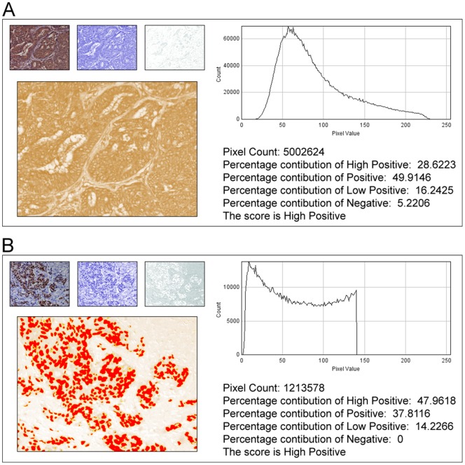
In anatomic pathology, immunohistochemistry (IHC) serves as a diagnostic and prognostic method for identification of disease markers in tissue samples that directly influences classification and grading the disease, influencing patient management. However, till today over most of the world, pathological analysis of tissue samples remained a time-consuming and subjective procedure, wherein the intensity of antibody staining is manually judged and thus scoring decision is directly influenced by visual bias. This instigated us to design a simple method of automated digital IHC image analysis algorithm for an unbiased, quantitative assessment of antibody staining intensity in tissue sections. As a first step, we adopted the spectral deconvolution method of DAB/hematoxylin color spectra by using optimized optical density vectors of the color deconvolution plugin for proper separation of the DAB color spectra. Then the DAB stained image is displayed in a new window wherein it undergoes pixel-by-pixel analysis, and displays the full profile along with its scoring decision. Based on the mathematical formula conceptualized, the algorithm is thoroughly tested by analyzing scores assigned to thousands (n = 1703) of DAB stained IHC images including sample images taken from human protein atlas web resource. The IHC Profiler plugin developed is compatible with the open resource digital image analysis software, ImageJ, which creates a pixel-by-pixel analysis profile of a digital IHC image and further assigns a score in a four tier system. A comparison study between manual pathological analysis and IHC Profiler resolved in a match of 88.6% (P<0.0001, CI = 95%). This new tool developed for clinical histopathological sample analysis can be adopted globally for scoring most protein targets where the marker protein expression is of cytoplasmic and/or nuclear type. We foresee that this method will minimize the problem of inter-observer variations across labs and further help in worldwide patient stratification potentially benefitting various multinational clinical trial initiatives.
Overview
The claims and statistics of this paper haven’t been checked yet.
Free: it reads the open-access text and checks the first 5 claims that cite a source we can check. Create a free account to check every claim.
Integrity signals
Registry
Cites
Statistics
Figures



Verification
Claims the paper makes
The claims and statistics of this paper haven’t been checked yet.
Free: it reads the open-access text and checks the first 5 claims that cite a source we can check. Create a free account to check every claim.
Claims other papers make about this one
Each is checked against this paper’s own text: does it say what the citing paper says it does?
In the citing paper
“For analysis of c-MYC expression in liver sections, random 20× non-tumor fields of the c-MYC IHC scans from liver sections of individual mice were output using NDP.view2 and blindly analyzed using ImageJ (https://imagej.nih.gov/ij/) with the plugin ‘IHC Profiler’62.”
From Tumor-specific cholinergic CD4+ T lymphocytes guide immunosurveillance of hepatocellular carcinoma (2023) · cites this paper as [62]
Self-check
Percentages, sums and reported differences re-computed from the paper’s own numbers.
The self-check needs the paper’s full text.
Citations
Cites
NIH Image to ImageJ: 25 years of image analysis
Schneider, C. A., Rasband, W. S., & Eliceiri, K. W. (2012) · Nature Methods · doi:10.1038/nmeth.2089 · Cited by 64,638 · cited 1×
“The zones were visually identified by using the threshold feature of Image menu of the ImageJ program [22].”
MethodHsu, S. M., Raine, L., & Fanger, H. (1981) · Journal of Histochemistry & Cytochemistry · doi:10.1177/29.4.6166661 · Cited by 14,140
Harvey, J. M., Clark, G. M., Osborne, C. K., & Allred, D. C. (1999) · Journal of Clinical Oncology · doi:10.1200/jco.1999.17.5.1474 · Cited by 2,056
Quantification of histochemical staining by color deconvolution.
Ruifrok, A. C. C., & Johnston, D. A. (2001) · PubMed · Cited by 2,029
Tuominen, V. J., Ruotoistenmäki, S., Viitanen, A., Jumppanen, M., & Isola, J. (2010) · Breast Cancer Research · doi:10.1186/bcr2615 · Cited by 472
McCarty, K. S., Szabo, E. M., Flowers, J. L., Cox, E. B., Leight, G. S., Miller, L. A., Konrath, J. G., Jt, S., Budwit, D. A., & Wt, C. (1986) · PubMed · Cited by 417
A global view of protein expression in human cells, tissues, and organs
Pontén, F., Gry, M., Fagerberg, L., Lundberg, E., Asplund, A., Berglund, L., Oksvold, P., Björling, E., Hober, S., Kampf, C., et al. (2009) · Molecular Systems Biology · doi:10.1038/msb.2009.93 · Cited by 222
Automated Selection of DAB-labeled Tissue for Immunohistochemical Quantification
Brey, E. M., Lalani, Z., Johnston, C., Wong, M., McIntire, L. V., Duke, P. J., & Patrick, C. W. (2003) · Journal of Histochemistry & Cytochemistry · doi:10.1177/002215540305100503 · Cited by 200
Quantitative image analysis of immunohistochemical stains using a CMYK color model
Pham, N.-A., Morrison, A., Schwock, J., Aviel-Ronen, S., Iakovlev, V., Tsao, M.-S., Ho, J., & Hedley, D. W. (2007) · Diagnostic Pathology · doi:10.1186/1746-1596-2-8 · Cited by 179
Quantification of renal pathology by image analysis (Methods in Renal Research)
RANGAN, G. K., & TESCH, G. H. (2007) · Nephrology · doi:10.1111/j.1440-1797.2007.00855.x · Cited by 176
Cited by
Items 101–200 of 1,000
Zheng, X. M., Gong, T., Luo, W., Hu, B., Gao, J., Li, Y., Liu, R. R., Xie, N., Yang, W., Xu, X., et al. (2024) · Science Advances · doi:10.1126/sciadv.ado0016 · Cited by 68
Park, D. Y., Min, B., Choi, B. H., Kim, Y. J., Kim, M., Suh‐Kim, H., & Kim, J. H. (2015) · The American Journal of Sports Medicine · doi:10.1177/0363546515605086 · Cited by 58
Sanz‐Martín, I., Rojo, E., Maldonado, E., Stroppa, G., Nart, J., & Sanz, M. (2018) · Clinical Oral Investigations · doi:10.1007/s00784-018-2516-9 · Cited by 58
Ka, N., Lim, G. Y., Hwang, S., Kim, S.-S., & Lee, M. (2021) · Cell Reports · doi:10.1016/j.celrep.2021.110138 · Cited by 66
Dong, J., Ping, L., Cao, T., Sun, L., Liu, D., Wang, S., Huo, G., & Li, B. (2022) · Frontiers in Immunology · doi:10.3389/fimmu.2022.947755 · Cited by 78
Abuyassin, B., Badran, M., Ayas, N., & Laher, I. (2018) · PLoS ONE · doi:10.1371/journal.pone.0192084 · Cited by 63
Prorok, T., Jana, M., Patel, D. R., & Pahan, K. (2019) · Neurochemical Research · doi:10.1007/s11064-018-02705-0 · Cited by 56
The deubiquitinase (DUB) USP13 promotes Mcl-1 stabilisation in cervical cancer
Morgan, E. L., Patterson, M. R., Barba‐Moreno, D., Scarth, J. A., Wilson, A., & Macdonald, A. (2021) · Oncogene · doi:10.1038/s41388-021-01679-8 · Cited by 60
Yang, S., Shang, J., Liu, L., Tang, Z., & Meng, X. (2022) · Food & Function · doi:10.1039/d2fo01577c · Cited by 58
Tenascin-C, a Prognostic Determinant of Esophageal Squamous Cell Carcinoma
Yang, Z., Yeo, S.-Y., Yin, Y.-X., Lin, Z., Lee, H., Xuan, Y., Cui, Y. R., & Kim, S. (2016) · PLoS ONE · doi:10.1371/journal.pone.0145807 · Cited by 57
WDR76 is a RAS binding protein that functions as a tumor suppressor via RAS degradation
Jeong, W.-J., Park, J., Kim, W.-S., Ro, E. J., Jeon, S. H., Lee, S., Park, Y. N., Min, D. S., & Choi, K. (2019) · Nature Communications · doi:10.1038/s41467-018-08230-6 · Cited by 56
Percutaneous liquid ablation agent for tumor treatment and drug delivery
Albadawi, H., Zhang, Z., Altun, İ., Hu, J., Jamal, L., Ibsen, K. N., Tanner, E. E. L., Mitragotri, S., & Öklü, R. (2021) · Science Translational Medicine · doi:10.1126/scitranslmed.abe3889 · Cited by 56
Approaches to Evaluate Lung Inflammation in Translational Research
Meyerholz, D. K., Sieren, J. C., Beck, A. P., & Flaherty, H. A. (2017) · Veterinary Pathology · doi:10.1177/0300985817726117 · Cited by 59
Ke, M., Xu, T., Fang, Y., Ye, Y., Li, Z.-J., Ren, F., Lu, S., Zhang, X., Wu, R., Lv, Y., et al. (2021) · Cancer Letters · doi:10.1016/j.canlet.2021.05.007 · Cited by 55
Noninvasive radiomics model reveals macrophage infiltration in glioma
Fan, X., Li, J., Huang, B., Lu, H., Lu, C., Pan, M., Wang, X., Zhang, H., You, Y., Wang, X., et al. (2023) · Cancer Letters · doi:10.1016/j.canlet.2023.216380 · Cited by 54
Lactate as a major epigenetic carbon source for histone acetylation via nuclear LDH metabolism
An, Y., Jo, S., Kim, J.-M., Kim, H. S., Kim, H., Jeon, S., Han, D., Yook, J. I., Kang, K. W., & Park, S. (2023) · Experimental & Molecular Medicine · doi:10.1038/s12276-023-01095-w · Cited by 59
He, M., Sun, L., Fu, X., McDonough, S. P., & Chu, C. (2018) · Acta Biomaterialia · doi:10.1016/j.actbio.2018.11.053 · Cited by 53
Basha, G., Ordobadi, M., Scott, W. R., Cottle, A., Liu, Y., Wang, H., & Cullis, P. R. (2016) · Molecular Therapy - Nucleic Acids · doi:10.1038/mtna.2016.68 · Cited by 52
Zhong, L., Yang, B., Zhang, Z., Wang, J., Wang, X., Guo, Y., Huang, W.-F., Wang, Q., Cai, G., Xia, F., et al. (2022) · Cell Death and Disease · doi:10.1038/s41419-022-04608-z · Cited by 51
Akimoto, T., Goto, O., Sasaki, M., Mizutani, M., Tsutsumi, K., Kiguchi, Y., Nakayama, A., Kato, M., Fujimoto, A., Ochiai, Y., et al. (2020) · Gastrointestinal Endoscopy · doi:10.1016/j.gie.2019.12.032 · Cited by 51
Aguiar, R. S., Pohl, F. B., de Morais, G. L., Nogueira, F. C. S., de Carvalho, J. B., Guida, L. D. C., Arge, L. W. P., Melo, A., Moreira, M. E. L., Cunha, D. P., et al. (2020) · Science Signaling · doi:10.1126/scisignal.aay6736 · Cited by 54
AI-guided discovery of the invariant host response to viral pandemics
Sahoo, D., Katkar, G. D., Khandelwal, S., Behroozikhah, M., Claire, A., Castillo, V., Tindle, C., Fuller, M., Taheri, S., Rogers, T. F., et al. (2021) · EBioMedicine · doi:10.1016/j.ebiom.2021.103390 · Cited by 53
Liver injury in non-alcoholic fatty liver disease is associated with urea cycle enzyme dysregulation
Gallego‐Durán, R., Ampuero, J., Pastor‐Ramírez, H., Álvarez‐Amor, L., Campo, J. A. D., Maya‐Miles, D., Montero‐Vallejo, R., Cárdenas-García, A., Pareja, M. J., Gato, S., et al. (2022) · Scientific Reports · doi:10.1038/s41598-022-06614-9 · Cited by 59
Kabel, A. M., & Elkhoely, A. A. (2017) · Biomedicine & Pharmacotherapy · doi:10.1016/j.biopha.2017.06.033 · Cited by 52
Wang, L., Liu, B., Cao, Y., Xu, W., Sun, D., Li, M., Shi, F.-X., Li, M., Tian, Q., Wang, J., et al. (2017) · Molecular Neurobiology · doi:10.1007/s12035-017-0676-2 · Cited by 53
Pham, D., Raut, P. K., Pandit, M., Chang, J., Katila, N., Choi, D., Jeong, J., & Park, P. (2020) · Cancers · doi:10.3390/cancers12030613 · Cited by 53
HMGA2-mediated tumorigenesis through angiogenesis in leiomyoma
Li, Y., Qiang, W., Griffin, B. B., Gao, T., Chakravarti, D., Bulun, S. E., Kim, J. J., & Wei, J. (2020) · Fertility and Sterility · doi:10.1016/j.fertnstert.2020.05.036 · Cited by 50
Impellizzeri, D., Siracusa, R., Cordaro, M., Peritore, A. F., Gugliandolo, E., D’Amico, R., Fusco, R., Crupi, R., Rizzarelli, E., Cuzzocrea, S., et al. (2020) · Biomedicine & Pharmacotherapy · doi:10.1016/j.biopha.2020.110023 · Cited by 49
RetractedMurata, D., Mineharu, Y., Arakawa, Y., Liu, B., Tanji, M., Yamaguchi, M., Fujimoto, K., Fukui, N., Terada, Y., Yokogawa, R., et al. (2017) · Journal of neurosurgery · doi:10.3171/2016.11.jns16991 · Cited by 49
S100A10, a novel biomarker in pancreatic ductal adenocarcinoma
Bydoun, M., Sterea, A. M., Liptay, H., Uzans, A., Huang, W., Rodrigues, G. J., Weaver, I. C. G., Gu, H., & Waisman, D. M. (2018) · Molecular Oncology · doi:10.1002/1878-0261.12356 · Cited by 52
Epstein-Barr Virus–Associated Pulmonary Carcinoma
Yeh, Y., Kao, H., Lee, K., Wu, M., Ho, H., & Chou, T. (2018) · The American Journal of Surgical Pathology · doi:10.1097/pas.0000000000001173 · Cited by 49
A novel tumor suppressor SPINK5 targets Wnt/β‐catenin signaling pathway in esophageal cancer
Wang, Q., Lv, Q., Bian, H., Yang, L., Guo, K., Ye, S., Dong, X., & Tao, L. (2019) · Cancer Medicine · doi:10.1002/cam4.2078 · Cited by 49
Huang, C., Zhang, X., Jiang, L., Zhang, L., Xiang, M., & Ren, H. (2019) · Molecular Therapy — Oncolytics · doi:10.1016/j.omto.2019.05.005 · Cited by 50
Mouratidis, P., Costa, M. M., Rivens, I., Repasky, E. A., & ter Haar, G. (2021) · Journal of The Royal Society Interface · doi:10.1098/rsif.2021.0266 · Cited by 52
Guo, K., Qian, K., Shi, Y., Sun, T., Chen, L., Mei, D., Dong, K., Gu, S., Liu, J., Lv, Z., et al. (2021) · Thyroid · doi:10.1089/thy.2021.0003 · Cited by 54
Rakić, M., Radunović, M., Petković-Ćurčin, A., Tatić, Z., Basta-Jovanović, G., & Sanz, M. (2022) · Clinical Oral Implants Research · doi:10.1111/clr.13928 · Cited by 55
Wu, F., Wang, M., Zhong, T., Xiao, C., Chen, X., Huang, Y., Wu, M., Yu, J., & Chen, D. (2023) · Experimental Hematology and Oncology · doi:10.1186/s40164-023-00428-9 · Cited by 50
Troyer, L. D., Zhao, P., Pastor, T., Baietti, M. F., Barra, J., Vendramin, R., Dok, R., Lechat, B., Najm, P., Haver, D. V., et al. (2020) · Nucleic Acids Research · doi:10.1093/nar/gkz1237 · Cited by 56
Ghorai, A., Mahaddalkar, T., Thorat, R. A., & Dutt, S. (2020) · Cancer Letters · doi:10.1016/j.canlet.2020.06.023 · Cited by 52
Liang, H., Zou, F., Liu, Q., Wang, B., Fu, L., Liang, X., Liu, J. L., & Liu, Q. (2021) · International Journal of Pharmaceutics · doi:10.1016/j.ijpharm.2021.120418 · Cited by 53
Yue, Y., Wang, Y., Xie, Q., Lv, X., Zhou, L., Smith, E. E., Cao, T., Zhang, Y., Li, B., Huo, G., et al. (2023) · Journal of Agricultural and Food Chemistry · doi:10.1021/acs.jafc.3c00421 · Cited by 54
Jin, C., Wu, S., Liang, Z., Zhang, J., Lei, X., Bai, H., Liang, G., Su, X., Chen, X., Wang, P., et al. (2024) · Microbiome · doi:10.1186/s40168-024-01760-w · Cited by 56
Targeting the m6A RNA methyltransferase METTL3 attenuates the development of kidney fibrosis
Jung, H. R., Lee, J., Hong, S.-P., Shin, N., Cho, A., Shin, D., Choi, J. W., Kim, J., Lee, J. P., & Cho, S. (2024) · Experimental & Molecular Medicine · doi:10.1038/s12276-024-01159-5 · Cited by 55
Evidence for Critical Role of Lymphocyte Cytosolic Protein 1 in Oral Cancer
Koide, N., Kasamatsu, A., Endo‐Sakamoto, Y., Ishida, S., Shimizu, T. S., Kimura, Y., Miyamoto, I., Yoshimura, S., Shiiba, M., Tanzawa, H., et al. (2017) · Scientific Reports · doi:10.1038/srep43379 · Cited by 47
Marcellin, L., Santulli, P., Chouzenoux, S., Cerles, O., Nicco, C., Dousset, B. E., Pallardy, M. J., Kerdine‐Römer, S., Just, P.-A., Chapron, C., et al. (2017) · Free Radical Biology and Medicine · doi:10.1016/j.freeradbiomed.2017.04.362 · Cited by 49
Li, K., Zhang, J., Tian, Y., He, Y., Xu, X., Pan, W., Gao, Y., Chen, F., & Wei, L. (2019) · Oncogene · doi:10.1038/s41388-019-1145-3 · Cited by 53
Wang, Y., Wang, B., Huang, Y., Li, Y., Yan, S., Xie, H., Zhang, Y., & Li, J. (2022) · Journal of Inflammation Research · doi:10.2147/jir.s356551 · Cited by 50
Emidio, G. D., Placidi, M., Rea, F., Rossi, G., Falone, S., Cristiano, L., Nottola, S. A., D’Alessandro, A. M., Amicarelli, F., Palmerini, M. G., et al. (2020) · Cells · doi:10.3390/cells9010209 · Cited by 48
Wang, J., Tan, L., Jia, B., Yu, X., Yao, R., Ouyang, N., Yu, X., Cao, X., Tong, J., Chen, T., et al. (2021) · International Journal of Biological Sciences · doi:10.7150/ijbs.58514 · Cited by 51
Cho, H., Shin, I., Yoon, H., Jeon, E., Lee, J., Kim, Y., Ryu, S., Song, C., Kwon, N. H., Moon, Y., et al. (2021) · Journal of Medicinal Chemistry · doi:10.1021/acs.jmedchem.1c00459 · Cited by 46
Kabel, A. M., Omar, M. S., & Elmaaboud, M. A. A. (2016) · International Immunopharmacology · doi:10.1016/j.intimp.2016.08.008 · Cited by 50
Song, J. W., Nam, H. S., Ahn, J. W., Park, H.-S., Kang, D. O., Kim, H. J., Kim, Y. H., Han, J., Choi, J. Y., Lee, S., et al. (2021) · Theranostics · doi:10.7150/thno.59759 · Cited by 47
Jafari, S. M. S., Shafighi, M., Beltraminelli, H., Geiser, T., Hunger, R. E., & Gazdhar, A. (2017) · Plastic & Reconstructive Surgery · doi:10.1097/prs.0000000000003259 · Cited by 46
Kabel, A. M., Al-Shehri, A. H., Al-Talhi, R. A., & Elmaaboud, M. A. A. (2017) · Chemico-Biological Interactions · doi:10.1016/j.cbi.2017.06.009 · Cited by 46
Li, Q., Wang, P., Huang, C., Chen, B., Liu, J., Zhao, M., & Zhao, J. (2019) · Journal of Molecular Neuroscience · doi:10.1007/s12031-019-01263-6 · Cited by 45
Sex-specific maternofetal innate immune responses triggered by group B Streptococci
Allard, M.-J., Giraud, A., Segura, M., & Sebire, G. (2019) · Scientific Reports · doi:10.1038/s41598-019-45029-x · Cited by 43
Effects of subcutaneous injection of ozone during wound healing in rats
Soares, C. D., Morais, T. M. D. L., Araújo, R. M. F. G., Meyer, P. F., de Oliveira, E. A. F., Silva, R. M. V., Carreiro, E. D. M., Carreiro, E. P., Bellocco, V., Mariz, B. A. L. A., et al. (2019) · Growth Factors · doi:10.1080/08977194.2019.1643339 · Cited by 55
Cheng, C.-C., Wooten, J., Gibbs, Z. A., McGlynn, K., Mishra, P., & Whitehurst, A. W. (2020) · eLife · doi:10.7554/elife.58108 · Cited by 44
PRMT1 driven PTX3 regulates ferritinophagy in glioma
Lathoria, K., Gowda, P., Umdor, S. B., Patrick, S., Suri, V. S., & Sen, E. (2023) · Autophagy · doi:10.1080/15548627.2023.2165757 · Cited by 47
PEA/Polydatin: Anti-Inflammatory and Antioxidant Approach to Counteract DNBS-Induced Colitis
Peritore, A. F., D’Amico, R., Cordaro, M., Siracusa, R., Fusco, R., Gugliandolo, E., Genovese, T., Crupi, R., Paola, R. D., Cuzzocrea, S., et al. (2021) · Antioxidants · doi:10.3390/antiox10030464 · Cited by 48
COVID-19 lung disease shares driver AT2 cytopathic features with Idiopathic pulmonary fibrosis
Sinha, S., Castillo, V., Espinoza, C. R., Tindle, C., Fonseca, A. G., Dan, J. M., Katkar, G. D., Das, S., Sahoo, D., & Ghosh, P. (2022) · EBioMedicine · doi:10.1016/j.ebiom.2022.104185 · Cited by 47
Lymph node metastasis of primary endometrial cancers: Associated proteins revealed by MALDI imaging
Mittal, P., Klingler‐Hoffmann, M., Arentz, G., Winderbaum, L., Lokman, N. A., Zhang, C., Anderson, L. A., Scurry, J., Leung, Y. C., Stewart, C. J. R., et al. (2016) · PROTEOMICS · doi:10.1002/pmic.201500455 · Cited by 42
Agrin has a pathological role in the progression of oral cancer
Rivera, C., Zandonadi, F. D. S., Sánchez‐Romero, C., Soares, C. D., Granato, D. C., González‐Arriagada, W. A., & Leme, A. F. P. (2018) · British Journal of Cancer · doi:10.1038/s41416-018-0135-5 · Cited by 44
Kabel, A. M., Estfanous, R. S., & Alrobaian, M. (2019) · Respiratory Physiology & Neurobiology · doi:10.1016/j.resp.2019.103316 · Cited by 42
Liu, W., Stachura, P., Xu, H. C., Ganesh, N. U., Cox, F. F., Wang, R., Lang, K. S., Gopalakrishnan, J., Häussinger, D., Homey, B., et al. (2020) · Journal of Experimental & Clinical Cancer Research · doi:10.1186/s13046-020-1539-7 · Cited by 45
Improved prime editors enable pathogenic allele correction and cancer modelling in adult mice
Liu, P., Liang, S.-Q., Zheng, C., Mintzer, E., Zhao, Y. G., Ponnienselvan, K., Mir, A., Sontheimer, E. J., Gao, G., Flotte, T. R., et al. (2020) · bioRxiv (Cold Spring Harbor Laboratory) · doi:10.1101/2020.12.15.422970 · Cited by 42
Taylor, C. R., Jadhav, A. P., Gholap, A., Kamble, G., Huang, J., Gown, A., Doshi, I., & Rimm, D. L. (2019) · Applied Immunohistochemistry & Molecular Morphology · doi:10.1097/pai.0000000000000737 · Cited by 41
Berbets, A. M., DAVYDENKO, I. S., Barbe, A. M., Konkov, D. G., Albota, O. M., & Yuzko, O. M. (2020) · Reproductive Sciences · doi:10.1007/s43032-020-00285-5 · Cited by 45
Klapper‐Goldstein, H., Verma, A., Elyagon, S., Gillis, R., Murninkas, M., Pittala, S., Paul, A., Shoshan‐Barmatz, V., & Etzion, Y. (2020) · Scientific Reports · doi:10.1038/s41598-020-79056-w · Cited by 45
Chang, M., Tsai, L., Nakagawa‐Goto, K., Lee, K., & Shyur, L. (2022) · Pharmacological Research · doi:10.1016/j.phrs.2022.106148 · Cited by 46
Targeting SOST using a small-molecule compound retards breast cancer bone metastasis
Sun, L., Zhang, Y., Chen, G., Ji, Y., Ma, Q., Qiao, X., Wu, S., Zhou, L. M., Bu, J., Zhu, X., et al. (2022) · Molecular Cancer · doi:10.1186/s12943-022-01697-4 · Cited by 48
Liu, S., Luo, L., Zuo, F., Huang, X., Zhong, L., Liu, S., Geng, Y., Ou, Y., Chen, D., Cai, W. C., et al. (2023) · Frontiers in Physiology · doi:10.3389/fphys.2023.1279051 · Cited by 51
Kabel, A. M., Omar, M. S., Alhadhrami, A., Alharthi, S. S., & Alrobaian, M. (2018) · Physiology & Behavior · doi:10.1016/j.physbeh.2018.01.028 · Cited by 42
Ryu, W., Lee, J. D., Park, J., Cha, P.-H., Cho, Y., Kim, J. Y., Sohn, J., Paik, S., & Choi, K. (2020) · Experimental & Molecular Medicine · doi:10.1038/s12276-020-0440-y · Cited by 43
Wang, Z., Liu, Y., Liu, J., Kong, N., Jiang, Y., Jiang, R., Zhen, X., Zhou, J., Li, C., Sun, H., et al. (2021) · Cell Death and Disease · doi:10.1038/s41419-021-03679-8 · Cited by 45
Kim, Y. S., Ko, B., Kim, D. J., Tak, J., Han, C. Y., Cho, J., Kim, W. K., & Kim, S. G. (2022) · Nature Communications · doi:10.1038/s41467-022-33749-0 · Cited by 44
Arrazuria, R., Pérez, V. P., Molina, E., Juste, R. A., Khafipour, E., & Elguezábal, N. (2018) · Scientific Reports · doi:10.1038/s41598-018-32484-1 · Cited by 47
Cardoso, P. G., Resende-de-Oliveira, R., & Rocha, E. (2019) · Environmental Pollution · doi:10.1016/j.envpol.2019.06.058 · Cited by 41
Gibbs, Z. A., Reza, L. C., Cheng, C.-C., Westcott, J. M., McGlynn, K., & Whitehurst, A. W. (2020) · eLife · doi:10.7554/elife.57679 · Cited by 41
Liu, X., Yuan, L., Gao, Y. P., Zhou, L.-M., Yang, J.-W., & Pei, Z. (2020) · Journal of Cancer · doi:10.7150/jca.44754 · Cited by 45
Characteristics of B lymphocyte infiltration in HPV+ head and neck squamous cell carcinoma
Si-wei, Z., Wang, B., Ma, F., Tong, F., Yan, B., Liu, T., Xie, H., Song, L., Yu, S., & Wei, L. (2021) · Cancer Science · doi:10.1111/cas.14834 · Cited by 43
Liu, Y., Xue, M., Han, Y., Li, Y., Xiao, B., Wang, W., Yu, J., & Ye, X. (2023) · Bioengineering & Translational Medicine · doi:10.1002/btm2.10500 · Cited by 42
Haub, P., & Meckel, T. (2015) · Scientific Reports · doi:10.1038/srep12096 · Cited by 38
Alteration of systemic and uterine endometrial immune populations in patients with endometriosis
Le, N., de Mola, J. R. L., Bremer, P., Groesch, K. A., Wilson, T. S., Díaz‐Sylvester, P., & Braundmeier‐Fleming, A. (2020) · American Journal of Reproductive Immunology · doi:10.1111/aji.13362 · Cited by 42
Loss of acinar cell VMP1 triggers spontaneous pancreatitis in mice
Wang, S., Chao, X., Jiang, X., Wang, T., Rodriguez, Y., Yang, L., Pacher, P., Ni, H., & Ding, W. (2021) · Autophagy · doi:10.1080/15548627.2021.1990672 · Cited by 40
Hsueh, P.-T., Wang, H.-H., Liu, C.-L., Ni, W.-F., Chen, Y.-L., & Liu, J.-K. (2017) · PLOS ONE · doi:10.1371/journal.pone.0179970 · Cited by 37
The Interaction of miR-378i-Skp2 Regulates Cell Senescence in Diabetic Nephropathy
Tsai, Y., Kuo, P., Kuo, M., Hung, W., Wu, L., Chang, W., Wu, P., Lee, S., Chen, H., & Hsu, Y. (2018) · Journal of Clinical Medicine · doi:10.3390/jcm7120468 · Cited by 38
Horibe, A., Eid, N. A. S., Ito, Y., Otsuki, Y., & Kondo, Y. (2019) · International Journal of Molecular Sciences · doi:10.3390/ijms20010184 · Cited by 39
Tian, P., Chen, Y., Qian, X., Zou, R., Zhu, H., Zhao, J., Zhang, H., Wang, G., & Chen, W. (2021) · Food & Function · doi:10.1039/d1fo01608c · Cited by 42
Marković, J., Stošić, M., Kojić, D. K., & Matavulj, M. (2017) · Acta Histochemica · doi:10.1016/j.acthis.2017.12.001 · Cited by 37
Chan, S., Wang, X., Wang, Z., Du, Y., Zuo, X., Chen, J., Sun, R., Zhang, Q. H., Li, L., Yang, Y., et al. (2023) · International Journal of Biological Sciences · doi:10.7150/ijbs.82000 · Cited by 43
Meybodi, M. A. M., Nilforoushzadeh, M. A., Khandandezfully, N., & Mansouri, P. (2024) · Life Sciences · doi:10.1016/j.lfs.2024.122915 · Cited by 36
Abboud, E., Chrayteh, D., Boussetta, N., Dalle, H., Malerba, M., Wu, T., Gall, M. L., Reelfs, O., Pourzand, C., Mellett, M., et al. (2024) · Nature Communications · doi:10.1038/s41467-024-50993-8 · Cited by 40
KLF6 depletion promotes NF-κB signaling in glioblastoma
Masilamani, A. P., Ferrarese, R., Kling, E., Thudi, N. K., Kim, H., Scholtens, D. M., Dai, F., Hadler, M., Unterkircher, T., Platania, L., et al. (2017) · Oncogene · doi:10.1038/onc.2016.507 · Cited by 36
AMPK-Independent LKB1 Activity Is Required for Efficient Epithelial Ovarian Cancer Metastasis
Buensuceso, A. V., Ramos-Valdes, Y., DiMattia, G. E., & Shepherd, T. G. (2019) · Molecular Cancer Research · doi:10.1158/1541-7786.mcr-19-0530 · Cited by 37
Xie, L., Zhai, R., Chen, T., Gao, C., Xue, R., Wang, N., Wang, J., Xu, Y., & Gui, D. (2020) · Drug Design Development and Therapy · doi:10.2147/dddt.s235491 · Cited by 36
Wilkie, T., Verma, A. K., Zhao, H., Charan, M., Ahirwar, D. K., Kant, S., Pancholi, V., Mishra, S., & Ganju, R. K. (2021) · Molecular Oncology · doi:10.1002/1878-0261.12975 · Cited by 43
Fibroblast growth factor 21 is a hepatokine involved in MASLD progression
Gallego‐Durán, R., Ampuero, J., Maya‐Miles, D., Pastor‐Ramírez, H., Montero‐Vallejo, R., Rivera‐Esteban, J., Álvarez‐Amor, L., Pareja, M. J., Rico, M. C., Millán, R., et al. (2024) · United European Gastroenterology Journal · doi:10.1002/ueg2.12534 · Cited by 41
Browe, D. C., Coleman, C. M., Barry, F., & Elliman, S. J. (2019) · Scientific Reports · doi:10.1038/s41598-019-49499-x · Cited by 37
Histo-genomics: digital pathology at the forefront of precision medicine
Barsoum, I. B., Tawedrous, E., Faragalla, H., & Yousef, G. M. (2018) · Diagnosis · doi:10.1515/dx-2018-0064 · Cited by 40
Metadata
- Authors
- Varghese, F., Bukhari, A. B., Malhotra, R., & De, A.
- Affiliations
- Tata Memorial Hospital (IN)
- Corresponding author
- De, Abhijit
- Venue
- PLoS ONE
- Publisher
- Public Library of Science (PLoS)
- Published
- 2014-05-06
- Volume
- 9
- Issue
- 5
- Pages
- e96801
- ISSN
- 1932-6203 (ISSN-L)
- PMC
- PMC4011881
- OpenAlex
- W1998734250
- Cited by
- 1,318
- References
- 30
- Citations by year
- 2026: 90 · 2025: 129 · 2024: 172 · 2023: 172 · 2022: 171 · 2021: 144 · 2020: 141 · 2019: 101 · 2018: 69 · 2017: 60 · 2016: 44 · 2015: 22 · 2014: 3
- FWCI
- 42.82
- Type
- journal article
- Language
- en
- Open access
- gold
- doi.org
- Locations
- PLoS ONE (publishedVersion, cc-by)
- Europe PMC (PubMed Central) (publishedVersion, cc-by)
- PubMed Central (publishedVersion, cc-by)
- PubMed (publishedVersion)
- RePEc: Research Papers in Economics (submittedVersion)
- DOAJ (DOAJ: Directory of Open Access Journals) (submittedVersion, cc-by-sa)
- Figshare (submittedVersion, cc-by)
- Funders
- Indian Council of Medical Research
- Advanced Centre for Treatment, Research and Education in Cancer
- Topics
- Physical Sciences › Computer Science › Artificial Intelligence › AI in cancer detection
- Life Sciences › Biochemistry, Genetics and Molecular Biology › Biophysics › Cell Image Analysis Techniques
- Physical Sciences › Computer Science › Computer Vision and Pattern Recognition › Digital Imaging for Blood Diseases
- Keywords
- immunohistochemistry image analysis, automated grading, ImageJ plugin, interobserver variability, digital pathology, human tissue samples, protein expression profiling
- MeSH terms
- Automation, Cell Nucleus, Cytoplasm, Humans, Image Processing, Computer-Assisted, Immunohistochemistry, Neoplasms, Software, Biomarkers, Tumor
About these checks
Registry, retraction, Cites and statistics results are computed, not judged, with one exception: a printed reference that no index lists is read once by a model before it counts as a citation error. Claims are extracted by a model and kept only when their quote appears verbatim in the text; each cited source is then checked, with a model’s help, for whether it says what the claim relies on. Those verdicts can be wrong — read the claim’s quote and the cited source before relying on one.
Statistics and arithmetic are recomputed, not judged.
Confidence bands are shown only for fields calibrated on our annotated test set. None is calibrated yet, so no band is shown.
What counts as a claim ›How we measure confidence ›How we check this ›
Are you an author, or do you see a problem with this page? Request a review
Data sources
Bibliographic data: Semantic Scholar, OpenAlex, Crossref.
Contains information from the Semantic Scholar Academic Graph Datasets, made available under the ODC Attribution License (ODC-BY 1.0).
Semantic Scholar’s data is described in: Kinney, R., et al. (2023). The Semantic Scholar Open Data Platform. arXiv:2301.10140.
