

IHC Profiler: An Open Source Plugin for the Quantitative Evaluation and Automated Scoring of Immunohistochemistry Images of Human Tissue Samples
Open accessThe DOI matches its Crossref record; 30 of the 30 references it cites were found in a registry or an index.
Abstract
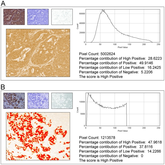
In anatomic pathology, immunohistochemistry (IHC) serves as a diagnostic and prognostic method for identification of disease markers in tissue samples that directly influences classification and grading the disease, influencing patient management. However, till today over most of the world, pathological analysis of tissue samples remained a time-consuming and subjective procedure, wherein the intensity of antibody staining is manually judged and thus scoring decision is directly influenced by visual bias. This instigated us to design a simple method of automated digital IHC image analysis algorithm for an unbiased, quantitative assessment of antibody staining intensity in tissue sections. As a first step, we adopted the spectral deconvolution method of DAB/hematoxylin color spectra by using optimized optical density vectors of the color deconvolution plugin for proper separation of the DAB color spectra. Then the DAB stained image is displayed in a new window wherein it undergoes pixel-by-pixel analysis, and displays the full profile along with its scoring decision. Based on the mathematical formula conceptualized, the algorithm is thoroughly tested by analyzing scores assigned to thousands (n = 1703) of DAB stained IHC images including sample images taken from human protein atlas web resource. The IHC Profiler plugin developed is compatible with the open resource digital image analysis software, ImageJ, which creates a pixel-by-pixel analysis profile of a digital IHC image and further assigns a score in a four tier system. A comparison study between manual pathological analysis and IHC Profiler resolved in a match of 88.6% (P<0.0001, CI = 95%). This new tool developed for clinical histopathological sample analysis can be adopted globally for scoring most protein targets where the marker protein expression is of cytoplasmic and/or nuclear type. We foresee that this method will minimize the problem of inter-observer variations across labs and further help in worldwide patient stratification potentially benefitting various multinational clinical trial initiatives.
Overview
The claims and statistics of this paper haven’t been checked yet.
Free: it reads the open-access text and checks the first 5 claims that cite a source we can check. Create a free account to check every claim.
Integrity signals
Registry
Cites
Statistics
Figures



Verification
Claims the paper makes
The claims and statistics of this paper haven’t been checked yet.
Free: it reads the open-access text and checks the first 5 claims that cite a source we can check. Create a free account to check every claim.
Claims other papers make about this one
Each is checked against this paper’s own text: does it say what the citing paper says it does?
In the citing paper
“For analysis of c-MYC expression in liver sections, random 20× non-tumor fields of the c-MYC IHC scans from liver sections of individual mice were output using NDP.view2 and blindly analyzed using ImageJ (https://imagej.nih.gov/ij/) with the plugin ‘IHC Profiler’62.”
From Tumor-specific cholinergic CD4+ T lymphocytes guide immunosurveillance of hepatocellular carcinoma (2023) · cites this paper as [62]
Self-check
Percentages, sums and reported differences re-computed from the paper’s own numbers.
The self-check needs the paper’s full text.
Citations
Cites
NIH Image to ImageJ: 25 years of image analysis
Schneider, C. A., Rasband, W. S., & Eliceiri, K. W. (2012) · Nature Methods · doi:10.1038/nmeth.2089 · Cited by 64,638 · cited 1×
“The zones were visually identified by using the threshold feature of Image menu of the ImageJ program [22].”
MethodHsu, S. M., Raine, L., & Fanger, H. (1981) · Journal of Histochemistry & Cytochemistry · doi:10.1177/29.4.6166661 · Cited by 14,140
Harvey, J. M., Clark, G. M., Osborne, C. K., & Allred, D. C. (1999) · Journal of Clinical Oncology · doi:10.1200/jco.1999.17.5.1474 · Cited by 2,056
Quantification of histochemical staining by color deconvolution.
Ruifrok, A. C. C., & Johnston, D. A. (2001) · PubMed · Cited by 2,029
Tuominen, V. J., Ruotoistenmäki, S., Viitanen, A., Jumppanen, M., & Isola, J. (2010) · Breast Cancer Research · doi:10.1186/bcr2615 · Cited by 472
McCarty, K. S., Szabo, E. M., Flowers, J. L., Cox, E. B., Leight, G. S., Miller, L. A., Konrath, J. G., Jt, S., Budwit, D. A., & Wt, C. (1986) · PubMed · Cited by 417
A global view of protein expression in human cells, tissues, and organs
Pontén, F., Gry, M., Fagerberg, L., Lundberg, E., Asplund, A., Berglund, L., Oksvold, P., Björling, E., Hober, S., Kampf, C., et al. (2009) · Molecular Systems Biology · doi:10.1038/msb.2009.93 · Cited by 222
Automated Selection of DAB-labeled Tissue for Immunohistochemical Quantification
Brey, E. M., Lalani, Z., Johnston, C., Wong, M., McIntire, L. V., Duke, P. J., & Patrick, C. W. (2003) · Journal of Histochemistry & Cytochemistry · doi:10.1177/002215540305100503 · Cited by 200
Quantitative image analysis of immunohistochemical stains using a CMYK color model
Pham, N.-A., Morrison, A., Schwock, J., Aviel-Ronen, S., Iakovlev, V., Tsao, M.-S., Ho, J., & Hedley, D. W. (2007) · Diagnostic Pathology · doi:10.1186/1746-1596-2-8 · Cited by 179
Quantification of renal pathology by image analysis (Methods in Renal Research)
RANGAN, G. K., & TESCH, G. H. (2007) · Nephrology · doi:10.1111/j.1440-1797.2007.00855.x · Cited by 176
Cited by
Items 601–700 of 1,000
ARHGAP25 expression in colorectal cancer as a biomarker associated with favorable prognosis
Zhang, Y., Lin, Y. H., Zhu, Y., Zhang, X., Li, T., & Yang, M. S. (2022) · Molecular and Clinical Oncology · doi:10.3892/mco.2022.2517 · Cited by 12
Tang, Z., Wang, L., Huang, Z., Guan, H., Leung, W., Chen, X., Xia, H., & Zhang, W. (2022) · Molecular Reproduction and Development · doi:10.1002/mrd.23569 · Cited by 12
Li, L., Deng, T., Zhang, L., Wang, Y., Zhou, Y., Liu, Y., Li, H., Dai, J., Yang, Y., Ling, N., et al. (2022) · International Journal of Biological Sciences · doi:10.7150/ijbs.71327 · Cited by 15
Zhang, X., Yin, Z., Li, C., Nie, L., & Chen, K. (2022) · Experimental Cell Research · doi:10.1016/j.yexcr.2022.113208 · Cited by 12
Prognostic Value and Biological Function of Galectins in Malignant Glioma
Zhu, H., Liu, D., Cheng, L., Liu, J., Wang, G., Li, H., Zhang, Y., Mi, H., Zhang, S., Shu, K., et al. (2022) · Frontiers in Oncology · doi:10.3389/fonc.2022.834307 · Cited by 14
Aerosol-Administered Adelmidrol Attenuates Lung Inflammation in a Murine Model of Acute Lung Injury
Interdonato, L., D’Amico, R., Cordaro, M., Siracusa, R., Fusco, R., Peritore, A. F., Gugliandolo, E., Crupi, R., Coaccioli, S., Genovese, T., et al. (2022) · Biomolecules · doi:10.3390/biom12091308 · Cited by 14
The Sialyl Lewis X Glycan Receptor Facilitates Infection of Subtype H7 Avian Influenza A Viruses
Guan, M., Olivier, A. K., Lu, X., Epperson, W. B., Zhang, X., Zhong, L., Waters, K., Mamaliger, N., Li, L., Wen, F., et al. (2022) · Journal of Virology · doi:10.1128/jvi.01344-22 · Cited by 12
Moxonidine ameliorates cardiac injury in rats with metabolic syndrome by regulating autophagy
El-Sayed, S. S., Rezq, S., Alsemeh, A. E., & Mahmoud, M. F. (2022) · Life Sciences · doi:10.1016/j.lfs.2022.121210 · Cited by 13
Elkhoely, A. A., Estfanous, R. S., Alrobaian, M., Borg, H. M., & Kabel, A. M. (2022) · Life Sciences · doi:10.1016/j.lfs.2022.121288 · Cited by 12
Yanuhar, U., & Khumaidi, A. (2017) · Jurnal Akuakultur Indonesia · doi:10.19027/jai.16.1.22-32 · Cited by 12
Hyperglycemia Alters the Protein Levels of Prominin-1 and VEGFA in the Retina of Albino Rats
Almasry, S. M., Habib, E. K., Elmansy, R. A., & Hassan, Z. A. (2017) · Journal of Histochemistry & Cytochemistry · doi:10.1369/0022155417737484 · Cited by 12
Frutos, J., Andrés, S., Yáñez-Ruíz, D. R., Benavides, J., López, S., Santos, A., Valladares, M. M., Rozada, F., & Giráldez, F. J. (2018) · animal · doi:10.1017/s1751731118000836 · Cited by 12
Lu, X., Chu, C., Liu, X., Gao, Y., Wu, M., Guo, F. F., Li, Y., Geng, C., Huang, Y., Zhang, Y., et al. (2019) · PLoS ONE · doi:10.1371/journal.pone.0222515 · Cited by 12
Eldomiaty, M. A., Elayat, A. A., Ali, S. S., Algaidi, S. A., & Elnaggar, M. N. (2019) · Folia Morphologica · doi:10.5603/fm.a2019.0131 · Cited by 13
Kang, K., Lee, S. J., Kim, J. H., Lee, B., Kim, S. J., Park, Y., & Kim, B. S. (2020) · BMC Cancer · doi:10.1186/s12885-020-07102-x · Cited by 12
Guo, J., Liu, T., Ma, L., Hao, W., Yan, H., Li, T., Yang, Y., Cai, J., Gao, F., Zhao, X., et al. (2020) · Oxidative Medicine and Cellular Longevity · doi:10.1155/2020/8905860 · Cited by 13
Winter, R., Antunes, Á. C. M., & de Oliveira, F. H. (2020) · Surgical Neurology International · doi:10.25259/sni_528_2020 · Cited by 12
Moiseev, A. A., Achkasova, K. A., Kiseleva, E. B., Сергеевич, Я. К., Potapov, A. L., Bederina, E. L., Kuznetsov, S. S., Sherstnev, E. P., Shabanov, D. V., Gelikonov, G. V., et al. (2022) · Biomedical Optics Express · doi:10.1364/boe.457467 · Cited by 13
Jameel, M., Chan, K. I., Mahadevappa, R., Wong, S. W., Wai, K. C., & Kwok, H. F. (2022) · International Journal of Biological Sciences · doi:10.7150/ijbs.69344 · Cited by 15
Kumar, G., Tajpara, P., Bukhari, A. B., Ramchandani, A. G., De, A., & Maru, G. B. (2014) · Toxicology Reports · doi:10.1016/j.toxrep.2014.11.008 · Cited by 12
Self-citationIntraperitoneal translocation of gut microbiota induces NETosis and promotes endometriosis
Wu, X., Wu, M., Li, H. W., Yang, Y., Shen, H., Siyuan, H., Pan, Y., Tao, L., Guo, S., Chen, J., et al. (2025) · Gut · doi:10.1136/gutjnl-2025-336185 · Cited by 11
Watanabe, S., Sugiura, H., & Arai, F. (2022) · IEEE Robotics and Automation Letters · doi:10.1109/lra.2022.3144766 · Cited by 12
Antitumor activity of a ROR1 × CD3 bispecific antibody in non-small cell lung cancer
Wang, Y., Zhang, Y., Sun, H., Chen, J., Yang, H., 钟占琼, Xiao, X., Li, Y., Tang, Y., Lu, H., et al. (2023) · International Immunopharmacology · doi:10.1016/j.intimp.2023.110686 · Cited by 11
Zhang, J., Kang, F., Gao, J., Jiao, J., Quan, Z., Ma, S., Li, Y., Guo, S., Li, Z., Jing, Y., et al. (2023) · Journal of Nuclear Medicine · doi:10.2967/jnumed.122.265001 · Cited by 13
Bai, Y., Lü, Z., Zhang, C., Guo, M., Xia, L., Tang, Z., Liu, Y., & Deng, S. (2023) · Oncogene · doi:10.1038/s41388-023-02866-5 · Cited by 14
Jeanmard, N., Bissanum, R., Sriplung, H., Charoenlappanit, S., Roytrakul, S., & Navakanitworakul, R. (2023) · PLoS ONE · doi:10.1371/journal.pone.0291574 · Cited by 11
Kim, Y., Ko, J. Y., Kong, H. K., Lee, M., Chung, W., Lim, S., Son, D., Oh, S., Park, J. W., Kim, D. Y., et al. (2024) · Cancers · doi:10.3390/cancers16091666 · Cited by 11
Yang, H., Wang, J., Zhou, X., Fu, D., Le, C., Wei, J., Qian, Y., Fang, Y., Jin, T., Kong, X., et al. (2024) · Nano Today · doi:10.1016/j.nantod.2024.102307 · Cited by 13
Liang, B., Lin, W., Tang, Y., Li, T., Chen, Q., Zhang, W., Zhou, X., Ma, J., Liu, B., Yu, Z., et al. (2024) · iScience · doi:10.1016/j.isci.2024.111036 · Cited by 14
Tang, M., Wang, X., Wang, S., Xing, C., Xu, Q., Mu, Y., Wu, X., Zhao, Z. A., & Li, F. (2024) · Biochemical Pharmacology · doi:10.1016/j.bcp.2024.116648 · Cited by 13
Yu, Y., Kang, N., Liu, X., Liang, Y., Jiao, Z., Zou, B., Cai, T., Yang, Z., Chen, W., Wu, T., et al. (2025) · Environmental Pollution · doi:10.1016/j.envpol.2025.125629 · Cited by 13
Cui, J., Wang, C., Zheng, Y., Zhang, Y., Luo, S., Ren, Z., Qin, X., Zhang, M., Gao, F., Huang, H., et al. (2025) · Communications Biology · doi:10.1038/s42003-025-08493-y · Cited by 13
Activation of Liver X Receptor α Sensitizes Mice to T‐Cell Mediated Hepatitis
Gao, L., Li, B., Wang, J., Shen, D., Yang, M., Sun, R., Tung, H., Xu, M., Ren, S., Zhang, M., et al. (2020) · Hepatology Communications · doi:10.1002/hep4.1584 · Cited by 12
Alsanosy, A. A., Noreldin, A. E., Elewa, Y. H. A., Mahmoud, S. F. E., Elnasharty, M., & Aboelnour, A. (2020) · Microscopy and Microanalysis · doi:10.1017/s1431927620024812 · Cited by 17
In vivo neural tissue engineering using adipose-derived mesenchymal stem cells and fibrin matrix
Chandrababu, K., Sreelatha, H. V., Sudhadevi, T., Anil, A., Sabareeswaran, A., & Krishnan, L. K. (2021) · Journal of Spinal Cord Medicine · doi:10.1080/10790268.2021.1930369 · Cited by 11
Xu, B., Liu, F., Gao, Y., Sun, J., Li, Y., Lin, Y., Liu, X., Wen, Y., Yi, S., Dang, J., et al. (2021) · Acta Dermato Venereologica · doi:10.2340/actadv.v101.570 · Cited by 11
Diffusion tensor imaging reveals brain structure changes in dogs after spinal cord injury
Li, D.-P., Li, J., Liu, C., Yang, D., Li, J., Qin, C., Zhang, X., & Liu, J. (2022) · Neural Regeneration Research · doi:10.4103/1673-5374.344839 · Cited by 11
Liu, Y., Liang, W., Chang, Y., He, Z., Wu, M., Zheng, H., Ke, X., Lv, M., Liu, Q., Liu, Q., et al. (2022) · Frontiers in Immunology · doi:10.3389/fimmu.2022.950884 · Cited by 11
Na, S., Cui, H., Guo, Z., Xiang, L., Sakran, K. A., Guo, X., Li, X., Xie, L., Zhu, Y., Qi, H. Y., et al. (2022) · Frontiers in Oncology · doi:10.3389/fonc.2022.1052375 · Cited by 11
Luo, M., Chen, Y., Xie, Y., Wang, Q., Xiang, Y., Long, N., Yang, W., Zhao, Y. M., & Zhou, J. (2022) · World Journal of Gastroenterology · doi:10.3748/wjg.v28.i47.6769 · Cited by 12
Onursal, C., Reel, B., Bintepe, C., Güzeloğlu, M., Ersoy, N., & Bağrıyanık, A. (2023) · Journal of Diabetes and its Complications · doi:10.1016/j.jdiacomp.2023.108421 · Cited by 12
Elmaaboud, M. A. A., Awad, M. M., El‐Shaer, R. A. A., & Kabel, A. M. (2023) · International Immunopharmacology · doi:10.1016/j.intimp.2023.110061 · Cited by 15
Jiang, Z., Shi, B., Zhang, Y., Yu, T., Cheng, Y., Zhu, J., Zhang, G., Zhong, M., Hu, S., & Ma, X. (2024) · iScience · doi:10.1016/j.isci.2024.108843 · Cited by 11
Spatial distribution of osteoblast activating peptide in the rat stomach
Noreldin, A. E., Sogabe, M., Yamano, Y., Uehara, M., Mahdy, M. A. A., Elnasharty, M., Sayed-Ahmed, A., Warita, K., & Hosaka, Y. Z. (2015) · Acta Histochemica · doi:10.1016/j.acthis.2015.12.001 · Cited by 11
A 2-Step Laemmli and Antigen Retrieval Method Improves Immunodetection
Scalia, C. R., Gendusa, R., & Cattoretti, G. (2015) · Applied immunohistochemistry & molecular morphology · doi:10.1097/pai.0000000000000203 · Cited by 11
Konrad, L., Hackethal, A., Oehmke, F., Berkes, E., Engel, J. B., & Tinneberg, H. (2016) · Reproductive Sciences · doi:10.1177/1933719116641756 · Cited by 12
Kasai, Y., Takeda, N., Kobayashi, S., Takagi, R., & Yamato, M. (2017) · FEBS Open Bio · doi:10.1002/2211-5463.12213 · Cited by 11
Pozor, M. A., Conley, A. J., Roser, J. F., Nolin, M., Zambrano, G. L., Runyon, S. P., Kelleman, A. A., & Macpherson, M. L. (2018) · Theriogenology · doi:10.1016/j.theriogenology.2018.05.009 · Cited by 11
In vivo COX-2 modulation and metabolite profiling of Pandanus tectorius leaves extracts
del Mundo, C. R., Castillo, A. L., An, S. S. A., & Tan, M. A. (2020) · 3 Biotech · doi:10.1007/s13205-020-2087-8 · Cited by 11
Bossart, S., Cazzaniga, S., Willenberg, T., RAMELET, A. A., Baumgartner, M., Hunger, R. E., & Jafari, S. M. S. (2020) · Journal of the European Academy of Dermatology and Venereology · doi:10.1111/jdv.16312 · Cited by 11
Schnoell, J., Jank, B. J., Kadletz‐Wanke, L., Stoiber, S., Spielvogel, C. P., Gurnhofer, E., Kenner, L., & Heiduschka, G. (2020) · Journal of Cancer Research and Clinical Oncology · doi:10.1007/s00432-020-03482-6 · Cited by 11
Olorundare, O. E., Adeneye, A. A., Akinsola, A. O., Ajayi, A. M., Agede, O. A., Soyemi, S. S., Mgbehoma, A. I., Okoye, I. I., Albrecht, R. M., Ntambi, J. M., et al. (2021) · Frontiers in Pharmacology · doi:10.3389/fphar.2020.610331 · Cited by 14
Ueda, R. M. R., de Souza, V. M., Magalhães, L. R., Chagas, P. H. N., Veras, A. S. C., Teixeira, G. R., & Nai, G. A. (2021) · Journal of Environmental Science and Health Part B · doi:10.1080/03601234.2021.1939622 · Cited by 11
Evaluation of protective effects of methylene blue on cisplatin-induced nephrotoxicity
Usefzay, O., Yari, S., Amiri, P., & Hasanein, P. (2022) · Biomedicine & Pharmacotherapy · doi:10.1016/j.biopha.2022.113023 · Cited by 12
Effects of Saikokaryukotsuboreito on Spermatogenesis and Fertility in Aging Male Mice
Zang, Z., Ji, S., Zhang, Y., Gao, Y., & Zhang, B. (2016) · Chinese Medical Journal · doi:10.4103/0366-6999.178972 · Cited by 13
Šošić‐Jurjević, B., Ajdžanović, V. Z., Filipović, B. R., Trifunović, S. L., Jarić, I., Ristić, N., & Milošević, V. (2016) · Acta Histochemica · doi:10.1016/j.acthis.2016.07.007 · Cited by 12
Lin, Q., Price, S. A., SKINNER, J. T., Hu, B., Fan, C., Yamaji-Kegan, K., & Johns, R. A. (2020) · PLoS ONE · doi:10.1371/journal.pone.0235546 · Cited by 12
Rakić, M., Canullo, L., Radovanović, S., Tatić, Z., Radunović, M., Souedain, A., Weiss, P., Struillou, X., & Vojvodić, D. (2023) · Dental Materials · doi:10.1016/j.dental.2023.10.003 · Cited by 10
Berndt‐Paetz, M., Han, S., Weimann, A., Reinhold, A., Nürnberger, S., & Neuhaus, J. (2023) · Biomedicines · doi:10.3390/biomedicines11112958 · Cited by 11
Ekici, M., Ateş, M. B., Ekici, H. B., & Özgür, A. (2023) · Reproductive BioMedicine Online · doi:10.1016/j.rbmo.2023.103778 · Cited by 13
Lin, Q., Ma, Y., & Chen, P. (2024) · ONCOLOGIE · doi:10.1515/oncologie-2023-0380 · Cited by 10
Li, X., Guan, W., Liu, H., Yuan, J., Wang, F., Guan, B., Chen, J., Lu, Q., Zhang, L., & Xu, G. (2024) · APOPTOSIS · doi:10.1007/s10495-024-01956-3 · Cited by 10
Peng, Z., Song, J., Zhu, W., Bao, H., Hu, Y., Shi, Y., Cheng, X., Jiang, M., Fang, F., Chen, J., et al. (2025) · Molecular Metabolism · doi:10.1016/j.molmet.2025.102109 · Cited by 11
TNF-α modulates cell proliferation via SOX4/TGF-β/Smad signaling in benign prostatic hyperplasia
Li, J., Chen, B., Huang, Y., Liao, X., You, J., Chen, Z., Ning, S., Reda, A. M., Zhao, J., Ran, B., et al. (2025) · Cell Death and Disease · doi:10.1038/s41419-025-07783-x · Cited by 22
Yamamoto, J., Kasamatsu, A., Okubo, Y., Nakashima, D., Fushimi, K., Minakawa, Y., Kasama, H., Shiiba, M., Tanzawa, H., & Uzawa, K. (2017) · Biochemical and Biophysical Research Communications · doi:10.1016/j.bbrc.2017.12.138 · Cited by 10
Effects of subchronic acrylamide treatment on the endocrine pancreas of juvenile male Wistar rats
Stošić, M., Matavulj, M., & Marković, J. (2018) · Biotechnic & Histochemistry · doi:10.1080/10520295.2017.1393562 · Cited by 10
Carril, P., da Silva, A. B., Tenreiro, R. P., & Cruz, C. (2020) · Frontiers in Plant Science · doi:10.3389/fpls.2020.00889 · Cited by 10
Prognostic significance of p53, Sox11, and Pax5 co-expression in mantle cell lymphoma
Jing, C., Zheng, Y., Yu, F., Cao, X., & Xu, C. (2021) · Scientific Reports · doi:10.1038/s41598-021-91433-7 · Cited by 10
Ram, A., Matuszewska, K., Petrik, J., Singh, A., & Oblak, M. L. (2021) · Frontiers in Veterinary Science · doi:10.3389/fvets.2021.758295 · Cited by 10
Molecular and histological traits of reduced lysosomal acid lipase activity in the fatty liver
Carotti, S., Lettieri‐Barbato, D., Piemonte, F., Ruggiero, S., Rosina, M., Zalfa, F., Zingariello, M., Arciprete, F., Valentini, F., Francesconi, M. A., et al. (2021) · Cell Death and Disease · doi:10.1038/s41419-021-04382-4 · Cited by 10
Mahmoud, M. F., Rezq, S., Alsemeh, A. E., Abdelfattah, M. A. O., El-Shazly, A. M., Daoud, R., Raey, M. A. E., & Sobeh, M. (2021) · Frontiers in Pharmacology · doi:10.3389/fphar.2021.799444 · Cited by 12
RetractedFeng, S., Peden, E. K., Guo, Q., Lee, T. H., Li, Q., Yuan, Y., Chen, C., Huang, F., & Cheng, J. (2022) · Journal of Biological Chemistry · doi:10.1016/j.jbc.2022.101816 · Cited by 10
Shi, S., Bonaccorsi‐Riani, E., Schurink, I. J., van den Bosch, T. P. P., Doukas, M., Lila, K. A., Roest, H. P., Xhema, D., Gianello, P., de Jonge, J., et al. (2022) · Frontiers in Immunology · doi:10.3389/fimmu.2022.890353 · Cited by 15
Huang, L., Palmieri, C., & Bertin, F. (2022) · Research in Veterinary Science · doi:10.1016/j.rvsc.2022.08.018 · Cited by 10
Luo, J., Wang, Y., Zhao, L., Wang, C., & Zhang, Z. (2022) · Cancers · doi:10.3390/cancers15010172 · Cited by 11
Li, S., Gong, F., Zhou, Z., & Gong, X. (2023) · International Journal of Nanomedicine · doi:10.2147/ijn.s377600 · Cited by 10
Tsai, K. H. ‐., Shi, H., Parungao, R., Naficy, S., Ding, X., Ding, X., Hew, J. J., Wang, X., Chrzanowski, W., Lavery, G. G., et al. (2023) · Burns & Trauma · doi:10.1093/burnst/tkac052 · Cited by 16
Šošić‐Jurjević, B., Lütjohann, D., Trifunović, S. L., Pavlović, S. Z., Borković-Mitić, S., Jovanović, L., Ristić, N., Marina, L., Ajdžanović, V. Z., & Filipović, B. R. (2023) · International Journal of Molecular Sciences · doi:10.3390/ijms241612624 · Cited by 14
Liu, X., Adamo, A. M., & Oteiza, P. I. (2023) · Journal of Nutrition · doi:10.1016/j.tjnut.2023.08.029 · Cited by 11
Wu, Q., Wang, P., Peng, Q., Kang, Z., Deng, Y., Li, J., Chen, Y., Li, J., & Ge, F. (2023) · International Journal of Molecular Sciences · doi:10.3390/ijms242316986 · Cited by 10
Involvement of calvarial stem cells in healing: A regional analysis of large cranial defects
Durham, E. L., Howie, R. N., Houck, R., Oakes, B., Grey, Z., Hall, S., Steed, M. B., LaRue, A. C., Muise‐Helmericks, R. C., & Cray, J. (2018) · Wound Repair and Regeneration · doi:10.1111/wrr.12658 · Cited by 10
Madka, V., Kumar, G., Pathuri, G., Zhang, Y., Lightfoot, S. A., Asch, A. S., Mohammed, A., Steele, V. E., & Rao, C. V. (2019) · Cancer Prevention Research · doi:10.1158/1940-6207.capr-19-0265 · Cited by 11
Comparison between placental and skeletal muscle ECM: in vivo implantation
Carvalho, C. M. F., Leonel, L., Cañada, R. R., Barreto, R. D. S. N., Maria, D. A., del Sol, M., Miglino, M. A., & Lobo, S. E. (2020) · Connective Tissue Research · doi:10.1080/03008207.2020.1834540 · Cited by 11
Mast cells and basic fibroblast growth factor in physiological aging of rat heart and kidney
Stamenov, N., Kotov, G. N., Iliev, A. A., Landzhov, B. V., Kirkov, V., & Stanchev, S. (2022) · Biotechnic & Histochemistry · doi:10.1080/10520295.2021.2024251 · Cited by 12
Yu, J., Zhang, X., He, R., Yang, X., Hu, J., Tan, F., Yao, J., Lei, X., & Wen, G. (2022) · Disease Markers · doi:10.1155/2022/6899777 · Cited by 11
Kim, J., Shin, W., Lee, S.-R., Kim, S., Choi, S.-Y., & Lee, S. (2022) · International Journal of Molecular Sciences · doi:10.3390/ijms232012195 · Cited by 11
Oligomers of human histone chaperone NPM1 alter p300/KAT3B folding to induce autoacetylation
Kaypee, S., Sahadevan, S. A., Sudarshan, D., Sinha, S. H., Patil, S., Senapati, P., Kodaganur, G. S., Mohiyuddin, A., Dasgupta, D., & Kundu, T. K. (2018) · Biochimica et Biophysica Acta (BBA) - General Subjects · doi:10.1016/j.bbagen.2018.05.003 · Cited by 11
Nasr, A. M., Rezq, S., Shaheen, A. M., & Elshazly, S. M. (2020) · Pharmacological Reports · doi:10.1007/s43440-020-00059-5 · Cited by 11
RetractedRodgers, J. I., Sundararaj, K. P., Bruner, E. T., Wolf, B. J., & Nowling, T. K. (2021) · Autoimmunity · doi:10.1080/08916934.2021.1897978 · Cited by 11
Yokobori, K., Kawasaki, Y., Sekine, Y., Nobusawa, S., Sakaki, T., Negishi, M., & Kakizaki, S. (2021) · Biochemical Pharmacology · doi:10.1016/j.bcp.2021.114794 · Cited by 10
Stachura, P., Liu, W., Xu, H. C., Wlodarczyk, A., Stencel, O., Pandey, P., Vogt, M., Bhatia, S., Picard, D. J., Remke, M., et al. (2023) · Molecular Cancer · doi:10.1186/s12943-023-01833-8 · Cited by 10
Wang, R., Huang, Y., Shao, K., Yan, J., & Sun, Q. (2024) · Inflammation · doi:10.1007/s10753-024-01995-7 · Cited by 9
Chen, P., Li, S., Xu, Z., & Cabral, H. (2024) · Nanoscale Horizons · doi:10.1039/d3nh00584d · Cited by 9
Ultrasound-Driven Nanomachine for Enhanced Sonodynamic Therapy of Non-Small-Cell Lung Cancer
Ping, W., Zhang, X., Zeng, H., Zhu, T., Zhang, N., & Yan, Q. (2024) · ACS Applied Materials & Interfaces · doi:10.1021/acsami.4c11546 · Cited by 10
Reinhold, A., Glasow, A., Nürnberger, S., Weimann, A., Telemann, L., Stolzenburg, J., Neuhaus, J., & Berndt‐Paetz, M. (2024) · Photodiagnosis and Photodynamic Therapy · doi:10.1016/j.pdpdt.2024.104459 · Cited by 9
Zhou, J., Ou, M., Yuan, B., Yan, B., Wang, X., Qiao, S., Huang, Y., Feng, L., Huang, L., & Luo, Y. (2025) · Photoacoustics · doi:10.1016/j.pacs.2025.100721 · Cited by 9
Tang, J., Chen, P., Huang, C., Wang, W., Wang, B., Shi, W., Tang, Y., Deng, Z., Zhang, Y., Li, J., et al. (2025) · Journal of Dermatological Science · doi:10.1016/j.jdermsci.2025.05.005 · Cited by 11
Jiang, H., Wang, X., Zhu, Z., Song, C., Li, D., Yun, Y., Hui, L., Bao, L., O’Connor, D. P., Ma, J., et al. (2025) · Cell Death and Disease · doi:10.1038/s41419-025-07977-3 · Cited by 9
Leanza, G., Faraj, M., Cannata, F., Viola, V., Pellegrini, N., Tramontana, F., Pedone, C., Vadalà, G., Piccoli, A., Strollo, R., et al. (2026) · Metabolism · doi:10.1016/j.metabol.2026.156492 · Cited by 12
Expression of Proton-Sensitive GPR31, GPR151, TASK1 and TASK3 in Common Skin Tumors
Förch, A., Wallner, S., Zeman, F., Ettl, T., Brochhausen, C., & Schreml, S. (2021) · Cells · doi:10.3390/cells11010027 · Cited by 9
Metadata
- Authors
- Varghese, F., Bukhari, A. B., Malhotra, R., & De, A.
- Affiliations
- Tata Memorial Hospital (IN)
- Corresponding author
- De, Abhijit
- Venue
- PLoS ONE
- Publisher
- Public Library of Science (PLoS)
- Published
- 2014-05-06
- Volume
- 9
- Issue
- 5
- Pages
- e96801
- ISSN
- 1932-6203 (ISSN-L)
- PMC
- PMC4011881
- OpenAlex
- W1998734250
- Cited by
- 1,318
- References
- 30
- Citations by year
- 2026: 90 · 2025: 129 · 2024: 172 · 2023: 172 · 2022: 171 · 2021: 144 · 2020: 141 · 2019: 101 · 2018: 69 · 2017: 60 · 2016: 44 · 2015: 22 · 2014: 3
- FWCI
- 42.82
- Type
- journal article
- Language
- en
- Open access
- gold
- doi.org
- Locations
- PLoS ONE (publishedVersion, cc-by)
- Europe PMC (PubMed Central) (publishedVersion, cc-by)
- PubMed Central (publishedVersion, cc-by)
- PubMed (publishedVersion)
- RePEc: Research Papers in Economics (submittedVersion)
- DOAJ (DOAJ: Directory of Open Access Journals) (submittedVersion, cc-by-sa)
- Figshare (submittedVersion, cc-by)
- Funders
- Indian Council of Medical Research
- Advanced Centre for Treatment, Research and Education in Cancer
- Topics
- Physical Sciences › Computer Science › Artificial Intelligence › AI in cancer detection
- Life Sciences › Biochemistry, Genetics and Molecular Biology › Biophysics › Cell Image Analysis Techniques
- Physical Sciences › Computer Science › Computer Vision and Pattern Recognition › Digital Imaging for Blood Diseases
- Keywords
- immunohistochemistry image analysis, automated grading, ImageJ plugin, interobserver variability, digital pathology, human tissue samples, protein expression profiling
- MeSH terms
- Automation, Cell Nucleus, Cytoplasm, Humans, Image Processing, Computer-Assisted, Immunohistochemistry, Neoplasms, Software, Biomarkers, Tumor
About these checks
Registry, retraction, Cites and statistics results are computed, not judged, with one exception: a printed reference that no index lists is read once by a model before it counts as a citation error. Claims are extracted by a model and kept only when their quote appears verbatim in the text; each cited source is then checked, with a model’s help, for whether it says what the claim relies on. Those verdicts can be wrong — read the claim’s quote and the cited source before relying on one.
Statistics and arithmetic are recomputed, not judged.
Confidence bands are shown only for fields calibrated on our annotated test set. None is calibrated yet, so no band is shown.
What counts as a claim ›How we measure confidence ›How we check this ›
Are you an author, or do you see a problem with this page? Request a review
Data sources
Bibliographic data: Semantic Scholar, OpenAlex, Crossref.
Contains information from the Semantic Scholar Academic Graph Datasets, made available under the ODC Attribution License (ODC-BY 1.0).
Semantic Scholar’s data is described in: Kinney, R., et al. (2023). The Semantic Scholar Open Data Platform. arXiv:2301.10140.
