

IHC Profiler: An Open Source Plugin for the Quantitative Evaluation and Automated Scoring of Immunohistochemistry Images of Human Tissue Samples
Open accessThe DOI matches its Crossref record; 30 of the 30 references it cites were found in a registry or an index.
Abstract
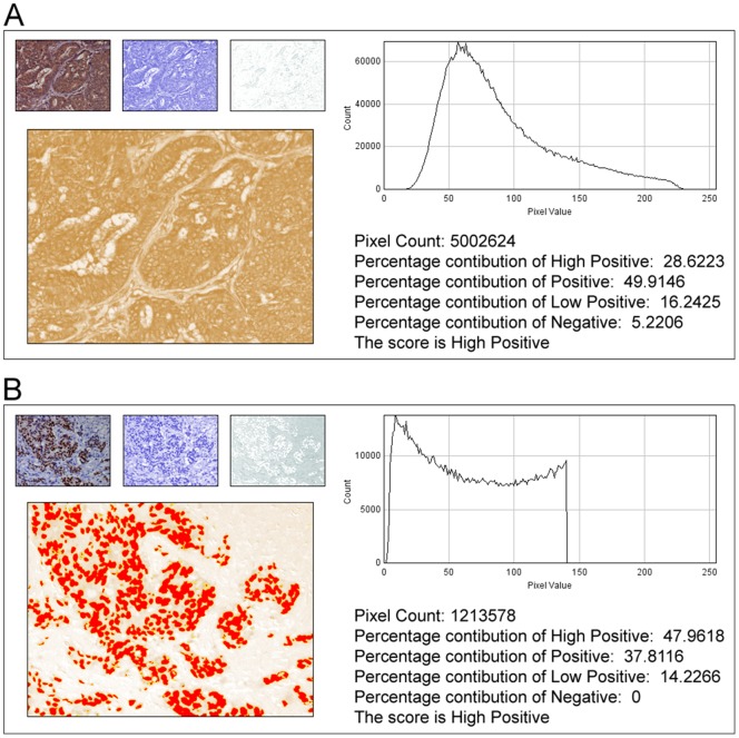
In anatomic pathology, immunohistochemistry (IHC) serves as a diagnostic and prognostic method for identification of disease markers in tissue samples that directly influences classification and grading the disease, influencing patient management. However, till today over most of the world, pathological analysis of tissue samples remained a time-consuming and subjective procedure, wherein the intensity of antibody staining is manually judged and thus scoring decision is directly influenced by visual bias. This instigated us to design a simple method of automated digital IHC image analysis algorithm for an unbiased, quantitative assessment of antibody staining intensity in tissue sections. As a first step, we adopted the spectral deconvolution method of DAB/hematoxylin color spectra by using optimized optical density vectors of the color deconvolution plugin for proper separation of the DAB color spectra. Then the DAB stained image is displayed in a new window wherein it undergoes pixel-by-pixel analysis, and displays the full profile along with its scoring decision. Based on the mathematical formula conceptualized, the algorithm is thoroughly tested by analyzing scores assigned to thousands (n = 1703) of DAB stained IHC images including sample images taken from human protein atlas web resource. The IHC Profiler plugin developed is compatible with the open resource digital image analysis software, ImageJ, which creates a pixel-by-pixel analysis profile of a digital IHC image and further assigns a score in a four tier system. A comparison study between manual pathological analysis and IHC Profiler resolved in a match of 88.6% (P<0.0001, CI = 95%). This new tool developed for clinical histopathological sample analysis can be adopted globally for scoring most protein targets where the marker protein expression is of cytoplasmic and/or nuclear type. We foresee that this method will minimize the problem of inter-observer variations across labs and further help in worldwide patient stratification potentially benefitting various multinational clinical trial initiatives.
Overview
The claims and statistics of this paper haven’t been checked yet.
Free: it reads the open-access text and checks the first 5 claims that cite a source we can check. Create a free account to check every claim.
Integrity signals
Registry
Cites
Statistics
Figures



Verification
Claims the paper makes
The claims and statistics of this paper haven’t been checked yet.
Free: it reads the open-access text and checks the first 5 claims that cite a source we can check. Create a free account to check every claim.
Claims other papers make about this one
Each is checked against this paper’s own text: does it say what the citing paper says it does?
In the citing paper
“For analysis of c-MYC expression in liver sections, random 20× non-tumor fields of the c-MYC IHC scans from liver sections of individual mice were output using NDP.view2 and blindly analyzed using ImageJ (https://imagej.nih.gov/ij/) with the plugin ‘IHC Profiler’62.”
From Tumor-specific cholinergic CD4+ T lymphocytes guide immunosurveillance of hepatocellular carcinoma (2023) · cites this paper as [62]
Self-check
Percentages, sums and reported differences re-computed from the paper’s own numbers.
The self-check needs the paper’s full text.
Citations
Cites
NIH Image to ImageJ: 25 years of image analysis
Schneider, C. A., Rasband, W. S., & Eliceiri, K. W. (2012) · Nature Methods · doi:10.1038/nmeth.2089 · Cited by 64,638 · cited 1×
“The zones were visually identified by using the threshold feature of Image menu of the ImageJ program [22].”
MethodHsu, S. M., Raine, L., & Fanger, H. (1981) · Journal of Histochemistry & Cytochemistry · doi:10.1177/29.4.6166661 · Cited by 14,140
Harvey, J. M., Clark, G. M., Osborne, C. K., & Allred, D. C. (1999) · Journal of Clinical Oncology · doi:10.1200/jco.1999.17.5.1474 · Cited by 2,056
Quantification of histochemical staining by color deconvolution.
Ruifrok, A. C. C., & Johnston, D. A. (2001) · PubMed · Cited by 2,029
Tuominen, V. J., Ruotoistenmäki, S., Viitanen, A., Jumppanen, M., & Isola, J. (2010) · Breast Cancer Research · doi:10.1186/bcr2615 · Cited by 472
McCarty, K. S., Szabo, E. M., Flowers, J. L., Cox, E. B., Leight, G. S., Miller, L. A., Konrath, J. G., Jt, S., Budwit, D. A., & Wt, C. (1986) · PubMed · Cited by 417
A global view of protein expression in human cells, tissues, and organs
Pontén, F., Gry, M., Fagerberg, L., Lundberg, E., Asplund, A., Berglund, L., Oksvold, P., Björling, E., Hober, S., Kampf, C., et al. (2009) · Molecular Systems Biology · doi:10.1038/msb.2009.93 · Cited by 222
Automated Selection of DAB-labeled Tissue for Immunohistochemical Quantification
Brey, E. M., Lalani, Z., Johnston, C., Wong, M., McIntire, L. V., Duke, P. J., & Patrick, C. W. (2003) · Journal of Histochemistry & Cytochemistry · doi:10.1177/002215540305100503 · Cited by 200
Quantitative image analysis of immunohistochemical stains using a CMYK color model
Pham, N.-A., Morrison, A., Schwock, J., Aviel-Ronen, S., Iakovlev, V., Tsao, M.-S., Ho, J., & Hedley, D. W. (2007) · Diagnostic Pathology · doi:10.1186/1746-1596-2-8 · Cited by 179
Quantification of renal pathology by image analysis (Methods in Renal Research)
RANGAN, G. K., & TESCH, G. H. (2007) · Nephrology · doi:10.1111/j.1440-1797.2007.00855.x · Cited by 176
Cited by
Items 801–900 of 1,000
WITHDRAWN: Developmental regulation and lateralization of GABA receptors in the rat hippocampus
Khoshdel‐Sarkarizi, H., Hami, J., Mohammadipour, A., Sadr‐Nabavi, A., Mahmoudi, M., Kheradmand, H., Peyvandi, M., Nourmohammadi, E., & Haghir, H. (2019) · International Journal of Developmental Neuroscience · doi:10.1016/j.ijdevneu.2019.01.002 · Cited by 7
RetractedYousef, E. M., El-Moneam, S. M. A., Yousof, S. M., Mohammed, S. A., Sultan, B. O., & Mansour, B. S. A. (2025) · Tissue and Cell · doi:10.1016/j.tice.2025.102934 · Cited by 7
Li, Y., Tang, X. A., Liu, Y., Chen, X., Wang, L., Su, Y., He, W., Li, J., Huang, Q., & Wu, P. Y. (2025) · Frontiers in Materials · doi:10.3389/fmats.2025.1573222 · Cited by 8
Piezoelectric-Driven Microneedle Array Delivery of Methotrexate for Enhanced Psoriasis Treatment
Li, L., Kai, Y., Wang, Y., & Chen, Z. (2025) · ACS Biomaterials Science & Engineering · doi:10.1021/acsbiomaterials.4c02378 · Cited by 6
Ouologuem, L., Kübler, A., Ouologuem, S., Hadzic, A., Stöckl, J. B., Siciliano, A. C., Forciniti, S., Nigro, S. L., Iuele, H., Onesto, V., et al. (2025) · Frontiers in Immunology · doi:10.3389/fimmu.2025.1668066 · Cited by 10
Wang, R., Zhang, Y., Meng, X., Zhao, J., Xing, Y., Ouyang, Q., Zhang, N., Chen, H., Miao, N., Song, E., et al. (2025) · Advanced Science · doi:10.1002/advs.202506518 · Cited by 10
ULK1 promotes metastatic progression in experimental models of epithelial ovarian cancer
Webb, J. D., Buensuceso, A. V., Tomas, E. J., Borrelli, M. J., Viola, L. H., Hovey, O. F. J., Valdés, Y. R., Singha, B., Li, S. S.-C., & Shepherd, T. G. (2026) · Oncogene · doi:10.1038/s41388-026-03702-2 · Cited by 7
Akinloye, D. I., Sojinu, S. O., Moses, C. A., Agemo, S., Taiwo, E. T., & Adene, T. S. (2022) · Clinical Nutrition Open Science · doi:10.1016/j.nutos.2022.06.004 · Cited by 6
Pan, L., Li, C., Wang, Z., Yang, L., & Zhang, L. (2022) · Biochemical Engineering Journal · doi:10.1016/j.bej.2022.108626 · Cited by 6
Shao, B., Ren, S., Wang, Z.-B., Wang, H.-D., Zhang, J., Qin, H., Zhu, Y., Sun, C., Xu, Y., Li, X., et al. (2023) · Frontiers in Immunology · doi:10.3389/fimmu.2023.1155090 · Cited by 6
Zhao, R., Xue, M., Lin, H., Smith, M. M., Liang, H. H., Weiler-Guettler, H., Griffin, J. H., & Jackson, C. (2023) · Wound Repair and Regeneration · doi:10.1111/wrr.13148 · Cited by 10
Ahmed, K., Sheikh, A., Fatima, S., Ghulam, T., Haider, G., Abbas, F., Sarría‐Santamera, A., Ghias, K. K., Mughal, N., & Abidi, S. H. (2024) · Scientific Reports · doi:10.1038/s41598-024-60538-0 · Cited by 9
Gâta, V. A., Paşca, A., Roman, A., Muntean, M. V., Morariu, D. Ș., Bonci, E.-A., Dina, C., & Ungureanu, L. (2024) · International Journal of Molecular Sciences · doi:10.3390/ijms25126377 · Cited by 6
A multivariate retrospective analysis of high‐grade gliomas: Survival and prognostic factors
Shi, W., Wang, X., Liu, S., Zheng, Z., Dong, L., & Jiang, X. M. (2024) · Cancer Medicine · doi:10.1002/cam4.7456 · Cited by 10
HIV-MTB Co-Infection Reduces CD4+ T Cells and Affects Granuloma Integrity
Huang, S., Liu, M., Zhang, H., Song, W., Guo, W., Feng, Y., Ma, X., Shi, X., Liu, J., Liu, L., et al. (2024) · Viruses · doi:10.3390/v16081335 · Cited by 10
ElKhooly, I. A., El‐Bassossy, H. M., Mohammed, H. O., Atwa, A. M., & Hassan, N. A. (2024) · Life Sciences · doi:10.1016/j.lfs.2024.123046 · Cited by 7
Shoier, N. O., Ghareib, S. A., Kothayer, H., Alsemeh, A. E., & El-Sayed, S. S. (2024) · Naunyn-Schmiedeberg s Archives of Pharmacology · doi:10.1007/s00210-024-03439-3 · Cited by 7
Xu, G., Yu, J., Lyu, J., Zhan, M., Xu, J., Huang, M., Zhao, R., Li, Y., Zhu, J., Feng, J., et al. (2024) · Advanced Science · doi:10.1002/advs.202401041 · Cited by 7
Zhang, S., Wang, Q., Xu, Y., Zhang, H., Mi, J., Lu, X., Fan, R., Lv, J., & Xu, G. (2024) · Behavioural Brain Research · doi:10.1016/j.bbr.2024.115364 · Cited by 5
Sheng, W., Zhang, C., Mohiuddin, T. M., Al-Rawe, M., Schmitz, R., Niebert, M., Konrad, L., Wagner, S., Zeppernick, F. A., Meinhold-Heerlein, I. M., et al. (2025) · Cells · doi:10.3390/cells14100691 · Cited by 7
Ahmad, Z., Alzubaidi, M. S., Al-Thelaya, K., Calì, C., Boughorbel, S., Schneider, J., & Agus, M. (2025) · Journal of Pathology Informatics · doi:10.1016/j.jpi.2025.100454 · Cited by 11
Li, X., Hua, D., Wu, D., Hong, W., Kang, Y., Tang, L., Yang, Q., Wang, X., Li, B., Li, R., et al. (2025) · Cancer Medicine · doi:10.1002/cam4.70987 · Cited by 8
Expression of Hypoxia-Inducible Factors in Different Stages of Pancreatic Tumor Progression
Jung, J. H., Sosnowska, D., Weaver, J., Parson, H. K., CASELLINI, C. M., & Vinik, A. I. (2020) · Reports — Medical Cases Images and Videos · doi:10.3390/reports3040030 · Cited by 6
Kumar, K., Moon, B., Datta, K., Fornace, A. J., & Suman, S. (2022) · Life Sciences in Space Research · doi:10.1016/j.lssr.2022.09.006 · Cited by 10
Pygo1 Regulates the Behavior of Human Non-Small-Cell Lung Cancer via the Wnt/β-Catenin Pathway
Wen, Y., Li, Y., Yang, B., Guo, X., Lin, L., Liang, J., Zhang, K., Xu, L., Jiang, Z., Wang, Y., et al. (2022) · Disease Markers · doi:10.1155/2022/6993994 · Cited by 7
Xie, T., Feng, W., He, M., Zhan, X., Liao, S., He, J., Qin, Z., Li, F., Xu, J., Liu, Y., et al. (2022) · Experimental Biology and Medicine · doi:10.1177/15353702221142607 · Cited by 9
Lee, J.-J., Hsu, Y., Huang, W., & Cheng, S. (2023) · Endocrine · doi:10.1007/s12020-023-03364-0 · Cited by 6
Huang, Q., Shen, Y.-J., Hsueh, C., Zhang, Y., Guo, Y., Yuan, X., Hu, C., Li, J.-Y., Tao, L., Gong, H., et al. (2023) · Heliyon · doi:10.1016/j.heliyon.2023.e17711 · Cited by 7
Prognostic value of mitochondrial CKMT2 in Pan-cancer and its tumor immune correlation analysis
Lin, W., Zhou, J., Ma, Y., Ge, L., Luo, Y., Wang, Y., & Zhou, S. (2024) · Scientific Reports · doi:10.1038/s41598-023-46468-3 · Cited by 9
Establishment and characterization of a highly metastatic hepatocellular carcinoma cell line
Huang, J., Sun, M., Wang, M., Yu, A., Zheng, H., Bu, C., Zhou, J., Zhang, Y., Qiao, Y., & Hu, Z. (2024) · Bioengineered · doi:10.1080/21655979.2023.2296775 · Cited by 6
Soltanpour, M., Boulanger, P., & Buck, B. H. (2024) · Journal of Medical Systems · doi:10.1007/s10916-024-02054-2 · Cited by 6
Anti-tumor efficacy of oncolytic reovirus against gastrointestinal stromal tumor cells
Inagaki, Y., Kubota, E., Mori, Y., Aoyama, M., Kataoka, H., Johnston, R. N., & Joh, T. (2017) · Oncotarget · doi:10.18632/oncotarget.23361 · Cited by 6
Nuclear localization and Overexpression of Smoothened in Pancreatic and Colorectal Cancers
Niyaz, M., Khan, M. S., Wani, R. A., Shah, O. J., Besina, S., & Mudassar, S. (2019) · Journal of Cellular Biochemistry · doi:10.1002/jcb.28477 · Cited by 7
Comparative distribution of somatostatin and somatostatin receptors in PTU-induced hypothyroidism
Singh, S., Somvanshi, R. K., Panda, V., & Kumar, U. (2020) · Endocrine · doi:10.1007/s12020-020-02309-1 · Cited by 6
An Automated Quantitative Method to Analyze Immunohistochemistry and Immunocytochemistry Images
Jin, Y., Shi, K., Gao, X., Langenbach, S. Y., Li, M., Harris, T., & Stewart, A. G. (2020) · Journal of Engineering and Science in Medical Diagnostics and Therapy · doi:10.1115/1.4048296 · Cited by 6
Techniques for digital histological morphometry of the pineal gland
Gheban, B., Colosi, H. A., Gheban-Roşca, I. A., Georgiu, C., Gheban, D., Crişan, D., & Crişan, M. (2022) · Acta Histochemica · doi:10.1016/j.acthis.2022.151897 · Cited by 7
Lee, K. H., Jeong, W.-J., Cha, P.-H., Lee, S. G., Min, D. S., & Choi, K. (2017) · Oncotarget · doi:10.18632/oncotarget.23313 · Cited by 7
Targeting CD47 and Angiogenesis Demonstrates Effective Anti-Tumor Effect in Bladder Cancer
Huang, X., Wang, Q., Nan, Y., Zhang, X., Ke Cheng, X. U., Ju, D., & Ding, W. (2024) · Biomedicines · doi:10.3390/biomedicines12092152 · Cited by 5
Teng, X., Tang, C. C., He, K., Chen, C., Tian, J., & Du, Y. (2025) · EJNMMI Research · doi:10.1186/s13550-025-01218-6 · Cited by 5
Benedetto, G. D., Varvarà, P., Drago, S. E., Cantone, A. F., Mauro, N., Gaudio, G., Burgaletto, C., Bellanca, C. M., Broggi, G., Caltabiano, R., et al. (2025) · International Journal of Pharmaceutics · doi:10.1016/j.ijpharm.2025.125729 · Cited by 7
Small-Molecule Inhibition of SOST Suppresses Breast Cancer Bone Metastasis
Khalil, Y. H., & Nassar, O. S. (2025) · Archive of International Journal of Cancer and Allied Science · doi:10.51847/xr3tpazzri · Cited by 5
Al-Rawi, N. H., Kawas, S. A., Ani, M. A., Alnuaimi, A. S., El‐Sayed, W., & Alrashdan, M. S. (2023) · European Journal of Dentistry · doi:10.1055/s-0042-1760301 · Cited by 5
Reduced YAP1 and FOLR1 in gliomas predict better response to chemotherapeutics
Patrick, S., Lathoria, K., Suri, V. S., & Sen, E. (2023) · Cellular Signalling · doi:10.1016/j.cellsig.2023.110738 · Cited by 7
Plá, R., Sanz, J., Noguerol, F., Vignoletti, F., Gamarra, P., & Sanz, M. (2023) · Bioengineering · doi:10.3390/bioengineering10070841 · Cited by 5
Andrade, D. L., Jalalizadeh, M., Salustiano, A. C. C., & Reis, L. O. (2023) · World Journal of Urology · doi:10.1007/s00345-023-04526-5 · Cited by 5
Wang, R., Yang, L., Zhen, Y., Li, X., Huang, S., Wen, H., & Sun, Q. (2024) · Experimental Dermatology · doi:10.1111/exd.14997 · Cited by 6
Erol, A. Y. G., Koçancı, F. G., Akıncı, E., Dora, D. D., Erendor, F., Şanlıoğlu, S., & Uysal, H. (2024) · European Journal of Neuroscience · doi:10.1111/ejn.16273 · Cited by 7
Unveiling the Role of Endoplasmic Reticulum Stress Pathways in Canine Demodicosis
Kelly, P., McHugo, G. P., Scaife, C., Peters, S., Stevenson, M. L., McKay, J. S., MacHugh, D. E., Lara‐Sáez, I., & Breathnach, R. (2024) · Parasite Immunology · doi:10.1111/pim.13033 · Cited by 5
Yen, H., Chen, P., Huang, R. Y., Jeng, J., & Lai, I. (2024) · The Journal of Pathology Clinical Research · doi:10.1002/2056-4538.12387 · Cited by 6
Act1 drives chemoresistance via regulation of antioxidant RNA metabolism and redox homeostasis
Hong, L., Herjan, T., Chen, X., Zagore, L. L., Bulek, K., Wang, H., Yang, C. J., Licatalosi, D. D., Li, X., & Li, X. (2024) · The Journal of Experimental Medicine · doi:10.1084/jem.20231442 · Cited by 5
Ma, Y., Duan, L., Reisch, B., Kimmig, R., Iannaccone, A., & Gellhaus, A. (2024) · Cells · doi:10.3390/cells13161372 · Cited by 5
Focusing on non-responders to infliximab with ulcerative colitis, what can we do first and next?
Pu, D., Wang, P., Wang, X., Tian, Y., Gong, H., Ma, X., Li, M., & Zhang, D. (2024) · International Immunopharmacology · doi:10.1016/j.intimp.2024.112943 · Cited by 5
A Novel Quinoline Inhibitor of the Canonical NF-κB Transcription Factor Pathway
Ntavaroukas, P., Michail, K., Tsiakalidou, R., Stampouloglou, E., Tsingene, A., Komiotis, D., Georgiou, N., Mavromoustakos, T., Manta, S., Aje, D., et al. (2024) · Biology · doi:10.3390/biology13110910 · Cited by 6
Spatial and planar profiling of Rac1/Cdc42 signaling in Alzheimer's disease brain
Akhtar, S. N., Tran, T. D., Chen, Y., & Lu, Q. (2024) · Journal of Alzheimer s Disease · doi:10.1177/13872877241291076 · Cited by 6
Hirase, Y., Arigami, T., Matsushita, D., Shimonosono, M., Tsuruda, Y., Sasaki, K., Baba, K., Kawasaki, Y., & Ohtsuka, T. (2025) · Cancer Immunology Immunotherapy · doi:10.1007/s00262-025-04084-2 · Cited by 6
Praseatsook, K., Vachiraarunwong, A., Sato, K., Dissook, S., Wanibuchi, H., Taya, S., Wongpoomchai, R., Dejkriengkraikul, P., Gi, M., & Yodkeeree, S. (2025) · International Journal of Molecular Sciences · doi:10.3390/ijms26135955 · Cited by 6
CANDiT: A machine learning framework for differentiation therapy in colorectal cancer
Sinha, S., Alcantara, J. A., Perry, K., Castillo, V., Ondersma, A. K., Banerjee, S., McLaren, E., Espinoza, C. R., Taheri, S., Vidales, E., et al. (2025) · Cell Reports Medicine · doi:10.1016/j.xcrm.2025.102421 · Cited by 9
Smith, A. H., Putta, P., Driscoll, E. C., Chaudhuri, P., Birnbaumer, L., Rosenbaum, M. A., & Graham, L. M. (2020) · JVS Vascular Science · doi:10.1016/j.jvssci.2020.07.002 · Cited by 5
Helicobacter pylori Infection and the Patterns of Gastric Mucin Expression in Children
Domșa, A.-M. T., Lupușoru, R., Gheban, D., Buruiană, A., Gheban, B., Lazăr, C., & Borzan, C. M. (2020) · Journal of Clinical Medicine · doi:10.3390/jcm9124030 · Cited by 5
Al-Rawi, N. H., Ani, M. A., Quadri, A., Hamdoon, Z., Awwad, A. A. A., Kawas, S. A., & Nuaimi, A. H. S. A. (2021) · PubMed · doi:10.14670/hh-18-363 · Cited by 5
Saha, D., Mitra, D., Alam, N., Sen, S., Mustafi, S. M., Mandal, S., Majumder, B., & Murmu, N. (2022) · International Journal of Experimental Pathology · doi:10.1111/iep.12430 · Cited by 5
Yan, K., Bai, B., Ren, Y., Cheng, B., Zhang, X., Zhou, H., Liang, Y., Chen, L., Zi, J., Yang, Q., et al. (2022) · Frontiers in Oncology · doi:10.3389/fonc.2022.848782 · Cited by 5
Wu, Y., Wang, H., & Wei, D. (2022) · Bioengineered · doi:10.1080/21655979.2022.2037214 · Cited by 5
ApoC3 is expressed in oocytes and increased expression is associated with PCOS progression
Zhou, J., Mo, H., Feng, Q., Li, L., & La, J. (2023) · Journal of Ovarian Research · doi:10.1186/s13048-023-01263-6 · Cited by 5
Akhtar, S. N., & Lu, Q. (2023) · Journal of Alzheimer s Disease · doi:10.3233/jad-230408 · Cited by 6
Zeng, J., Gao, W., Tang, Y., Wang, Y., Liu, X., Yin, J., Su, X., Zhang, M., Kang, E., Tian, Y., et al. (2023) · Cell Reports · doi:10.1016/j.celrep.2023.113442 · Cited by 8
Camillo, L., Esposto, E., Gironi, L. C., Airoldi, C., Alhamed, S. A., Boldorini, R. L., Zavattaro, E., & Savoia, P. (2023) · Journal of Clinical Medicine · doi:10.3390/jcm12227137 · Cited by 6
Asad, S. B., Qian, X., Wang, J., Asad, W., Gao, Q., Cao, Y., Huang, Y., Jardan, Y. A. B., Shah, T. A., Wondmie, G. F., et al. (2023) · Frontiers in Medicine · doi:10.3389/fmed.2023.1283302 · Cited by 6
Chen, S., Xu, X., Yang, L., Yao, Y., Yang, Y., & Meng, W. (2024) · BMC Oral Health · doi:10.1186/s12903-023-03780-8 · Cited by 5
Sun, H. Q., Kawano, M., Iwasaki, T., Itonaga, I., Kubota, Y., Tsumura, H., & Tanaka, K. (2024) · Oncology Research Featuring Preclinical and Clinical Cancer Therapeutics · doi:10.32604/or.2023.044085 · Cited by 7
Tomaszewska, E., Hułas‐Stasiak, M., Dobrowolski, P., Świątkiewicz, M., Muszyński, S., Tomczyk‐Warunek, A., Blicharski, T., Donaldson, J., Arciszewski, M. B., Świetlicki, M., et al. (2024) · International Journal of Molecular Sciences · doi:10.3390/ijms25041989 · Cited by 8
Lu, X., Xu, X., Zhou, M., Ge, J., Chen, L., Yu, W., & Wang, H. (2024) · Cytokine · doi:10.1016/j.cyto.2024.156676 · Cited by 5
Li, X., Feng, J., Cheng, H., Jin, N., Jin, S., Liu, Z., Xu, J., & Xie, J. (2024) · Acta Biochimica et Biophysica Sinica · doi:10.3724/abbs.2024207 · Cited by 5
Sun, Y., Wang, M., Jing, L., Wu, Q., Han, G., & Zhou, J. (2024) · Human Cell · doi:10.1007/s13577-024-01164-w · Cited by 8
Wen, F., Ling, H., Ran, R., Li, X., Wang, H., Liu, Q., Li, M., & Yu, T. (2024) · Translational Oncology · doi:10.1016/j.tranon.2024.102256 · Cited by 9
Tewary, S., Arun, I., Ahmed, R., Chatterjee, S., & Chakraborty, C. (2017) · Analytical Methods · doi:10.1039/c7ay02302b · Cited by 5
Ghiciuc, C. M., Strat, A. L., Ochiuz, L., Lupușoru, C. E., Ignat, M., Vasile, A., Grigorovici, A., Stoleriu, I., & Solcan, C. (2017) · Molecules · doi:10.3390/molecules22112014 · Cited by 5
Nguyen, V. T., Bhalla, R., Cowin, G., Stimson, D. H. R., Song, X., Chong, S., Jackson, A. R., Trigg, W., Tieng, Q. M., Mardon, K., et al. (2020) · Nuclear Medicine and Biology · doi:10.1016/j.nucmedbio.2020.07.005 · Cited by 4
Tejeda, M. E., Canto, P., Tenorio‐Torres, A., Orozco-Arguelles, L., Coral‐Vázquez, R. M., Zentella‐Dehesa, A., Leal-García, M., Vega-García, C. C., Bautista‐Piña, V., & Méndez, J. P. (2021) · Journal of Clinical Pathology · doi:10.1136/jclinpath-2020-207249 · Cited by 9
Chakraborti, S., Paul, R. R., Pal, M., Chatterjee, J., & Das, R. K. (2021) · Medical Molecular Morphology · doi:10.1007/s00795-021-00304-7 · Cited by 6
Platelet-activating factor acetyl hydrolase IB2 dysregulated cell proliferation in ovarian cancer
He, Y., He, Z., Zhang, X., & Liu, S. (2021) · Cancer Cell International · doi:10.1186/s12935-021-02406-9 · Cited by 5
Optimizing rat and human blood cells sampling for in silico morphometric analysis
Zamora-Bello, I., Hernández-Baltazar, D., Rodríguez‐Landa, J. F., & Rivadeneyra-Domínguez, E. (2022) · Acta Histochemica · doi:10.1016/j.acthis.2022.151917 · Cited by 10
Bouzid, A., Ani, M. A., de la Fuente, D., Shareef, Z. A., Quadri, A., Hamoudi, R., & Al-Rawi, N. H. (2023) · Frontiers in Oncology · doi:10.3389/fonc.2023.1128753 · Cited by 5
Iliev, A. A., Gaydarski, L., Kotov, G. N., Landzhov, B. V., Kirkov, V., & Stanchev, S. (2024) · The Anatomical Record · doi:10.1002/ar.25453 · Cited by 5
Perspektif Pengalaman Konsumen Terhadap Kesuksesan Event Lari Borobudur Marathon 2019
Larassary, A. (2020) · JUARA Jurnal Olahraga · doi:10.33222/juara.v5i2.889 · Cited by 5
Identification of differentially methylated genes in first-trimester placentas with trisomy 16
Tolmacheva, E. N., Vasilyev, S. А., Tv, N., Lytkina, E. S., Sazhenova, E. A., Жигалина, Д. И., Vasilyeva, O. Y., Markov, A., Demeneva, V. V., Таширева, Л. А., et al. (2022) · Scientific Reports · doi:10.1038/s41598-021-04107-9 · Cited by 5
Wang, Y., Shi, H., Zhang, Y., Zeng, Q., Chen, T., & Chai, C. (2022) · Frontiers in Oncology · doi:10.3389/fonc.2022.848206 · Cited by 5
Kim, J. S., Kim, M. W., Jeong, H. Y., Kang, S. J., Park, S. I., Lee, Y. K., Kim, H. S., Kim, K. S., & Park, Y. S. (2016) · The Journal of Gene Medicine · doi:10.1002/jgm.2884 · Cited by 5
Plumbagin targets the GLUT1/MMP-2 axis to inhibit oral squamous cell carcinoma progression
He, F., Wang, W., Elayah, S. A., Xie, L., Yu, M., Gong, Y., Cui, H., Xiang, L., Tu, J., Han, Y., et al. (2025) · Cancer Cell International · doi:10.1186/s12935-025-03915-7 · Cited by 4
Abdel-Rahman, R. F., Alsugoor, M. H., Alsuhaymi, N., Fayed, H. M., Mahmoud, S. S., Ibrahim, F. A., & Elbaset, M. A. (2025) · Saudi Pharmaceutical Journal · doi:10.1007/s44446-025-00039-x · Cited by 5
Liu, R., Geng, Y., Luo, H., Zhang, Q., Yang, Z., Li, S., Sun, S., Liu, Z., Dong, M., Du, T., et al. (2024) · Journal of Cancer · doi:10.7150/jca.91559 · Cited by 5
El-Saidy, S. A., El-Feki, A. S., El‐Khodary, G. M., Hassan, A. A., Elgendy, D. I., & Gawaan, Y. M. (2024) · Journal of Parasitic Diseases · doi:10.1007/s12639-024-01737-4 · Cited by 5
Penugurti, V., Dey, O., Kalali, S., Kashyap, D., Khumukcham, S. S., Kizhuvedath, A., Satpathy, S., Kiran, M., Row, A. T., Surapaneni, T., et al. (2025) · Cellular Signalling · doi:10.1016/j.cellsig.2025.111633 · Cited by 4
Zhang, Z., Liu, S., Liang, S., Bai, X., He, Y., Zhong, J., Liang, W., Hou, P., Zhong, H., Wu, T., et al. (2025) · European Journal of Nuclear Medicine and Molecular Imaging · doi:10.1007/s00259-025-07171-9 · Cited by 4
Li, J., Zeng, S., Sun, Y., Zou, J., Zhang, E., Yan, Q., Chen, L., & Li, J. (2025) · Phytomedicine · doi:10.1016/j.phymed.2025.156825 · Cited by 5
Özbek, M., Beyaz, F., Öztop, M., Karaca, H., Cabir, A., & Kiryar, B. F. (2025) · The Anatomical Record · doi:10.1002/ar.25682 · Cited by 5
Xie, X., Zhao, Y., Liu, J., Li, H., Zhu, X., Liu, Y., Sang, Y., Xu, K., Du, F., Ren, H., et al. (2025) · Cell Death and Disease · doi:10.1038/s41419-025-07889-2 · Cited by 6
Gao, C., Chen, F., Gong, N., Hu, S., Zhu, M., Zhou, Q., & Gao, X. (2025) · Journal of Functional Foods · doi:10.1016/j.jff.2025.106985 · Cited by 4
Sun, J., Shi, J., Zhang, J., Sun, X., Du, S., Wang, X., Liu, G., Li, X., Feng, X., Wang, J., et al. (2025) · Journal of Ethnopharmacology · doi:10.1016/j.jep.2025.120696 · Cited by 5
Bai, Y., Gao, Y., He, X., Gao, F., Sun, J., Bao, P., Zhao, J., & Jiang, T. (2025) · Genomics · doi:10.1016/j.ygeno.2025.111147 · Cited by 4
Metadata
- Authors
- Varghese, F., Bukhari, A. B., Malhotra, R., & De, A.
- Affiliations
- Tata Memorial Hospital (IN)
- Corresponding author
- De, Abhijit
- Venue
- PLoS ONE
- Publisher
- Public Library of Science (PLoS)
- Published
- 2014-05-06
- Volume
- 9
- Issue
- 5
- Pages
- e96801
- ISSN
- 1932-6203 (ISSN-L)
- PMC
- PMC4011881
- OpenAlex
- W1998734250
- Cited by
- 1,318
- References
- 30
- Citations by year
- 2026: 90 · 2025: 129 · 2024: 172 · 2023: 172 · 2022: 171 · 2021: 144 · 2020: 141 · 2019: 101 · 2018: 69 · 2017: 60 · 2016: 44 · 2015: 22 · 2014: 3
- FWCI
- 42.82
- Type
- journal article
- Language
- en
- Open access
- gold
- doi.org
- Locations
- PLoS ONE (publishedVersion, cc-by)
- Europe PMC (PubMed Central) (publishedVersion, cc-by)
- PubMed Central (publishedVersion, cc-by)
- PubMed (publishedVersion)
- RePEc: Research Papers in Economics (submittedVersion)
- DOAJ (DOAJ: Directory of Open Access Journals) (submittedVersion, cc-by-sa)
- Figshare (submittedVersion, cc-by)
- Funders
- Indian Council of Medical Research
- Advanced Centre for Treatment, Research and Education in Cancer
- Topics
- Physical Sciences › Computer Science › Artificial Intelligence › AI in cancer detection
- Life Sciences › Biochemistry, Genetics and Molecular Biology › Biophysics › Cell Image Analysis Techniques
- Physical Sciences › Computer Science › Computer Vision and Pattern Recognition › Digital Imaging for Blood Diseases
- Keywords
- immunohistochemistry image analysis, automated grading, ImageJ plugin, interobserver variability, digital pathology, human tissue samples, protein expression profiling
- MeSH terms
- Automation, Cell Nucleus, Cytoplasm, Humans, Image Processing, Computer-Assisted, Immunohistochemistry, Neoplasms, Software, Biomarkers, Tumor
About these checks
Registry, retraction, Cites and statistics results are computed, not judged, with one exception: a printed reference that no index lists is read once by a model before it counts as a citation error. Claims are extracted by a model and kept only when their quote appears verbatim in the text; each cited source is then checked, with a model’s help, for whether it says what the claim relies on. Those verdicts can be wrong — read the claim’s quote and the cited source before relying on one.
Statistics and arithmetic are recomputed, not judged.
Confidence bands are shown only for fields calibrated on our annotated test set. None is calibrated yet, so no band is shown.
What counts as a claim ›How we measure confidence ›How we check this ›
Are you an author, or do you see a problem with this page? Request a review
Data sources
Bibliographic data: Semantic Scholar, OpenAlex, Crossref.
Contains information from the Semantic Scholar Academic Graph Datasets, made available under the ODC Attribution License (ODC-BY 1.0).
Semantic Scholar’s data is described in: Kinney, R., et al. (2023). The Semantic Scholar Open Data Platform. arXiv:2301.10140.
