

IHC Profiler: An Open Source Plugin for the Quantitative Evaluation and Automated Scoring of Immunohistochemistry Images of Human Tissue Samples
Open accessThe DOI matches its Crossref record; 30 of the 30 references it cites were found in a registry or an index.
Abstract
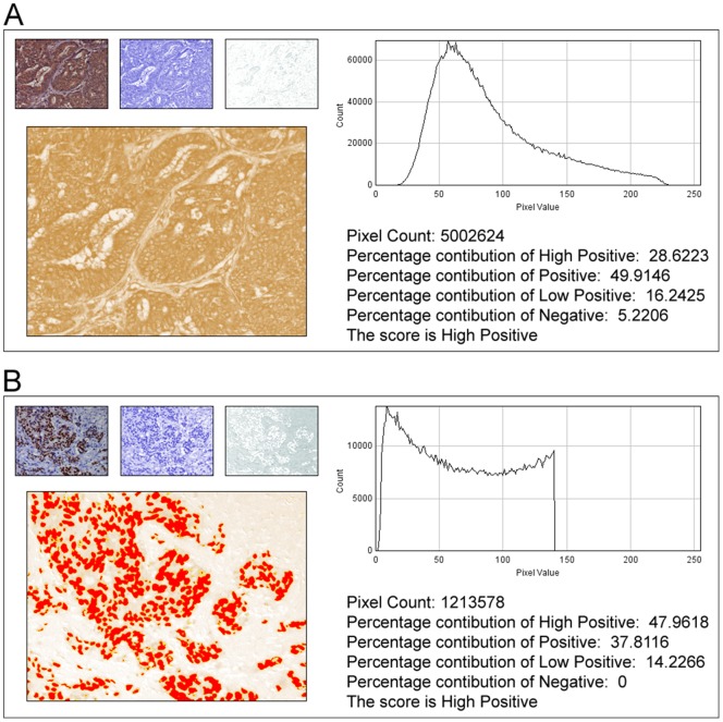
In anatomic pathology, immunohistochemistry (IHC) serves as a diagnostic and prognostic method for identification of disease markers in tissue samples that directly influences classification and grading the disease, influencing patient management. However, till today over most of the world, pathological analysis of tissue samples remained a time-consuming and subjective procedure, wherein the intensity of antibody staining is manually judged and thus scoring decision is directly influenced by visual bias. This instigated us to design a simple method of automated digital IHC image analysis algorithm for an unbiased, quantitative assessment of antibody staining intensity in tissue sections. As a first step, we adopted the spectral deconvolution method of DAB/hematoxylin color spectra by using optimized optical density vectors of the color deconvolution plugin for proper separation of the DAB color spectra. Then the DAB stained image is displayed in a new window wherein it undergoes pixel-by-pixel analysis, and displays the full profile along with its scoring decision. Based on the mathematical formula conceptualized, the algorithm is thoroughly tested by analyzing scores assigned to thousands (n = 1703) of DAB stained IHC images including sample images taken from human protein atlas web resource. The IHC Profiler plugin developed is compatible with the open resource digital image analysis software, ImageJ, which creates a pixel-by-pixel analysis profile of a digital IHC image and further assigns a score in a four tier system. A comparison study between manual pathological analysis and IHC Profiler resolved in a match of 88.6% (P<0.0001, CI = 95%). This new tool developed for clinical histopathological sample analysis can be adopted globally for scoring most protein targets where the marker protein expression is of cytoplasmic and/or nuclear type. We foresee that this method will minimize the problem of inter-observer variations across labs and further help in worldwide patient stratification potentially benefitting various multinational clinical trial initiatives.
Overview
The claims and statistics of this paper haven’t been checked yet.
Free: it reads the open-access text and checks the first 5 claims that cite a source we can check. Create a free account to check every claim.
Integrity signals
Registry
Cites
Statistics
Figures



Verification
Claims the paper makes
The claims and statistics of this paper haven’t been checked yet.
Free: it reads the open-access text and checks the first 5 claims that cite a source we can check. Create a free account to check every claim.
Claims other papers make about this one
Each is checked against this paper’s own text: does it say what the citing paper says it does?
In the citing paper
“For analysis of c-MYC expression in liver sections, random 20× non-tumor fields of the c-MYC IHC scans from liver sections of individual mice were output using NDP.view2 and blindly analyzed using ImageJ (https://imagej.nih.gov/ij/) with the plugin ‘IHC Profiler’62.”
From Tumor-specific cholinergic CD4+ T lymphocytes guide immunosurveillance of hepatocellular carcinoma (2023) · cites this paper as [62]
Self-check
Percentages, sums and reported differences re-computed from the paper’s own numbers.
The self-check needs the paper’s full text.
Citations
Cites
NIH Image to ImageJ: 25 years of image analysis
Schneider, C. A., Rasband, W. S., & Eliceiri, K. W. (2012) · Nature Methods · doi:10.1038/nmeth.2089 · Cited by 64,638 · cited 1×
“The zones were visually identified by using the threshold feature of Image menu of the ImageJ program [22].”
MethodHsu, S. M., Raine, L., & Fanger, H. (1981) · Journal of Histochemistry & Cytochemistry · doi:10.1177/29.4.6166661 · Cited by 14,140
Harvey, J. M., Clark, G. M., Osborne, C. K., & Allred, D. C. (1999) · Journal of Clinical Oncology · doi:10.1200/jco.1999.17.5.1474 · Cited by 2,056
Quantification of histochemical staining by color deconvolution.
Ruifrok, A. C. C., & Johnston, D. A. (2001) · PubMed · Cited by 2,029
Tuominen, V. J., Ruotoistenmäki, S., Viitanen, A., Jumppanen, M., & Isola, J. (2010) · Breast Cancer Research · doi:10.1186/bcr2615 · Cited by 472
McCarty, K. S., Szabo, E. M., Flowers, J. L., Cox, E. B., Leight, G. S., Miller, L. A., Konrath, J. G., Jt, S., Budwit, D. A., & Wt, C. (1986) · PubMed · Cited by 417
A global view of protein expression in human cells, tissues, and organs
Pontén, F., Gry, M., Fagerberg, L., Lundberg, E., Asplund, A., Berglund, L., Oksvold, P., Björling, E., Hober, S., Kampf, C., et al. (2009) · Molecular Systems Biology · doi:10.1038/msb.2009.93 · Cited by 222
Automated Selection of DAB-labeled Tissue for Immunohistochemical Quantification
Brey, E. M., Lalani, Z., Johnston, C., Wong, M., McIntire, L. V., Duke, P. J., & Patrick, C. W. (2003) · Journal of Histochemistry & Cytochemistry · doi:10.1177/002215540305100503 · Cited by 200
Quantitative image analysis of immunohistochemical stains using a CMYK color model
Pham, N.-A., Morrison, A., Schwock, J., Aviel-Ronen, S., Iakovlev, V., Tsao, M.-S., Ho, J., & Hedley, D. W. (2007) · Diagnostic Pathology · doi:10.1186/1746-1596-2-8 · Cited by 179
Quantification of renal pathology by image analysis (Methods in Renal Research)
RANGAN, G. K., & TESCH, G. H. (2007) · Nephrology · doi:10.1111/j.1440-1797.2007.00855.x · Cited by 176
Cited by
Fedchenko, N., & Reifenrath, J. (2014) · Diagnostic Pathology · doi:10.1186/s13000-014-0221-9 · Cited by 808
Lifetime-engineered NIR-II nanoparticles unlock multiplexed in vivo imaging
Fan, Y., Wang, P., Lu, Y., Wang, R., Zhou, L., Zheng, X., Li, X., Piper, J. A., & Zhang, F. (2018) · Nature Nanotechnology · doi:10.1038/s41565-018-0221-0 · Cited by 795
Liu, J., Eckert, M. A., Harada, B. T., Liu, S.-M., Lu, Z., Yu, K., Tienda, S. M., Chryplewicz, A., Zhu, A. C., Yang, Y., et al. (2018) · Nature Cell Biology · doi:10.1038/s41556-018-0174-4 · Cited by 778
Crowe, A., & Yue, W. (2019) · BIO-PROTOCOL · doi:10.21769/bioprotoc.3465 · Cited by 771
Hong, D., Kurzrock, R., Kim, Y., Woessner, R., Younes, A., Nemunaitis, J., Fowler, N., Zhou, T., Schmidt, J., Jo, M., et al. (2015) · Science Translational Medicine · doi:10.1126/scitranslmed.aac5272 · Cited by 445
Rho, H., Terry, A. R., Chronis, C., & Hay, N. (2023) · Cell Metabolism · doi:10.1016/j.cmet.2023.06.013 · Cited by 404
Aeffner, F., Zarella, M. D., Buchbinder, N., Bui, M. M., Goodman, M. R., Hartman, D. J., Lujan, G. M., Molani, M. A., Parwani, A. V., Lillard, K., et al. (2019) · Journal of Pathology Informatics · doi:10.4103/jpi.jpi_82_18 · Cited by 403
Principles and approaches for reproducible scoring of tissue stains in research
Meyerholz, D. K., & Beck, A. P. (2018) · Laboratory Investigation · doi:10.1038/s41374-018-0057-0 · Cited by 341
Suarez, E. R., Chang, D.-K., Sun, J., Sui, J., Freeman, G. J., Signoretti, S., Zhu, Q., & Marasco, W. A. (2016) · Oncotarget · doi:10.18632/oncotarget.9114 · Cited by 340
Shah, K. N., Bhatt, R., Rotow, J., Rohrberg, J., Olivas, V., Wang, V. E., Hemmati, G., Martins, M. M., Maynard, A., Kuhn, J., et al. (2019) · Nature Medicine · doi:10.1038/s41591-018-0264-7 · Cited by 286
Logue, S. E., McGrath, E. P., Cleary, P., Greene, S., Mnich, K., Almanza, A., Chevet, E., Dwyer, R. M., Oommen, A., Legembre, P., et al. (2018) · Nature Communications · doi:10.1038/s41467-018-05763-8 · Cited by 283
Complement Signals Determine Opposite Effects of B Cells in Chemotherapy-Induced Immunity
Lu, Y., Zhao, Q., Liao, J.-Y., Song, E., Xia, Q., Pan, J., Li, Y., Li, J., Zhou, B., Ye, Y., et al. (2020) · Cell · doi:10.1016/j.cell.2020.02.015 · Cited by 280
Improved prime editors enable pathogenic allele correction and cancer modelling in adult mice
Liu, P., Liang, S.-Q., Zheng, C., Mintzer, E., Zhao, Y. G., Ponnienselvan, K., Mir, A., Sontheimer, E. J., Gao, G., Flotte, T. R., et al. (2021) · Nature Communications · doi:10.1038/s41467-021-22295-w · Cited by 273
Mapping spatial heterogeneity in the tumor microenvironment: a new era for digital pathology
Heindl, A., Nawaz, S., & Yuan, Y. (2015) · Laboratory Investigation · doi:10.1038/labinvest.2014.155 · Cited by 216
Urea cycle dysregulation in non-alcoholic fatty liver disease
De Chiara, F., Heebøll, S., Marrone, G., Montoliu, C., Hamilton-Dutoit, S., Ferrandez, A., Andreola, F., Rombouts, K., Grønbæk, H., Felipo, V., et al. (2018) · Journal of Hepatology · doi:10.1016/j.jhep.2018.06.023 · Cited by 208
YAP-independent mechanotransduction drives breast cancer progression
Lee, J. Y., Chang, J. K., Dominguez, A. A., Lee, H.-P., Nam, S., Chang, J., Varma, S., Qi, L. S., West, R. B., & Chaudhuri, O. (2019) · Nature Communications · doi:10.1038/s41467-019-09755-0 · Cited by 199
Hu, Q., Wei, W., Wu, D., Huang, F., Li, M., Li, W., Yin, J., Peng, Y., Lu, Y., Zhao, Q., et al. (2022) · Frontiers in Cell and Developmental Biology · doi:10.3389/fcell.2022.810327 · Cited by 187
Nisbet, R. M., Van der Jeugd, A., Leinenga, G., Evans, H. T., Janowicz, P. W., & Götz, J. (2017) · Brain · doi:10.1093/brain/awx052 · Cited by 187
Zhang, Z., Zhou, C., Li, X., Barnes, S. D., Deng, S., Hoover, E., Chen, C.-C., Lee, Y. S., Zhang, Y., Wang, C., et al. (2020) · Cancer Cell · doi:10.1016/j.ccell.2020.03.001 · Cited by 166
Huang, Z., Zhang, M., Plec, A. A., Estill, S. J., Cai, L., Repa, J. J., McKnight, S. L., & Tu, B. P. (2018) · Proceedings of the National Academy of Sciences · doi:10.1073/pnas.1806635115 · Cited by 158
Siracusa, R., Scuto, M. C., Fusco, R., Salinaro, A. T., Ontario, M. L., Crea, R., Paola, R. D., Cuzzocrea, S., & Calabrese, V. (2020) · Antioxidants · doi:10.3390/antiox9090824 · Cited by 159
Wang, G., Chow, R. D.-W., Zhu, L., Bai, Z., Ye, L., Zhang, F., Renauer, P. A., Dong, M. B., Dai, X., Zhang, X., et al. (2020) · Cancer Discovery · doi:10.1158/2159-8290.cd-19-1448 · Cited by 148
Fiber Reinforced Cartilage ECM Functionalized Bioinks for Functional Cartilage Tissue Engineering
Rathan, S., Dejob, L., Schipani, R., Haffner, B., Möbius, M. E., & Kelly, D. J. (2019) · Advanced Healthcare Materials · doi:10.1002/adhm.201801501 · Cited by 140
Effects of Autologous Platelet-Rich Plasma on Regeneration of Damaged Endometrium in Female Rats
Jang, H.-Y., Myoung, S., Choe, J. M., Kim, T., Cheon, Y., Kim, Y. M., & Park, H. (2017) · Yonsei Medical Journal · doi:10.3349/ymj.2017.58.6.1195 · Cited by 126
Yan, R., Cui, W., Ma, W., Li, J., Liu, Z., & Lin, Y. (2023) · ACS Nano · doi:10.1021/acsnano.3c02102 · Cited by 136
El‐Agamy, D. S., El-Harbi, K. M., Khoshhal, S., Ahmed, N., Elkablawy, M. A., Shaaban, A. A., & Abo‐Haded, H. M. (2018) · Cancer Management and Research · doi:10.2147/cmar.s186696 · Cited by 136
Rahman, M. S., & Rahman, M. S. (2020) · Environmental Research · doi:10.1016/j.envres.2020.110428 · Cited by 129
Jafari, S. M. S., & Hunger, R. E. (2016) · Applied immunohistochemistry & molecular morphology · doi:10.1097/pai.0000000000000370 · Cited by 121
Investigation on the effects of the atmospheric pressure plasma on wound healing in diabetic rats
Fathollah, S., Mirpour, S., Mansouri, P., Dehpour, A. R., GHORANNEVISS, M., Rahimi, N., Naraghi, Z. S., Chalangari, R., & Martits‐Chalangari, K. (2016) · Scientific Reports · doi:10.1038/srep19144 · Cited by 121
Lipophagy Impairment Is Associated With Disease Progression in NAFLD
Carotti, S., Aquilano, K., Zalfa, F., Ruggiero, S., Valentini, F., Zingariello, M., Francesconi, M., Perrone, G., Alletto, F., Antonelli-Incalzi, R., et al. (2020) · Frontiers in Physiology · doi:10.3389/fphys.2020.00850 · Cited by 120
Cucco, F., Barrans, S., Sha, C., Clipson, A., Crouch, S., Dobson, R. C., Chen, Z., Thompson, J. S., Care, M. A., Cummin, T., et al. (2019) · Leukemia · doi:10.1038/s41375-019-0691-6 · Cited by 126
Čížková, K., Foltynkova, T., Gachechiladze, M. A., & Tauber, Z. (2021) · ACTA HISTOCHEMICA ET CYTOCHEMICA · doi:10.1267/ahc.20-00032 · Cited by 130
Siracusa, R., Fusco, R., Peritore, A. F., Cordaro, M., D’Amico, R., Genovese, T., Gugliandolo, E., Crupi, R., Smeriglio, A., Mandalari, G., et al. (2020) · Nutrients · doi:10.3390/nu12030834 · Cited by 126
Cao, C., Vasilatos, S. N., Bhargava, R., Fine, J. L., Oesterreich, S., Davidson, N. E., & Huang, Y. (2016) · Oncogene · doi:10.1038/onc.2016.186 · Cited by 114
A positive feedback loop between mTORC1 and cathelicidin promotes skin inflammation in rosacea
Deng, Z., Chen, M., Liu, Y., Xu, S., Ouyang, Y., Shi, W., Jian, D., Wang, B., Liu, F., Li, J., et al. (2021) · EMBO Molecular Medicine · doi:10.15252/emmm.202013560 · Cited by 124
Zheng, X., Liu, R., Zhou, C., Yu, H., Luo, W., Zhu, J., Liu, J., Zhang, Z., Xie, N., Peng, X., et al. (2021) · Cancer Research · doi:10.1158/0008-5472.can-21-2273 · Cited by 116
TSC2 regulates lysosome biogenesis via a non-canonical RAGC and TFEB-dependent mechanism
Alesi, N., Akl, E. W., Khabibullin, D., Liu, H.-J., Nidhiry, A. S., Garner, E. R., Filippakis, H., Lam, H. C., Shi, W., Viswanathan, S. R., et al. (2021) · Nature Communications · doi:10.1038/s41467-021-24499-6 · Cited by 103
Ammonia Scavenging Prevents Progression of Fibrosis in Experimental Nonalcoholic Fatty Liver Disease
De Chiara, F., Thomsen, K. L., Habtesion, A., Jones, H., Davies, N., Gracia‐Sancho, J., Manicardi, N., Hall, A., Andreola, F., Paish, H. L., et al. (2020) · Hepatology · doi:10.1002/hep.30890 · Cited by 103
Charan, M., Das, S., Mishra, S., Chatterjee, N., Varikuti, S., Kaul, K., Misri, S., Ahirwar, D. K., Satoskar, A. R., & Ganju, R. K. (2020) · Cell Death and Disease · doi:10.1038/s41419-020-02992-y · Cited by 105
Xie, L., Cai, S., Lu, H.-Y., Tang, F., Zhu, R., Tian, Y., & Li, M. (2024) · Gut Microbes · doi:10.1080/19490976.2024.2347722 · Cited by 122
Ahmed, O. M., Fahim, H. I., Ahmed, H. Y., Al-Muzafar, H. M., Ahmed, R. R., Amin, K. A., El‐Nahass, E., & Abdelazeem, W. H. (2019) · Oxidative Medicine and Cellular Longevity · doi:10.1155/2019/2745352 · Cited by 100
Subcellular mRNA Localization Regulates Ribosome Biogenesis in Migrating Cells
(2020) · doi:10.1016/j.devcel.2020.10.006 · Cited by 96
Morgan, E. L., Scarth, J. A., Patterson, M. R., Wasson, C. W., Hemingway, G. C., Barba‐Moreno, D., & Macdonald, A. (2020) · Cell Death and Differentiation · doi:10.1038/s41418-020-00693-9 · Cited by 102
miRNA-32 Drives Brown Fat Thermogenesis and Trans-activates Subcutaneous White Fat Browning in Mice
Ng, R., Hussain, N. A., Zhang, Q., Chang, C., Li, H., Fu, Y., Cao, L., Han, W., Stunkel, W., & Xu, F. (2017) · Cell Reports · doi:10.1016/j.celrep.2017.04.035 · Cited by 93
ARID2 is a pomalidomide-dependent CRL4CRBN substrate in multiple myeloma cells
Yamamoto, J., Suwa, T., Murase, Y., Tateno, S., Mizutome, H., Asatsuma‐Okumura, T., Shimizu, N., Kishi, T., Momose, S., Kizaki, M., et al. (2020) · Nature Chemical Biology · doi:10.1038/s41589-020-0645-3 · Cited by 98
Liu, H., Zeng, X., Ren, X., Zhang, Y., Huang, M., Tan, L., Dai, Z., Lai, J., Xie, W., Chen, Z., et al. (2022) · Gut · doi:10.1136/gutjnl-2022-327230 · Cited by 106
Design of a mucin-selective protease for targeted degradation of cancer-associated mucins
Pedram, K., Shon, D. J., Tender, G. S., Mantuano, N. R., Northey, J. J., Metcalf, K. J., Wisnovsky, S., Riley, N. M., Forcina, G. C., Malaker, S. A., et al. (2023) · Nature Biotechnology · doi:10.1038/s41587-023-01840-6 · Cited by 95
He, J., Chai, X., Zhang, Q., Wang, Y., Wang, Y., Yang, X., Wu, J., Feng, B., Sun, J. P., Rui, W., et al. (2025) · Nature Immunology · doi:10.1038/s41590-024-02068-5 · Cited by 107
Ahmed, O. M., Elkomy, M. H., Fahim, H. I., Ashour, M. B., Naguib, I. A., Alghamdi, B. S. A., Mahmoud, H. U. R., & Ahmed, N. A. (2022) · Oxidative Medicine and Cellular Longevity · doi:10.1155/2022/2710607 · Cited by 89
Zhang, X., Su, T., Wu, Y., Cai, Y., Wang, L., Liang, C., Zhou, L., Wang, S., Li, X., Peng, S., et al. (2024) · Cancer Research · doi:10.1158/0008-5472.can-23-1916 · Cited by 107
Privat‐Maldonado, A., Gorbanev, Y. Y., Dewilde, S., Smits, E., & Bogaerts, A. (2018) · Cancers · doi:10.3390/cancers10110394 · Cited by 89
Therapeutic Mechanisms of Human Adipose-Derived Mesenchymal Stem Cells in a Rat Tendon Injury Model
Lee, S. Y., Kwon, B., Lee, K., Son, Y. H., & Chung, S. G. (2017) · The American Journal of Sports Medicine · doi:10.1177/0363546517689874 · Cited by 84
Kabel, A. M. (2017) · Biomedicine & Pharmacotherapy · doi:10.1016/j.biopha.2017.10.144 · Cited by 84
Wu, Q., Tian, A.-L., Li, B., Leduc, M., Forveille, S., Hamley, P., Galloway, W., Xie, W., Liu, P., Zhao, L., et al. (2021) · Journal for ImmunoTherapy of Cancer · doi:10.1136/jitc-2021-002722 · Cited by 83
Impellizzeri, D., Siracusa, R., Cordaro, M., Crupi, R., Peritore, A. F., Gugliandolo, E., D'Amico, R., Petrosino, S., Evangelista, M., Di Paola, R., et al. (2019) · Neurobiology of Disease · doi:10.1016/j.nbd.2019.01.007 · Cited by 82
Li, Y., Xu, C., Wang, B., Xu, F., Ma, F., Qu, Y., Jiang, D., Li, K., Feng, J., Tian, S., et al. (2022) · Nature Communications · doi:10.1038/s41467-022-33282-0 · Cited by 80
Yang, Y., Zheng, W., Tan, W., Wu, X., Dai, Z., Li, Z., Yan, Z., Ji, Y., Wang, Y., Su, W., et al. (2022) · Acta Biomaterialia · doi:10.1016/j.actbio.2022.11.065 · Cited by 85
Rizza, S., Montagna, C., Cardaci, S., Maiani, E., Giacomo, G. D., Sánchez‐Quiles, V., Blagoev, B., Rasola, A., Zio, D. D., Stamler, J. S., et al. (2016) · Cancer Research · doi:10.1158/0008-5472.can-15-2637 · Cited by 81
Hussain, M. A., Abogresha, N. M., Kader, G. A., Hassan, R., Abdelaziz, E. Z., & Greish, S. M. (2021) · Oxidative Medicine and Cellular Longevity · doi:10.1155/2021/8841726 · Cited by 84
Guimarães, G. R., Maklouf, G. R., Teixeira, C. E., Santos, L. D. O., Tessarollo, N. G., de Toledo, N. E., Serain, A. F., de Lanna, C. A., Pretti, M. A. M., da Cruz, J. G. V., et al. (2024) · Nature Communications · doi:10.1038/s41467-024-49916-4 · Cited by 97
Circadian regulator CLOCK promotes tumor angiogenesis in glioblastoma
Pang, L., Dunterman, M., Xuan, W., Gonzalez, A. M., Lin, Y., Hsu, W.-H., Khan, F., Hagan, R. S., Müller, W. A., Heimberger, A. B., et al. (2023) · Cell Reports · doi:10.1016/j.celrep.2023.112127 · Cited by 86
D’Amico, R., Fusco, R., Cordaro, M., Siracusa, R., Peritore, A. F., Gugliandolo, E., Crupi, R., Scuto, M., Cuzzocrea, S., Di Paola, R., et al. (2020) · International Journal of Molecular Sciences · doi:10.3390/ijms21062144 · Cited by 76
Tumor cells impair immunological synapse formation via central nervous system-enriched metabolite
Li, Y., Huang, M., Wang, M., Wang, Y., Deng, P., Li, C., Huang, J., Chen, H., Wei, Z., Ouyang, Q., et al. (2024) · Cancer Cell · doi:10.1016/j.ccell.2024.05.006 · Cited by 84
Tumor-specific cholinergic CD4+ T lymphocytes guide immunosurveillance of hepatocellular carcinoma
Zheng, C., Snow, B. E., Elia, A. J., Nechanitzky, R., Dominguez-Brauer, C., Liu, S., Tong, Y., Cox, M. A., Focaccia, E., Wakeham, A. C., et al. (2023) · Nature Cancer · doi:10.1038/s43018-023-00624-w · Cited by 75
“For analysis of c-MYC expression in liver sections, random 20× non-tumor fields of the c-MYC IHC scans from liver sections of individual mice were output using NDP.view2 and blindly analyzed using ImageJ (https://imagej.nih.gov/ij/) with the plugin ‘IHC Profiler’ 62 .”
MethodVitamin D3 Reduces Tissue Damage and Oxidative Stress Caused by Exhaustive Exercise
Ke, C.-Y., Yang, F.-L., Wu, W.-T., Chung, C.-H., Lee, R.-P., Yang, W.-T., Subeq, Y.-M., & Liao, K.-W. (2016) · International Journal of Medical Sciences · doi:10.7150/ijms.13746 · Cited by 73
Pang, L., Dunterman, M., Guo, S., Khan, F., Liu, Y., Taefi, E., Bahrami, A., Geula, C., Hsu, W.-H., Horbinski, C., et al. (2023) · Nature Immunology · doi:10.1038/s41590-023-01605-y · Cited by 80
Upregulation of Myt1 Promotes Acquired Resistance of Cancer Cells to Wee1 Inhibition
Lewis, C. W., Bukhari, A. B., Xiao, E. J., Choi, W.-S., Smith, J. D., Homola, E. M., Mackey, J. R., Campbell, S. D., Gamper, A. M., & Chan, G. K. T. (2019) · Cancer Research · doi:10.1158/0008-5472.can-19-1961 · Cited by 74
Self-citationSawant, S., Gokulan, R. C., Dongre, H., Vaidya, M. M., Chaukar, D. A., Prabhash, K., Ingle, A., Joshi, S., Dange, P. P., Joshi, S., et al. (2015) · Clinical Oral Investigations · doi:10.1007/s00784-015-1476-6 · Cited by 72
Shin, G., Kang, H. S., Lee, A. R., & Kim, K. (2016) · Autophagy · doi:10.1080/15548627.2016.1239002 · Cited by 73
Browe, D. C., Díaz‐Payno, P. J., Freeman, F. E., Schipani, R., Burdis, R., Ahern, D. P., Nulty, J. M., Guler, S., Randall, L. D., Buckley, C. T., et al. (2022) · Acta Biomaterialia · doi:10.1016/j.actbio.2022.03.009 · Cited by 79
Titanium particles: An emerging risk factor for peri-implant bone loss
Berryman, Z., Bridger, L., Hussaini, H. M., Rich, A. M., Atieh, M. A., & Tawse‐Smith, A. (2019) · The Saudi Dental Journal · doi:10.1016/j.sdentj.2019.09.008 · Cited by 80
Impellizzeri, D., Peritore, A. F., Cordaro, M., Gugliandolo, E., Siracusa, R., Crupi, R., D'Amico, R., Fusco, R., Evangelista, M., Cuzzocrea, S., et al. (2019) · The FASEB Journal · doi:10.1096/fj.201900538r · Cited by 69
Xu, G., Huang, R., Wumaier, R., Lyu, J., Huang, M., Zhang, Y., Chen, Q., Liu, W., Tao, M., Li, J., et al. (2024) · Cancer Research · doi:10.1158/0008-5472.can-23-3998 · Cited by 78
Hassan, R. A., Hozayen, W. G., Sree, H. T. A., Al-Muzafar, H. M., Amin, K. A., & Ahmed, O. M. (2021) · Oxidative Medicine and Cellular Longevity · doi:10.1155/2021/9990091 · Cited by 67
Niu, M., Guo, H.-X., Cai, J.-W., & Meng, X. (2022) · Nutrients · doi:10.3390/nu14183671 · Cited by 72
Wu, Q., Li, L., Miao, C., Hasnat, M., Sun, L., Jiang, Z., & Zhang, L. (2022) · Cell Death and Disease · doi:10.1038/s41419-022-04806-9 · Cited by 77
Dynamically remodeled hepatic extracellular matrix predicts prognosis of early-stage cirrhosis
Wu, Y., Cao, Y., Xu, K., Zhu, Y., Qiao, Y., Wu, Y., Chen, J., Li, C., Zeng, R., & Ge, G. (2021) · Cell Death and Disease · doi:10.1038/s41419-021-03443-y · Cited by 73
Chen, Z., Xiong, L., Jin, H., Yu, J., Li, X., Fu, H., Wen, L., Qi, H., Tong, C., Saffery, R., et al. (2021) · Aging Cell · doi:10.1111/acel.13417 · Cited by 74
Cao, C., Huang, P., Prasopthum, A., Parsons, A. J., Ai, F., & Yang, J. (2022) · Biomaterials Science · doi:10.1039/d1bm01645h · Cited by 65
Tatmatsu-Rocha, J. C., Tim, C. R., de Avó, L. R. D. S., Bernardes‐Filho, R., Brassolatti, P., Kido, H. W., Hamblin, M. R., & Parizotto, N. A. (2018) · Journal of Photochemistry and Photobiology B Biology · doi:10.1016/j.jphotobiol.2018.07.032 · Cited by 70
Singh, V. P., Aggarwal, R., Singh, S., Banik, A., Ahmad, T., PATTNAIK, B. R., Nappanveettil, G., Pratap, K., Aggarwal, M. L., Ghosh, B., et al. (2015) · PLoS ONE · doi:10.1371/journal.pone.0129850 · Cited by 69
Zhu, F., Li, H., Liu, Y., Tan, C., Liu, X., Fan, H., Wu, H., Dong, Y., Yu, T., Chu, S., et al. (2020) · Biomedicine & Pharmacotherapy · doi:10.1016/j.biopha.2020.109909 · Cited by 63
Conditioned medium-electrospun fiber biomaterials for skin regeneration
Chen, L., Cheng, L., Wang, Z., Zhang, J., Mao, X., Liu, Z., Zhang, Y., Cui, W., & Sun, X. (2021) · Bioactive Materials · doi:10.1016/j.bioactmat.2020.08.022 · Cited by 64
A guide to integrating immunohistochemistry and chemical imaging
Bishop, D., Cole, N., Zhang, T., Doble, P., & Hare, D. J. (2018) · Chemical Society Reviews · doi:10.1039/c7cs00610a · Cited by 72
Seo, Y., Kim, J., Park, S. J., Park, J. J., Cheon, J. H., Kim, W. H., & Kim, T. I. (2020) · Cancers · doi:10.3390/cancers12092554 · Cited by 64
Automated analysis of images for molecular quantification in immunohistochemistry
Guirado, R., Carceller, H., Castillo-Gómez, E., Castrén, E., & Nacher, J. (2018) · Heliyon · doi:10.1016/j.heliyon.2018.e00669 · Cited by 63
“These methodologies have been especially used in clinical pathology of different diseases, including cancer and Alzheimer disease (Varghese et al., 2014).”
MethodYabo, Y. A., Moreno‐Sanchez, P. M., Pires‐Afonso, Y., Kaoma, T., Nosirov, B., Scafidi, A., Ermini, L., Lipsa, A., Oudin, A., Kyriakis, D., et al. (2024) · Genome Medicine · doi:10.1186/s13073-024-01321-8 · Cited by 66
Claudin reduction may relate to an impaired skin barrier in rosacea
Deng, Z., Chen, M., Xie, H., Jian, D., Xu, S., Peng, Q., Sha, K., Liu, Y., Zhang, Y., Shi, W., et al. (2019) · The Journal of Dermatology · doi:10.1111/1346-8138.14792 · Cited by 63
Bansod, S., Doijad, N. S., & Godugu, C. (2020) · Toxicology and Applied Pharmacology · doi:10.1016/j.taap.2020.115162 · Cited by 71
Sato, K., Masuda, T., Hu, Q., Tobo, T., Gillaspie, S., Niida, A., Thornton, M. M., Kuroda, Y., Eguchi, H., Nakagawa, T., et al. (2019) · EBioMedicine · doi:10.1016/j.ebiom.2019.05.058 · Cited by 75
Aryl hydrocarbon receptor utilises cellular zinc signals to maintain the gut epithelial barrier
Hu, X., Xiao, W., Lei, Y., Green, A., Lee, X., Maradana, M. R., Gao, Y., Xie, X., Wang, R., Chennell, G., et al. (2023) · Nature Communications · doi:10.1038/s41467-023-41168-y · Cited by 67
Inhibition of LSD1 epigenetically attenuates oral cancer growth and metastasis
Alsaqer, S. F., Tashkandi, M. M., Kartha, V. K., Yang, Y., Alkheriji, Y., Salama, A., Varelas, X., Kukuruzinska, M. A., Monti, S., & Bais, M. V. (2017) · Oncotarget · doi:10.18632/oncotarget.19637 · Cited by 62
Mane, D. R., Kale, A. D., & Belaldavar, C. (2017) · Journal of Oral and Maxillofacial Pathology · doi:10.4103/jomfp.jomfp_234_16 · Cited by 61
APC loss induces Warburg effect via increased PKM2 transcription in colorectal cancer
Cha, P.-H., Hwang, J., Kwak, D.-K., Koh, E., Kim, K., & Choi, K. (2020) · British Journal of Cancer · doi:10.1038/s41416-020-01118-7 · Cited by 67
Management of Acute Lung Injury: Palmitoylethanolamide as a New Approach
Peritore, A. F., D’Amico, R., Siracusa, R., Cordaro, M., Fusco, R., Gugliandolo, E., Genovese, T., Crupi, R., Paola, R. D., Cuzzocrea, S., et al. (2021) · International Journal of Molecular Sciences · doi:10.3390/ijms22115533 · Cited by 62
Xie, W., Fu, X., Tang, F., Mo, Y., Cheng, J., Wang, H., & Chen, X. (2019) · Journal of Materials Chemistry B · doi:10.1039/c8tb02938e · Cited by 62
Apaya, M. K., Hsiao, P., Yang, Y., & Shyur, L. (2020) · Cancers · doi:10.3390/cancers12010199 · Cited by 60
Zhao, Y. C., Cao, Y., Chen, Y., Wu, L., Hang, H., Jiang, C., & Zhou, X. (2021) · Immunology · doi:10.1111/imm.13384 · Cited by 67
Meisenberg, C., Ashour, M. E., El-Shafie, L., Liao, C., Hodgson, A., Pilborough, A., Khurram, S. A., Downs, J. A., Ward, S. E., & El-Khamisy, S. F. (2016) · Nucleic Acids Research · doi:10.1093/nar/gkw1026 · Cited by 59
D’Amico, R., Salinaro, A. T., Fusco, R., Cordaro, M., Impellizzeri, D., Scuto, M. C., Ontario, M. L., Dico, G. M. L., Cuzzocrea, S., Paola, R. D., et al. (2021) · Antioxidants · doi:10.3390/antiox10060898 · Cited by 63
Metadata
- Authors
- Varghese, F., Bukhari, A. B., Malhotra, R., & De, A.
- Affiliations
- Tata Memorial Hospital (IN)
- Corresponding author
- De, Abhijit
- Venue
- PLoS ONE
- Publisher
- Public Library of Science (PLoS)
- Published
- 2014-05-06
- Volume
- 9
- Issue
- 5
- Pages
- e96801
- ISSN
- 1932-6203 (ISSN-L)
- PMC
- PMC4011881
- OpenAlex
- W1998734250
- Cited by
- 1,318
- References
- 30
- Citations by year
- 2026: 90 · 2025: 129 · 2024: 172 · 2023: 172 · 2022: 171 · 2021: 144 · 2020: 141 · 2019: 101 · 2018: 69 · 2017: 60 · 2016: 44 · 2015: 22 · 2014: 3
- FWCI
- 42.82
- Type
- journal article
- Language
- en
- Open access
- gold
- doi.org
- Locations
- PLoS ONE (publishedVersion, cc-by)
- Europe PMC (PubMed Central) (publishedVersion, cc-by)
- PubMed Central (publishedVersion, cc-by)
- PubMed (publishedVersion)
- RePEc: Research Papers in Economics (submittedVersion)
- DOAJ (DOAJ: Directory of Open Access Journals) (submittedVersion, cc-by-sa)
- Figshare (submittedVersion, cc-by)
- Funders
- Indian Council of Medical Research
- Advanced Centre for Treatment, Research and Education in Cancer
- Topics
- Physical Sciences › Computer Science › Artificial Intelligence › AI in cancer detection
- Life Sciences › Biochemistry, Genetics and Molecular Biology › Biophysics › Cell Image Analysis Techniques
- Physical Sciences › Computer Science › Computer Vision and Pattern Recognition › Digital Imaging for Blood Diseases
- Keywords
- immunohistochemistry image analysis, automated grading, ImageJ plugin, interobserver variability, digital pathology, human tissue samples, protein expression profiling
- MeSH terms
- Automation, Cell Nucleus, Cytoplasm, Humans, Image Processing, Computer-Assisted, Immunohistochemistry, Neoplasms, Software, Biomarkers, Tumor
About these checks
Registry, retraction, Cites and statistics results are computed, not judged, with one exception: a printed reference that no index lists is read once by a model before it counts as a citation error. Claims are extracted by a model and kept only when their quote appears verbatim in the text; each cited source is then checked, with a model’s help, for whether it says what the claim relies on. Those verdicts can be wrong — read the claim’s quote and the cited source before relying on one.
Statistics and arithmetic are recomputed, not judged.
Confidence bands are shown only for fields calibrated on our annotated test set. None is calibrated yet, so no band is shown.
What counts as a claim ›How we measure confidence ›How we check this ›
Are you an author, or do you see a problem with this page? Request a review
Data sources
Bibliographic data: Semantic Scholar, OpenAlex, Crossref.
Contains information from the Semantic Scholar Academic Graph Datasets, made available under the ODC Attribution License (ODC-BY 1.0).
Semantic Scholar’s data is described in: Kinney, R., et al. (2023). The Semantic Scholar Open Data Platform. arXiv:2301.10140.
