Cited by 57

The DOI matches its Crossref record; 1 of the 1 references it cites were found in a registry or an index.

Novel Apo E-Derived ABCA1 Agonist Peptide (CS-6253) Promotes Reverse Cholesterol Transport and Induces Formation of preβ-1 HDL In Vitro

Hafiane, Anouar, Bielicki, John K., Johansson, Jan O., Genest, Jacques·2015·PLOS ONE

Overview

The claims and statistics of this paper haven’t been checked yet.

Run the check — free, no account needed. It reads the open-access text and checks the sources it cites.

A few free checks a day · papers already verified open instantly · results are public

Integrity signals

Registry

Crossref · 10.1371/journal.pone.0131997

Cites

1 of 1 references verified0 citation errors · 0 not checkable

Statistics

Figures

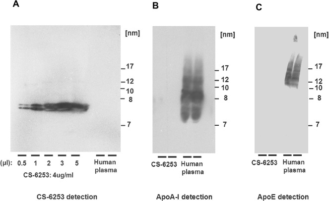
Fig 1 Cross reactivity characterisation of anti-CS-6253 antibody. Non denaturing gradient gel electrophoresis (4–20%) is used to separate CS-6253 complexes, apo A-I and apo E. CS-6253 was immunoblotted using antibody against human apo A-I, human apo E and against CS-6253. Normolipidemic human plasma was used as positive control. Molecular weight markers were revealed by Ponceau S. Left panel shows dose response relationship with an antibody directed against CS-6253. Middle panel shows anti-apo A-I antibody does not cross-react with CS-6253; similarly, right panel shows antibody against apo E does not react against CS-6253.
Fig 1 Cross reactivity characterisation of anti-CS-6253 antibody. Non denaturing gradient gel electrophoresis (4–20%) is used to separate CS-6253 complexes, apo A-I and apo E. CS-6253 was immunoblotted using antibody against human apo A-I, human apo E and against CS-6253. Normolipidemic human plasma was used as positive control. Molecular weight markers were revealed by Ponceau S. Left panel shows dose response relationship with an antibody directed against CS-6253. Middle panel shows anti-apo A-I antibody does not cross-react with CS-6253; similarly, right panel shows antibody against apo E does not react against CS-6253.CC BY · PubMed Central
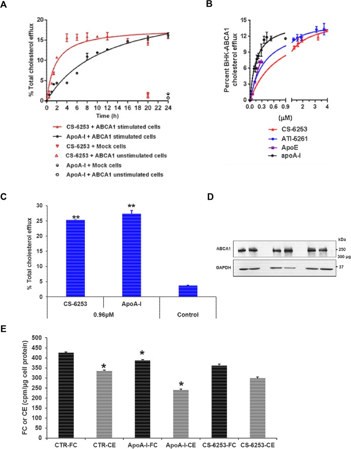
Fig 2 Effect of CS-6253 treatment on cholesterol efflux. A. Time-course activity of ABCA1-mediated cholesterol efflux to lipid-free CS-6253 (30 μg/ml) and apo A-I (10 μg/ml), using BHK cells expressing ABCA1, and control cells (unstimulated BHK-ABCA1 and BHK-mock cells). Cholesterol efflux from either ABCA1 cells or control cells to apo A-I (closed circles), and CS-6253 (closed triangles) was determined at the indicated time points. B. Dose dependent ABCA1 mediated cholesterol efflux in BHK-ABCA1 induced with mifepristone. Kinetic parameters for ABCA1-mediated cholesterol efflux from BHK-ABCA1 cells to apo A-I: Km = 4.53±0.67 μg/ml (0.15±0.02 μM), Vmax = 14.85±0.02% efflux/4h, and relative catalytic efficiency: Vmax/Km = 3.27. CS-6253: Km = 2.27±0.16 μg/ml (0.73±0.05 μM), Vmax = 15.25±0.05% efflux/4h, and relative catalytic efficiency: Vmax/Km = 6.71. ATI-5261: Km = 1.04 ± 0.16 μg/ml (0.37 ± 0.04 μM), Vmax = 14.48 ± 0.29% efflux/ h, and relative catalytic efficiency: Vmax/Km = 13.92. Apo E: Km = 10.30±2.84 μg/ml (0.21±0.05 μM), Vmax = 11.45±1.16% efflux/4h, and relative catalytic efficiency: Vmax/Km = 1.11. C. Ability of CS-6253 to stimulate cholesterol efflux from human macrophages THP-1. Foam cells were incubated with CS-6253 or apoA-I at equimolar ratio (0.96 μM) for 4h. Cholesterol efflux induced by CS-6253 and apoA-I were compared to control cells incubated alone. P<0.001 by Student’s t-test. D. Membrane ABCA1 levels assessment by Western blotting assays. ABCA1 was detected from THP-1 foam cells lysis at 4°C with lysis buffer containing 0.5% n-dodecylmaltoside in the presence of a protease inhibitor mixture followed by low speed centrifugation to remove cell debris. Protein concentration was determined by standard assay (Bio-Rad). The supernatants were then separated by SDS-PAGE (4–22.5%) in duplicate. After electrophoresis, ABCA1 was detected by an anti-ABCA1 antibody. GAPDH was used as loading control. E. Quantification of FC and CE in foam cells after interaction with CS-6253 and apoA-I. After a period of 4h cholesterol efflux, THP-1 foam cell lipids were extracted with hexane: isopropanol (3v/2v) as under ‘‘Material and Methods’. 3[H]-FC and 3[H]-CE were separated by TLC and located by exposure to iodine vapor, and were scraped off into liquid scintillating vials and assayed for radioactivity. Results are from a single experiment using triplicate wells and the mean (±SD) is presented. P<0.0001 versus control sample untreated THP-1 cells for cholesterol efflux. P<0.05 versus FC or CE in untreated cells, by Student’s t-test.
Fig 2 Effect of CS-6253 treatment on cholesterol efflux. A. Time-course activity of ABCA1-mediated cholesterol efflux to lipid-free CS-6253 (30 μg/ml) and apo A-I (10 μg/ml), using BHK cells expressing ABCA1, and control cells (unstimulated BHK-ABCA1 and BHK-mock cells). Cholesterol efflux from either ABCA1 cells or control cells to apo A-I (closed circles), and CS-6253 (closed triangles) was determined at the indicated time points. B. Dose dependent ABCA1 mediated cholesterol efflux in BHK-ABCA1 induced with mifepristone. Kinetic parameters for ABCA1-mediated cholesterol efflux from BHK-ABCA1 cells to apo A-I: Km = 4.53±0.67 μg/ml (0.15±0.02 μM), Vmax = 14.85±0.02% efflux/4h, and relative catalytic efficiency: Vmax/Km = 3.27. CS-6253: Km = 2.27±0.16 μg/ml (0.73±0.05 μM), Vmax = 15.25±0.05% efflux/4h, and relative catalytic efficiency: Vmax/Km = 6.71. ATI-5261: Km = 1.04 ± 0.16 μg/ml (0.37 ± 0.04 μM), Vmax = 14.48 ± 0.29% efflux/ h, and relative catalytic efficiency: Vmax/Km = 13.92. Apo E: Km = 10.30±2.84 μg/ml (0.21±0.05 μM), Vmax = 11.45±1.16% efflux/4h, and relative catalytic efficiency: Vmax/Km = 1.11. C. Ability of CS-6253 to stimulate cholesterol efflux from human macrophages THP-1. Foam cells were incubated with CS-6253 or apoA-I at equimolar ratio (0.96 μM) for 4h. Cholesterol efflux induced by CS-6253 and apoA-I were compared to control cells incubated alone. P<0.001 by Student’s t-test. D. Membrane ABCA1 levels assessment by Western blotting assays. ABCA1 was detected from THP-1 foam cells lysis at 4°C with lysis buffer containing 0.5% n-dodecylmaltoside in the presence of a protease inhibitor mixture followed by low speed centrifugation to remove cell debris. Protein concentration was determined by standard assay (Bio-Rad). The supernatants were then separated by SDS-PAGE (4–22.5%) in duplicate. After electrophoresis, ABCA1 was detected by an anti-ABCA1 antibody. GAPDH was used as loading control. E. Quantification of FC and CE in foam cells after interaction with CS-6253 and apoA-I. After a period of 4h cholesterol efflux, THP-1 foam cell lipids were extracted with hexane: isopropanol (3v/2v) as under ‘‘Material and Methods’. 3[H]-FC and 3[H]-CE were separated by TLC and located by exposure to iodine vapor, and were scraped off into liquid scintillating vials and assayed for radioactivity. Results are from a single experiment using triplicate wells and the mean (±SD) is presented. P<0.0001 versus control sample untreated THP-1 cells for cholesterol efflux. P<0.05 versus FC or CE in untreated cells, by Student’s t-test.CC BY · PubMed Central
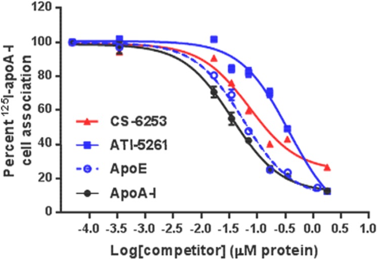
Fig 3 Competitive binding of CS-6253 peptide to ABCA1 cells expressing ABCA1. BHK-ABCA1 cells were plated in 24-well plates and stimulated for 20 h. Cells were then incubated with 2 μg/ml of 125I-apo A-I for 2 h at 37°C with increasing molar concentrations relative to apo A-I of either CS-6253 peptide, ATI-5261, human apo E, and unlabelled apo A-I (0, 0.0035, 0.017, 0.035, 0.071, 0.17, 0.35, 1.79 μM). Cells were then washed rapidly three times with ice-cold PBS/BSA and then PBS alone. 125I-apo A-I cell associated was determined as described under “Experimental Procedures.” The values shown represent the mean ± S.D from triplicate wells. The 100% of control value measured in the absence of competitors was 0.21 ng of apo A-I /μg cell protein. Values of IC50 shown were determined using the Graph Pad Prism 6 software.
Fig 3 Competitive binding of CS-6253 peptide to ABCA1 cells expressing ABCA1. BHK-ABCA1 cells were plated in 24-well plates and stimulated for 20 h. Cells were then incubated with 2 μg/ml of 125I-apo A-I for 2 h at 37°C with increasing molar concentrations relative to apo A-I of either CS-6253 peptide, ATI-5261, human apo E, and unlabelled apo A-I (0, 0.0035, 0.017, 0.035, 0.071, 0.17, 0.35, 1.79 μM). Cells were then washed rapidly three times with ice-cold PBS/BSA and then PBS alone. 125I-apo A-I cell associated was determined as described under “Experimental Procedures.” The values shown represent the mean ± S.D from triplicate wells. The 100% of control value measured in the absence of competitors was 0.21 ng of apo A-I /μg cell protein. Values of IC50 shown were determined using the Graph Pad Prism 6 software.CC BY · PubMed Central
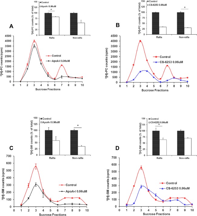
Fig 4 Apo A-I (left panels) and CS-6253 (right panels) desorb phospholipids species from raft and nonraft domains. BHK-ABCA1 cells were labeled with 3[H]choline for 48 h, followed by stimulation with mifepristone for 18–20h as described in “experimental procedures”. Cells were then incubated for 12h with apo A-I or CS-6253 (0.96 μM). After lipid extraction by Folch, 3[H]phosphatidyl choline (3[H]PC) (A and B), or 3[H]sphingomyelin (3[H]SM) (C and D) in each fraction was assessed for radioactivity. Radioactivity appearing in fractions corresponding to raft (1–5) and nonraft (6–10) material was pooled, and desorption of 3[H]PC (A and B inset) and 3[H]SM (C and D inset), from raft versus nonraft in the presence of apo A-I or CS-6253 was expressed as a percentage of control (100%, in the absence of acceptor). 3[H]PC and 3[H]SM distributions in sucrose gradient fractions after 12h incubation with apo A-I or CS-6253 were assessed by TLC respectively. Results shown are representative of three independent experiments. *P < 0.05 by Student's t-test.
Fig 4 Apo A-I (left panels) and CS-6253 (right panels) desorb phospholipids species from raft and nonraft domains. BHK-ABCA1 cells were labeled with 3[H]choline for 48 h, followed by stimulation with mifepristone for 18–20h as described in “experimental procedures”. Cells were then incubated for 12h with apo A-I or CS-6253 (0.96 μM). After lipid extraction by Folch, 3[H]phosphatidyl choline (3[H]PC) (A and B), or 3[H]sphingomyelin (3[H]SM) (C and D) in each fraction was assessed for radioactivity. Radioactivity appearing in fractions corresponding to raft (1–5) and nonraft (6–10) material was pooled, and desorption of 3[H]PC (A and B inset) and 3[H]SM (C and D inset), from raft versus nonraft in the presence of apo A-I or CS-6253 was expressed as a percentage of control (100%, in the absence of acceptor). 3[H]PC and 3[H]SM distributions in sucrose gradient fractions after 12h incubation with apo A-I or CS-6253 were assessed by TLC respectively. Results shown are representative of three independent experiments. *P < 0.05 by Student's t-test.CC BY · PubMed Central
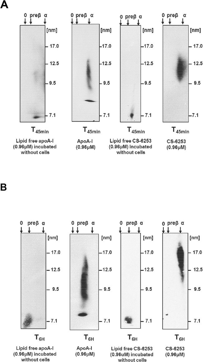
Fig 5 Time course ability of CS-6253 to generate nHDL-mimetic like particles. (A and B): nHDL-CS-6253 and nHDL-apo A-I released to the medium at specific time points (45 min and 6 h) from BHK cells expressing ABCA1 were analyzed by 2D-PAGGE. Apo A-I was detected by polyclonal anti-apo A-I, CS-6253 peptides was detected by anti-CS-6253 and revealed by chemiluminescence. Molecular markers are indicated.
Fig 5 Time course ability of CS-6253 to generate nHDL-mimetic like particles. (A and B): nHDL-CS-6253 and nHDL-apo A-I released to the medium at specific time points (45 min and 6 h) from BHK cells expressing ABCA1 were analyzed by 2D-PAGGE. Apo A-I was detected by polyclonal anti-apo A-I, CS-6253 peptides was detected by anti-CS-6253 and revealed by chemiluminescence. Molecular markers are indicated.CC BY · PubMed Central
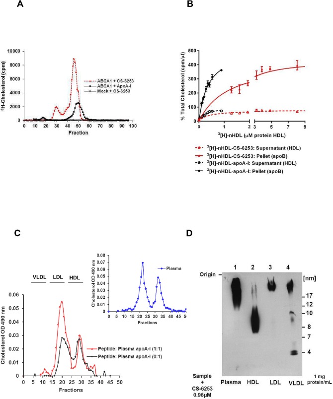
Fig 6 Dynamics of transfer and redistribution of BHK-ABCA1 cell-derived cholesterol content of nHDL-CS-6253. nHDL-CS-6253 particles were labelled with cell derived 3[H]cholesterol and incubated with normolipidemic plasma for various time periods at 37°C (1μg peptide: 10μg plasma apo A-I). A. Characterization of lipid particles released to the medium in the presence of CS-6253. 3[H]-cholesterol-labelled BHK cells expressing ABCA1 or not were incubated in the presence of 0.96 μM apo A-I (solid line) or CS-6253 (dashed line) for 45min at 37°C. Concentrated medium from cells was analysed by FPLC, and radioactivity associated with each fraction was determined. B. Transfer of total 3[H]cholesterol from 3[H]nHDL-CS-6253 to plasma lipoproteins. nHDL-CS-6253 or nHDL-apo A-I were generated after incubation (4h) of BHK cells expressing ABCA1 with increased doses of lipid free CS-6253 or apo A-I. Media cell culture containing 3[H]-nHDL-particles was incubated in human plasma for 1h, 37°C. After ApoB precipitation, fractions were dialysed and counted for radioactivity. Kinetic parameters for cholesterol transfer to apoB particles in plasma: nHDL-CS-6253 (439±0.20μM), Vmax (439± 19.74%cpm/μl) vs nHDL-apo A-I (0.37 ± 0.035), Vmax (517±24.45).C. Effects of ex vivo incubation of CS-6253 on plasma cholesterol lipoproteins. FPLC cholesterol profiles (OD 490 nm) of plasma apo A-I: peptide ratio (1:1) (dashed line) and plasma apo A-I: peptide ratios (1:0; i.e. apo A-I alone) (solid line) are obtained after 5 min incubation at 37°C. In all experiment counts were made in triplicate, (*P<0.05 by Student‘s t-test). For reference purposes, the inset shows an FPLC profile of normal plasma. D. Effect of ex vivo incubation of CS-6253 on plasma lipoproteins after separation by ND-PAGGE (5–35%) electrophoresis. CS-6253 (0.96 μM final dose) was incubated for 1h at 37°C with HDL, LDL or VLDL isolated by ultracentrifugation or with human plasma and then separated by electrophoresis at concentration of 1 mg/protein/mL. Gel is followed by Western blot analysis with anti-CS-6253 antibody. Internal control was run on the left of the gel and detected by Ponceau S.
Fig 6 Dynamics of transfer and redistribution of BHK-ABCA1 cell-derived cholesterol content of nHDL-CS-6253. nHDL-CS-6253 particles were labelled with cell derived 3[H]cholesterol and incubated with normolipidemic plasma for various time periods at 37°C (1μg peptide: 10μg plasma apo A-I). A. Characterization of lipid particles released to the medium in the presence of CS-6253. 3[H]-cholesterol-labelled BHK cells expressing ABCA1 or not were incubated in the presence of 0.96 μM apo A-I (solid line) or CS-6253 (dashed line) for 45min at 37°C. Concentrated medium from cells was analysed by FPLC, and radioactivity associated with each fraction was determined. B. Transfer of total 3[H]cholesterol from 3[H]nHDL-CS-6253 to plasma lipoproteins. nHDL-CS-6253 or nHDL-apo A-I were generated after incubation (4h) of BHK cells expressing ABCA1 with increased doses of lipid free CS-6253 or apo A-I. Media cell culture containing 3[H]-nHDL-particles was incubated in human plasma for 1h, 37°C. After ApoB precipitation, fractions were dialysed and counted for radioactivity. Kinetic parameters for cholesterol transfer to apoB particles in plasma: nHDL-CS-6253 (439±0.20μM), Vmax (439± 19.74%cpm/μl) vs nHDL-apo A-I (0.37 ± 0.035), Vmax (517±24.45).C. Effects of ex vivo incubation of CS-6253 on plasma cholesterol lipoproteins. FPLC cholesterol profiles (OD 490 nm) of plasma apo A-I: peptide ratio (1:1) (dashed line) and plasma apo A-I: peptide ratios (1:0; i.e. apo A-I alone) (solid line) are obtained after 5 min incubation at 37°C. In all experiment counts were made in triplicate, (*P<0.05 by Student‘s t-test). For reference purposes, the inset shows an FPLC profile of normal plasma. D. Effect of ex vivo incubation of CS-6253 on plasma lipoproteins after separation by ND-PAGGE (5–35%) electrophoresis. CS-6253 (0.96 μM final dose) was incubated for 1h at 37°C with HDL, LDL or VLDL isolated by ultracentrifugation or with human plasma and then separated by electrophoresis at concentration of 1 mg/protein/mL. Gel is followed by Western blot analysis with anti-CS-6253 antibody. Internal control was run on the left of the gel and detected by Ponceau S.CC BY · PubMed Central
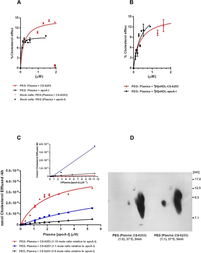
Fig 7 Dose dependent effects of pre-incubation of plasma with lipid free CS-6253, or nHDL-CS-6253 on ABCA1 cholesterol efflux. A. Increased doses of lipid free CS-6253 or apo A-I were added to normolipidemic plasma and incubated for 1h at 37°C. A volume of 2.8% of HDL fractions isolated by PEG was used for the measurements of cholesterol efflux from BHK cells expressing ABCA1 and mock cells. Kinetic parameters for ABCA1-mediated cholesterol efflux from BHK expressing ABCA1 cells to apo A-I in plasma: Km = 0.03±0.008 μM, Vmax = 9.26±0.29% efflux/4h. CS-6253: Km = 0.20±0.05 μM, Vmax = 14.71±0.39% efflux/4h. Kinetic parameters for cholesterol efflux from BHK-mock cells to apo A-I in plasma: Km = 0.07±0.95 μM, Vmax = 0.30±0.15% efflux/6h. CS-6253: Km = 0.65±0.63 μM, Vmax = 0.84±0.17% efflux/4h. B. nHDL-CS-6253 and nHDL-apo A-I were generated as indicated above. nHDL particles were incubated in plasma for 1h at 37°C, and specific HDL fractions isolated by PEG were subjected to cholesterol efflux in 3[H]-radiolabelled BHK cells. Kinetic parameters for ABCA1-mediated cholesterol efflux from BHK expressing ABCA1 cells to nHDL-apo A-I in plasma: Km = 0.25±0.08 μM, Vmax = 15.73±2.14% efflux/4h. nHDL-CS-6253: Km = 0.32±0.15 μM, Vmax = 15.01±0.84% efflux/4h. C. Mobilization of cholesterol efflux of increasing concentrations of peptide: plasma apo A-I molar ratios: (1:0) (closed circles), (1:10) (upright closed triangles), and (1:1) (closed squares). (C inset). The double reciprocal-plot was used to calculate the apparent appKm of the efflux activity in peptide: plasma with indicated ratios. Kinetics values are as follows, molar ratio of plasma apo A-I: CS-6253 (1:0); appKm (28±0.07 μM). Molar ratio of plasma apo A-I: CS-6253 (1:1); appKm (4.17±0.47 μM). Molar ratio of plasma apo A-I: CS-6253 (1:10); appKm (0.71±0.46 μM). D. Lipid free CS-6253 was incubated with normolipidemic plasma, using peptide: apo A-I mole ratio of (1:1) for 5 min at 37°C versus plasma alone. Isolated HDL fractions by PEG were separated by 2D-PAGGE and HDL subpopulations were detected by apo A-I Western blot. Molecular size markers are shown. In all experiments, efflux of 3[H]cholesterol is shown as means ± SD of triplicate experiments.
Fig 7 Dose dependent effects of pre-incubation of plasma with lipid free CS-6253, or nHDL-CS-6253 on ABCA1 cholesterol efflux. A. Increased doses of lipid free CS-6253 or apo A-I were added to normolipidemic plasma and incubated for 1h at 37°C. A volume of 2.8% of HDL fractions isolated by PEG was used for the measurements of cholesterol efflux from BHK cells expressing ABCA1 and mock cells. Kinetic parameters for ABCA1-mediated cholesterol efflux from BHK expressing ABCA1 cells to apo A-I in plasma: Km = 0.03±0.008 μM, Vmax = 9.26±0.29% efflux/4h. CS-6253: Km = 0.20±0.05 μM, Vmax = 14.71±0.39% efflux/4h. Kinetic parameters for cholesterol efflux from BHK-mock cells to apo A-I in plasma: Km = 0.07±0.95 μM, Vmax = 0.30±0.15% efflux/6h. CS-6253: Km = 0.65±0.63 μM, Vmax = 0.84±0.17% efflux/4h. B. nHDL-CS-6253 and nHDL-apo A-I were generated as indicated above. nHDL particles were incubated in plasma for 1h at 37°C, and specific HDL fractions isolated by PEG were subjected to cholesterol efflux in 3[H]-radiolabelled BHK cells. Kinetic parameters for ABCA1-mediated cholesterol efflux from BHK expressing ABCA1 cells to nHDL-apo A-I in plasma: Km = 0.25±0.08 μM, Vmax = 15.73±2.14% efflux/4h. nHDL-CS-6253: Km = 0.32±0.15 μM, Vmax = 15.01±0.84% efflux/4h. C. Mobilization of cholesterol efflux of increasing concentrations of peptide: plasma apo A-I molar ratios: (1:0) (closed circles), (1:10) (upright closed triangles), and (1:1) (closed squares). (C inset). The double reciprocal-plot was used to calculate the apparent appKm of the efflux activity in peptide: plasma with indicated ratios. Kinetics values are as follows, molar ratio of plasma apo A-I: CS-6253 (1:0); appKm (28±0.07 μM). Molar ratio of plasma apo A-I: CS-6253 (1:1); appKm (4.17±0.47 μM). Molar ratio of plasma apo A-I: CS-6253 (1:10); appKm (0.71±0.46 μM). D. Lipid free CS-6253 was incubated with normolipidemic plasma, using peptide: apo A-I mole ratio of (1:1) for 5 min at 37°C versus plasma alone. Isolated HDL fractions by PEG were separated by 2D-PAGGE and HDL subpopulations were detected by apo A-I Western blot. Molecular size markers are shown. In all experiments, efflux of 3[H]cholesterol is shown as means ± SD of triplicate experiments.CC BY · PubMed Central
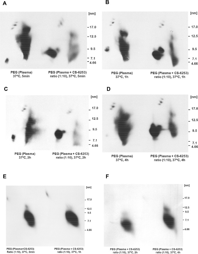
Fig 8 Time dependent effects of incubation of lipid free CS-6253 with human plasma, on HDL subparticles. (A, B, C and D). Lipid free CS-6253 was incubated with normolipidemic plasma, using peptide: apo A-I molar ratio of (10:1) and (0:1 i.e. no peptide) for (5 min, 1h, 2h, and 4h) at 37°C. HDL subpopulations were detected by an anti-apo A-I antibody. E and F Samples were then PEG precipitated to remove apoB-containing lipoproteins and separated by 2D-PAGGE. HDL subfractions were detected by an antibody against CS-6253. Molecular size markers are shown.
Fig 8 Time dependent effects of incubation of lipid free CS-6253 with human plasma, on HDL subparticles. (A, B, C and D). Lipid free CS-6253 was incubated with normolipidemic plasma, using peptide: apo A-I molar ratio of (10:1) and (0:1 i.e. no peptide) for (5 min, 1h, 2h, and 4h) at 37°C. HDL subpopulations were detected by an anti-apo A-I antibody. E and F Samples were then PEG precipitated to remove apoB-containing lipoproteins and separated by 2D-PAGGE. HDL subfractions were detected by an antibody against CS-6253. Molecular size markers are shown.CC BY · PubMed Central
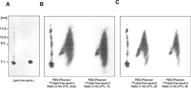
Fig 9 Time dependent effects of incubation of lipid free CS-6253 with human plasma, on HDL subparticles. (A, B, C). Radiolabeled Lipid free apoA-I was prepared as indicated in “Material and Methods”. 125I-lipid free apoA-I in incubated with normolipidemic plasma, using molar ratio (10 μg Lipid free apoA-I or peptide: 1 μg plasma apoA-I) at 37°C for increasing time for (5 min, 1h, 2h, and 4h) at 37°C. Samples were separated by 2D-PAGGE, and 125I-apoA-I was detected by autoradiography. Lipid-free 125I-apoA-I (C) incubated in PBS for 4 h at 37°C is shown as controls. Molecular size markers are shown.
Fig 9 Time dependent effects of incubation of lipid free CS-6253 with human plasma, on HDL subparticles. (A, B, C). Radiolabeled Lipid free apoA-I was prepared as indicated in “Material and Methods”. 125I-lipid free apoA-I in incubated with normolipidemic plasma, using molar ratio (10 μg Lipid free apoA-I or peptide: 1 μg plasma apoA-I) at 37°C for increasing time for (5 min, 1h, 2h, and 4h) at 37°C. Samples were separated by 2D-PAGGE, and 125I-apoA-I was detected by autoradiography. Lipid-free 125I-apoA-I (C) incubated in PBS for 4 h at 37°C is shown as controls. Molecular size markers are shown.CC BY · PubMed Central
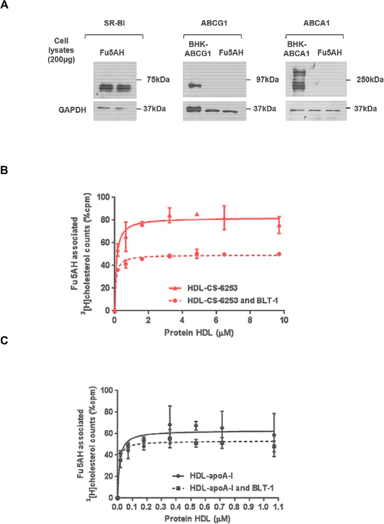
Fig 10 Uptake of cholesterol to hepatic cells expressing SR-BI from plasma containing HDL-CS-6253 or HDL-apo A-I. A. Fu5AH liver hepatoma cells expression of SR-BI transporter ability. Fu5AH cells were grown as described in Experimental procedures, cells were lysed at 4°C with (20mM Tris, 5mM EDTA, and 5 mM EGTA; pH 7.5) containing 0.5% n-dodecylmaltoside. The suspension was subjected for further lysis in the presence of protease inhibitor mixture followed by low speed centrifugation to remove insoluble material. Protein concentration was determined by standard assay (Bio-Rad). Cells were separated by SDS-PAGE (4–22.5%), and immunoblotted using antibodies against human SR-BI, ABCG1 and ABCA1. Cells lysate from BHK cells expressing ABCG1 and ABCG1 was used as positive control. Molecular weight (Bio-Rad) is shown on the left of the gel. B and C. Radiolabelled nHDL-CS-6253 or nHDL-apoA-I particles were generated after incubation of J774 cells with lipid free CS-6253 or apo A-I at increased concentrations as showed in experimental procedures. nHDL particles with CS-6253 or apoA-I were then incubated in plasma to induce lipidation. Cholesterol uptake by SR-BI Fu5AH cells was determined by incubation for 6h with 20% apoB depleted plasma as described in experimental procedures. BLT-1 was used to specifically inhibit SR-BI. The values shown represent the mean ± S.D from triplicate wells.
Fig 10 Uptake of cholesterol to hepatic cells expressing SR-BI from plasma containing HDL-CS-6253 or HDL-apo A-I. A. Fu5AH liver hepatoma cells expression of SR-BI transporter ability. Fu5AH cells were grown as described in Experimental procedures, cells were lysed at 4°C with (20mM Tris, 5mM EDTA, and 5 mM EGTA; pH 7.5) containing 0.5% n-dodecylmaltoside. The suspension was subjected for further lysis in the presence of protease inhibitor mixture followed by low speed centrifugation to remove insoluble material. Protein concentration was determined by standard assay (Bio-Rad). Cells were separated by SDS-PAGE (4–22.5%), and immunoblotted using antibodies against human SR-BI, ABCG1 and ABCA1. Cells lysate from BHK cells expressing ABCG1 and ABCG1 was used as positive control. Molecular weight (Bio-Rad) is shown on the left of the gel. B and C. Radiolabelled nHDL-CS-6253 or nHDL-apoA-I particles were generated after incubation of J774 cells with lipid free CS-6253 or apo A-I at increased concentrations as showed in experimental procedures. nHDL particles with CS-6253 or apoA-I were then incubated in plasma to induce lipidation. Cholesterol uptake by SR-BI Fu5AH cells was determined by incubation for 6h with 20% apoB depleted plasma as described in experimental procedures. BLT-1 was used to specifically inhibit SR-BI. The values shown represent the mean ± S.D from triplicate wells.CC BY · PubMed Central
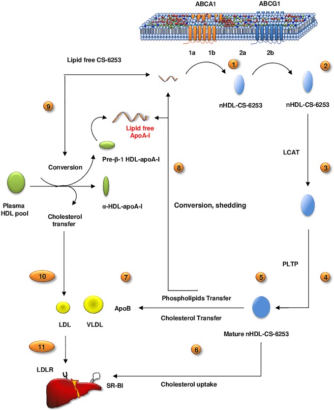
Fig 11 A proposed in-vitro model for lipid free CS-6253 lipidation and remodelling in RCT pathway. 1). ABCA1 interaction: a. ABCA1 oligomerization, b. Binding to ABCA-1. 2). Lipid efflux process: a. PM microdomains lipid desorption, b. ABCA1 and ABCG1 induction of lipid efflux and nHDL-CS-6253 formation, 3–4) HDL-CS-6253 remodelling by LCAT/PLTP in plasma, 5) generation of mature HDL-CS-6253, 6) Cholesterol uptake by liver via SR-BI, and 7) lipid transfer to both apoB-containing particles and the plasma HDL resident pool. 8) Although the mechanisms underlying this process is presently ambiguous, it is possible that nHDL-CS-6253 remodelling may lead to loss or ‘shedding’ of CS-6253 from HDL-CS-6253 in plasma as they are actively delipidated of phospholipids by PLTP to yield lipid poor apo A-I precursor of RCT activation. 9) Alternatively lipid free CS-6253 associates with αHDL-apo A-I species leading to preβ1-apo A-I formation, that can also close the loop and initiate again RCT. 10) in this process, the peptide transfer cholesterol to apoB particles, and 11) cholesterol is delivered to liver by the LDLR.
Fig 11 A proposed in-vitro model for lipid free CS-6253 lipidation and remodelling in RCT pathway. 1). ABCA1 interaction: a. ABCA1 oligomerization, b. Binding to ABCA-1. 2). Lipid efflux process: a. PM microdomains lipid desorption, b. ABCA1 and ABCG1 induction of lipid efflux and nHDL-CS-6253 formation, 3–4) HDL-CS-6253 remodelling by LCAT/PLTP in plasma, 5) generation of mature HDL-CS-6253, 6) Cholesterol uptake by liver via SR-BI, and 7) lipid transfer to both apoB-containing particles and the plasma HDL resident pool. 8) Although the mechanisms underlying this process is presently ambiguous, it is possible that nHDL-CS-6253 remodelling may lead to loss or ‘shedding’ of CS-6253 from HDL-CS-6253 in plasma as they are actively delipidated of phospholipids by PLTP to yield lipid poor apo A-I precursor of RCT activation. 9) Alternatively lipid free CS-6253 associates with αHDL-apo A-I species leading to preβ1-apo A-I formation, that can also close the loop and initiate again RCT. 10) in this process, the peptide transfer cholesterol to apoB particles, and 11) cholesterol is delivered to liver by the LDLR.CC BY · PubMed Central

Verification

Claims the paper makes

The claims and statistics of this paper haven’t been checked yet.

Run the check — free, no account needed. It reads the open-access text and checks the sources it cites.

A few free checks a day · papers already verified open instantly · results are public

Claims other papers make about this one

Each is checked against this paper’s own text: does it say what the citing paper says it does?

Self-check

Percentages, sums and reported differences re-computed from the paper’s own numbers.

The self-check needs the paper’s full text.

Citations

Cites

Show all 1 references

Research works printed but not linked: 83

  1. DJ Rader, 2009, The role of reverse cholesterol transport in animals and humans and relationship to atherosclerosis, J Lipid Res, 50, S189, 10.1194/jlr.R800088-JLR200 · DOI
  2. LJ Leman, 2014, Molecules that mimic apolipoprotein A-I: potential agents for treating atherosclerosis, J Med Chem, 57, 2169, 10.1021/jm4005847 · DOI
  3. GS Getz, 2010, Biological properties of apolipoprotein a-I mimetic peptides, Curr Atheroscler Rep, 12, 96, 10.1007/s11883-010-0097-4 · DOI
  4. A Hafiane, 2014, Treatment options for low high-density lipoproteins, Current opinion in endocrinology, diabetes, and obesity, 21, 134, 10.1097/MED.0000000000000049 · DOI
  5. GS Getz, 2009, Apoprotein A-I mimetic peptides and their potential anti-atherogenic mechanisms of action, Curr Opin Lipidol, 20, 171, 10.1097/MOL.0b013e32832ac051 · DOI
  6. AT Remaley, 2008, HDL-replacement therapy: mechanism of action, types of agents and potential clinical indications, Expert Rev Cardiovasc Ther, 6, 1203, 10.1586/14779072.6.9.1203 · DOI
  7. JK Bielicki, 2010, A new HDL mimetic peptide that stimulates cellular cholesterol efflux with high efficiency greatly reduces atherosclerosis in mice, J Lipid Res, 51, 1496, 10.1194/jlr.M003665 · DOI
  8. AT Remaley, 2003, Synthetic amphipathic helical peptides promote lipid efflux from cells by an ABCA1-dependent and an ABCA1-independent pathway, J Lipid Res, 44, 828, 10.1194/jlr.M200475-JLR200 · DOI
  9. MJ Amar, 2010, 5A apolipoprotein mimetic peptide promotes cholesterol efflux and reduces atherosclerosis in mice, J Pharmacol Exp Ther, 334, 634, 10.1124/jpet.110.167890 · DOI
  10. Y Zheng, 2011, HDL mimetic peptide ATI-5261 forms an oligomeric assembly in solution that dissociates to monomers upon dilution, Biochemistry, 50, 4068, 10.1021/bi2002955 · DOI
  11. A Hafiane, 2014, Apolipoprotein E derived HDL mimetic peptide ATI-5261 promotes nascent HDL formation and reverse cholesterol transport in vitro, Biochim Biophys Acta, 1842, 1498, 10.1016/j.bbalip.2014.07.018 · DOI
  12. JF Oram, 2001, ATP-binding cassette transporter A1 mediates cellular secretion of alpha-tocopherol, J Biol Chem, 276, 39898, 10.1074/jbc.M106984200 · DOI
  13. AM Vaughan, 2006, ABCA1 and ABCG1 or ABCG4 act sequentially to remove cellular cholesterol and generate cholesterol-rich HDL, J Lipid Res, 47, 2433, 10.1194/jlr.M600218-JLR200 · DOI
  14. L Kritharides, 1998, Cholesterol metabolism and efflux in human THP-1 macrophages, Arterioscler Thromb Vasc Biol, 18, 1589, 10.1161/01.ATV.18.10.1589 · DOI
  15. M Denis, 2004, Characterization of oligomeric human ATP binding cassette transporter A1. Potential implications for determining the structure of nascent high density lipoprotein particles, J Biol Chem, 279, 41529, 10.1074/jbc.M406881200 · DOI
  16. L Krimbou, 2004, Molecular interactions between apoE and ABCA1: impact on apoE lipidation, J Lipid Res, 45, 839, 10.1194/jlr.M300418-JLR200 · DOI · Paper page
  17. OH Lowry, 1951, Protein measurement with the Folin phenol reagent, J Biol Chem, 193, 265, 10.1016/S0021-9258(19)52451-6 · DOI
  18. A Hafiane, 2014, High-density lipoprotein mediated cellular cholesterol efflux in acute coronary syndromes, Am J Cardiol, 113, 249, 10.1016/j.amjcard.2013.09.006 · DOI
  19. RJ Doonan, 2014, Cholesterol efflux capacity, carotid atherosclerosis, and cerebrovascular symptomatology, Arterioscler Thromb Vasc Biol, 34, 921, 10.1161/ATVBAHA.113.302590 · DOI
  20. D Bailey, 2010, Analysis of lipid transfer activity between model nascent HDL particles and plasma lipoproteins: implications for current concepts of nascent HDL maturation and genesis, J Lipid Res, 51, 785, 10.1194/jlr.M001875 · DOI
  21. HH Hassan, 2008, Quantitative analysis of ABCA1-dependent compartmentalization and trafficking of apolipoprotein A-I: implications for determining cellular kinetics of nascent high density lipoprotein biogenesis, J Biol Chem, 283, 11164, 10.1074/jbc.M707720200 · DOI
  22. FT Hatch, 1968, Practical methods for plasma lipoprotein analysis, Adv Lipid Res, 6, 1
  23. L Krimbou, 1997, Characterization of human plasma apolipoprotein E-containing lipoproteins in the high density lipoprotein size range: focus on pre-beta1-LpE, pre-beta2-LpE, and alpha-LpE, J Lipid Res, 38, 35, 10.1016/S0022-2275(20)37273-4 · DOI · Paper page
  24. M Denis, 2004, Molecular and cellular physiology of apolipoprotein A-I lipidation by the ATP-binding cassette transporter A1 (ABCA1), J Biol Chem, 279, 7384, 10.1074/jbc.M306963200 · DOI
  25. F Zimetti, 2006, Measurement of cholesterol bidirectional flux between cells and lipoproteins, J Lipid Res, 47, 605, 10.1194/jlr.M500466-JLR200 · DOI
  26. A Hafiane, 2014, Apolipoprotein E derived HDL mimetic peptide ATI-5261 promotes nascent HDL formation and reverse cholesterol transport in vitro, Biochim Biophys Acta
  27. BK Nordskog, 1999, Sterol synthesis is up-regulated in cholesterol-loaded pigeon macrophages during induction of cholesterol efflux, J Lipid Res, 40, 1806, 10.1016/S0022-2275(20)34897-5 · DOI
  28. ET Alexander, 2011, Influence of apolipoprotein A-I domain structure on macrophage reverse cholesterol transport in mice, Arterioscler Thromb Vasc Biol, 31, 320, 10.1161/ATVBAHA.110.216226 · DOI
  29. PG Yancey, 2000, High density lipoprotein phospholipid composition is a major determinant of the bi-directional flux and net movement of cellular free cholesterol mediated by scavenger receptor BI, J Biol Chem, 275, 36596, 10.1074/jbc.M006924200 · DOI
  30. TJ Nieland, 2002, Discovery of chemical inhibitors of the selective transfer of lipids mediated by the HDL receptor SR-BI, Proc Natl Acad Sci U S A, 99, 15422, 10.1073/pnas.222421399 · DOI
  31. AA Sethi, 2008, Asymmetry in the lipid affinity of bihelical amphipathic peptides. A structural determinant for the specificity of ABCA1-dependent cholesterol efflux by peptides, J Biol Chem, 283, 32273, 10.1074/jbc.M804461200 · DOI
  32. A Hafiane, 2013, HDL, Atherosclerosis, and Emerging Therapies, Cholesterol, 2013, 891403, 10.1155/2013/891403 · DOI
  33. JD Smith, 2010, Apolipoprotein A-I and its mimetics for the treatment of atherosclerosis, Curr Opin Investig Drugs, 11, 989
  34. I Iatan, 2011, Membrane microdomains modulate oligomeric ABCA1 function: impact on apoAI-mediated lipid removal and phosphatidylcholine biosynthesis, J Lipid Res, 52, 2043, 10.1194/jlr.M016196 · DOI
  35. L Krimbou, 2005, Biogenesis and speciation of nascent apoA-I-containing particles in various cell lines, J Lipid Res, 46, 1668, 10.1194/jlr.M500038-JLR200 · DOI
  36. HH Hassan, 2006, Cellular cholesterol homeostasis in vascular endothelial cells, Can J Cardiol, 22, 35B, 10.1016/S0828-282X(06)70985-0 · DOI
  37. R Liu, 2007, Macrophage phospholipid transfer protein deficiency and ApoE secretion: impact on mouse plasma cholesterol levels and atherosclerosis, Arterioscler Thromb Vasc Biol, 27, 190, 10.1161/01.ATV.0000249721.96666.e5 · DOI
  38. Y Liu, 2012, Regulation of ABCA1 functions by signaling pathways, Biochim Biophys Acta, 1821, 522, 10.1016/j.bbalip.2011.08.015 · DOI
  39. S Diditchenko, 2013, Novel formulation of a reconstituted high-density lipoprotein (CSL112) dramatically enhances ABCA1-dependent cholesterol efflux, Arterioscler Thromb Vasc Biol, 33, 2202, 10.1161/ATVBAHA.113.301981 · DOI
  40. C Tardy, 2014, CER-001, a HDL-mimetic, stimulates the reverse lipid transport and atherosclerosis regression in high cholesterol diet-fed LDL-receptor deficient mice, Atherosclerosis, 232, 110, 10.1016/j.atherosclerosis.2013.10.018 · DOI
  41. MP Adorni, 2007, The roles of different pathways in the release of cholesterol from macrophages, J Lipid Res, 48, 2453, 10.1194/jlr.M700274-JLR200 · DOI
  42. C Vedhachalam, 2007, The C-terminal lipid-binding domain of apolipoprotein E is a highly efficient mediator of ABCA1-dependent cholesterol efflux that promotes the assembly of high-density lipoproteins, Biochemistry, 46, 2583, 10.1021/bi602407r · DOI
  43. T Mazzone, 1996, Apolipoprotein E secretion by macrophages: its potential physiological functions, Curr Opin Lipidol, 7, 303, 10.1097/00041433-199610000-00008 · DOI
  44. S Nandi, 2009, ABCA1-mediated cholesterol efflux generates microparticles in addition to HDL through processes governed by membrane rigidity, J Lipid Res, 50, 456, 10.1194/jlr.M800345-JLR200 · DOI
  45. GD Wool, 2008, Apolipoprotein A-I mimetic peptide helix number and helix linker influence potentially anti-atherogenic properties, J Lipid Res, 49, 1268, 10.1194/jlr.M700552-JLR200 · DOI
  46. DO Sviridov, 2013, Hydrophobic amino acids in the hinge region of the 5A apolipoprotein mimetic peptide are essential for promoting cholesterol efflux by the ABCA1 transporter, J Pharmacol Exp Ther, 344, 50, 10.1124/jpet.112.198143 · DOI
  47. GS Getz, 2014, The structure/function of apoprotein A-I mimetic peptides: an update, Curr Opin Endocrinol Diabetes Obes, 21, 129, 10.1097/MED.0000000000000045 · DOI
  48. PG Yancey, 1995, Efflux of cellular cholesterol and phospholipid to lipid-free apolipoproteins and class A amphipathic peptides, Biochemistry, 34, 7955, 10.1021/bi00024a021 · DOI
  49. AR Tall, 2002, Regulation and mechanisms of macrophage cholesterol efflux, J Clin Invest, 110, 899, 10.1172/JCI0216391 · DOI
  50. JF Oram, 1996, Apolipoprotein-mediated removal of cellular cholesterol and phospholipids, J Lipid Res, 37, 2473, 10.1016/S0022-2275(20)37453-8 · DOI
  51. XH Liu, 2010, Contribution of D4-F to ABCA1 expression and cholesterol efflux in THP-1 macrophage-derived foam cells, J Cardiovasc Pharmacol, 56, 309, 10.1097/FJC.0b013e3181edaf69 · DOI
  52. AJ Lusis, 2000, Atherosclerosis, Nature, 407, 233, 10.1038/35025203 · DOI
  53. H Tavori, 2015, Macrophage apoAI protects against dyslipidemia-induced dermatitis and atherosclerosis without affecting HDL, J Lipid Res
  54. CK Tang, 2004, Effect of apolipoprotein A-I on ATP binding cassette transporter A1 degradation and cholesterol efflux in THP-1 macrophage-derived foam cells, Acta Biochim Biophys Sin (Shanghai), 36, 218, 10.1093/abbs/36.3.218 · DOI
  55. KO Nagata, 2013, ABCA1 dimer-monomer interconversion during HDL generation revealed by single-molecule imaging, Proc Natl Acad Sci U S A, 110, 5034, 10.1073/pnas.1220703110 · DOI
  56. D Trompier, 2006, Transition from dimers to higher oligomeric forms occurs during the ATPase cycle of the ABCA1 transporter, J Biol Chem, 281, 20283, 10.1074/jbc.M601072200 · DOI
  57. MG Sorci-Thomas, 2012, High density lipoprotein biogenesis, cholesterol efflux, and immune cell function, Arterioscler Thromb Vasc Biol, 32, 2561, 10.1161/ATVBAHA.112.300135 · DOI
  58. AT Remaley, 2001, Apolipoprotein specificity for lipid efflux by the human ABCAI transporter, Biochem Biophys Res Commun, 280, 818, 10.1006/bbrc.2000.4219 · DOI
  59. R Arakawa, 2004, Phosphorylation and stabilization of ATP binding cassette transporter A1 by synthetic amphiphilic helical peptides, J Biol Chem, 279, 6217, 10.1074/jbc.C300553200 · DOI
  60. MC Phillips, 2013, New insights into the determination of HDL structure by apolipoproteins: Thematic review series: high density lipoprotein structure, function, and metabolism, J Lipid Res, 54, 2034, 10.1194/jlr.R034025 · DOI
  61. C Vedhachalam, 2007, Mechanism of ATP-binding cassette transporter A1-mediated cellular lipid efflux to apolipoprotein A-I and formation of high density lipoprotein particles, J Biol Chem, 282, 25123, 10.1074/jbc.M704590200 · DOI
  62. CR White, 2012, Regulation of pattern recognition receptors by the apolipoprotein A-I mimetic peptide 4F, Arteriosclerosis, thrombosis, and vascular biology, 32, 2631, 10.1161/ATVBAHA.112.300167 · DOI
  63. LE Smythies, 2010, Apolipoprotein A-I mimetic 4F alters the function of human monocyte-derived macrophages, Am J Physiol Cell Physiol, 298, C1538, 10.1152/ajpcell.00467.2009 · DOI
  64. MG Sorci-Thomas, 2012, Nascent high density lipoproteins formed by ABCA1 resemble lipid rafts and are structurally organized by three apoA-I monomers, J Lipid Res, 53, 1890, 10.1194/jlr.M026674 · DOI
  65. S Lund-Katz, 2013, Mechanisms responsible for the compositional heterogeneity of nascent high density lipoprotein, J Biol Chem, 288, 23150, 10.1074/jbc.M113.495523 · DOI
  66. C Cavelier, 2006, Lipid efflux by the ATP-binding cassette transporters ABCA1 and ABCG1, Biochim Biophys Acta, 1761, 655, 10.1016/j.bbalip.2006.04.012 · DOI
  67. M de la Llera-Moya, 2010, The ability to promote efflux via ABCA1 determines the capacity of serum specimens with similar high-density lipoprotein cholesterol to remove cholesterol from macrophages, Arterioscler Thromb Vasc Biol, 30, 796, 10.1161/ATVBAHA.109.199158 · DOI
  68. O Miyazaki, 2014, Evidence for the presence of lipid-free monomolecular apolipoprotein A-1 in plasma, J Lipid Res, 55, 214, 10.1194/jlr.M041038 · DOI
  69. E Favari, 2009, Small discoidal pre-beta1 HDL particles are efficient acceptors of cell cholesterol via ABCA1 and ABCG1, Biochemistry, 48, 11067, 10.1021/bi901564g · DOI · Paper page
  70. L Calabresi, 2010, Lecithin:cholesterol acyltransferase, high-density lipoproteins, and atheroprotection in humans, Trends Cardiovasc Med, 20, 50, 10.1016/j.tcm.2010.03.007 · DOI · Paper page
  71. AD Dergunov, 2013, Sequence-specific apolipoprotein A-I effects on lecithin:cholesterol acyltransferase activity, Mol Cell Biochem, 378, 283, 10.1007/s11010-013-1619-5 · DOI
  72. GM Anantharamaiah, 1985, Studies of synthetic peptide analogs of the amphipathic helix. Structure of complexes with dimyristoyl phosphatidylcholine, J Biol Chem, 260, 10248, 10.1016/S0021-9258(17)39238-4 · DOI
  73. DW Garber, 1992, Turnover of synthetic class A amphipathic peptide analogues of exchangeable apolipoproteins in rats. Correlation with physical properties, Arterioscler Thromb, 12, 886, 10.1161/01.ATV.12.8.886 · DOI
  74. X Chen, 2009, An apoA-I mimetic peptide increases LCAT activity in mice through increasing HDL concentration, International journal of biological sciences, 5, 489, 10.7150/ijbs.5.489 · DOI
  75. DJ Bolin, 1996, Sphingomyelin inhibits the lecithin-cholesterol acyltransferase reaction with reconstituted high density lipoproteins by decreasing enzyme binding, J Biol Chem, 271, 19152, 10.1074/jbc.271.32.19152 · DOI
  76. JA de Souza, 2008, Metabolic syndrome features small, apolipoprotein A-I-poor, triglyceride-rich HDL3 particles with defective anti-apoptotic activity, Atherosclerosis, 197, 84, 10.1016/j.atherosclerosis.2007.08.009 · DOI
  77. JS Troutt, 2008, An apolipoprotein A-I mimetic dose-dependently increases the formation of prebeta1 HDL in human plasma, J Lipid Res, 49, 581, 10.1194/jlr.M700385-JLR200 · DOI
  78. M Kawano, 1993, Quantitation of pre beta-HDL-dependent and nonspecific components of the total efflux of cellular cholesterol and phospholipid, Biochemistry, 32, 5025, 10.1021/bi00070a008 · DOI
  79. R Waksman, 2010, A first-in-man, randomized, placebo-controlled study to evaluate the safety and feasibility of autologous delipidated high-density lipoprotein plasma infusions in patients with acute coronary syndrome, J Am Coll Cardiol, 55, 2727, 10.1016/j.jacc.2009.12.067 · DOI
  80. P Kee, 2002, Metabolism of apoA-I as lipid-free protein or as component of discoidal and spherical reconstituted HDLs: studies in wild-type and hepatic lipase transgenic rabbits, Arterioscler Thromb Vasc Biol, 22, 1912, 10.1161/01.ATV.0000038485.94020.7F · DOI
  81. M Wroblewska, 2011, The origin and metabolism of a nascent pre-beta high density lipoprotein involved in cellular cholesterol efflux, Acta Biochim Pol, 58, 275, 10.18388/abp.2011_2237 · DOI
  82. X Song, 2009, An apoA-I mimetic peptide facilitates off-loading cholesterol from HDL to liver cells through scavenger receptor BI, Int J Biol Sci, 5, 637, 10.7150/ijbs.5.637 · DOI
  83. SW Law, 1983, cDNA cloning of human apoA-I: amino acid sequence of preproapoA-I, Biochem Biophys Res Commun, 112, 257, 10.1016/0006-291X(83)91824-7 · DOI

Cited by

Sorted by citations

Metadata

Authors
Hafiane, A., Bielicki, J. K., Johansson, J. O., & Genest, J.
Venue
PLOS ONE
Published
2015-07-24
Volume
10
Issue
7
Pages
e0131997
ISSN
1932-6203
Cited by
57
References
1
Language
en

About these checks

Registry, retraction, Cites and statistics results are computed, not judged, with one exception: a printed reference that no index lists is read once by a model before it counts as a citation error. Claims are extracted by a model and kept only when their quote appears verbatim in the text; each cited source is then checked, with a model’s help, for whether it says what the claim relies on. Those verdicts can be wrong — read the claim’s quote and the cited source before relying on one.

Statistics and arithmetic are recomputed, not judged.

Confidence bands are shown only for fields calibrated on our annotated test set. None is calibrated yet, so no band is shown.

What counts as a claim ›How we measure confidence ›How we check this ›

Are you an author, or do you see a problem with this page? Request a review