

The DOI matches its Crossref record; 43 of the 44 references it cites were found in a registry or an index.
Regional early and progressive loss of brain pericytes but not vascular smooth muscle cells in adult mice with disrupted platelet-derived growth factor receptor-β signaling
Open accessAbstract
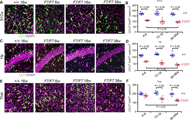
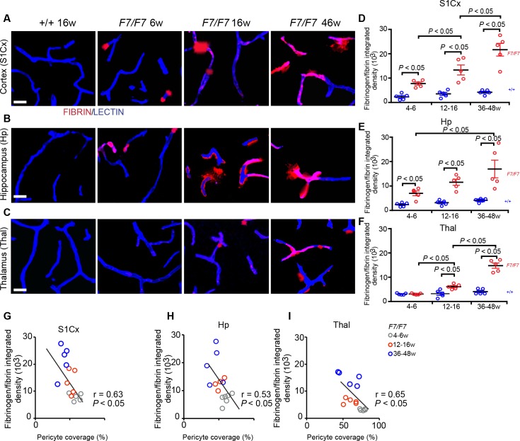
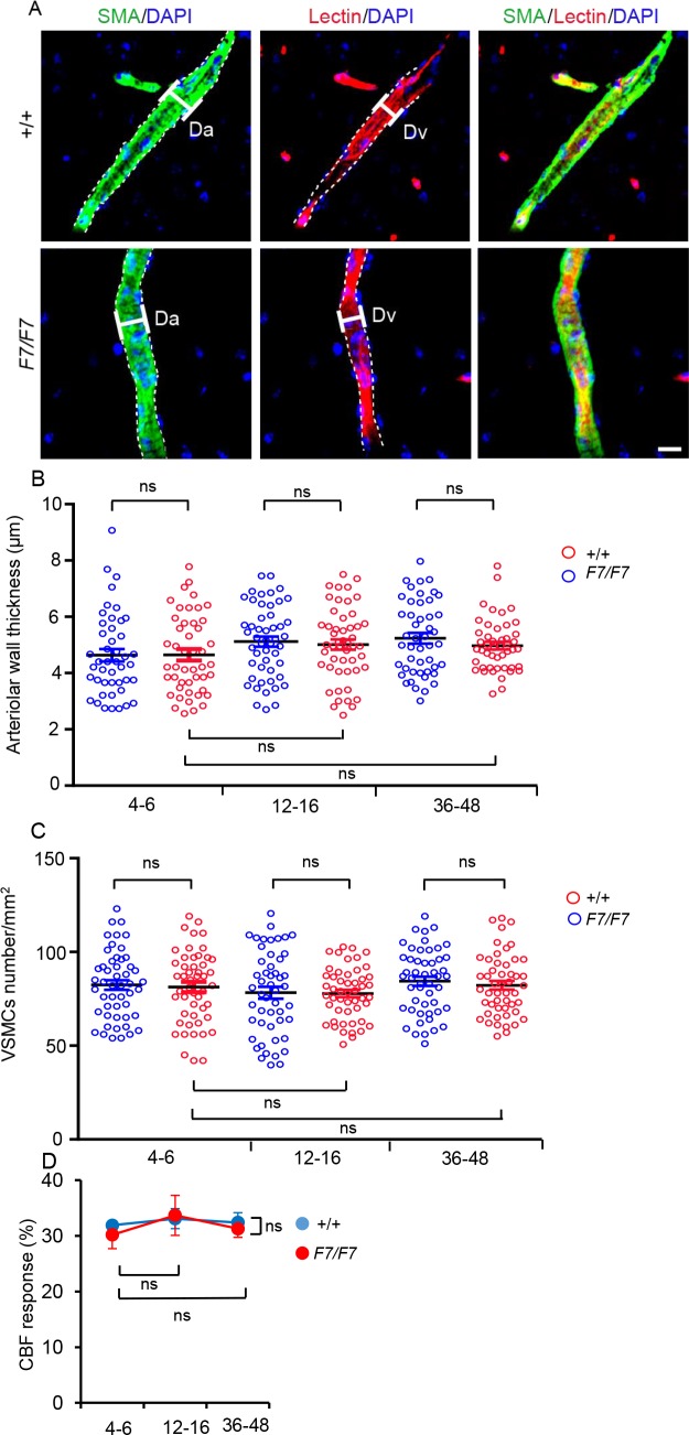
Pericytes regulate key neurovascular functions of the brain. Studies in pericyte-deficient transgenic mice with aberrant signaling between endothelial-derived platelet-derived growth factor BB (PDGF-BB) and platelet-derived growth factor receptor β (PDGFRβ) in pericytes have contributed to better understanding of the role of pericytes in the brain. Here, we studied PdgfrβF7/F7 mice, which carry seven point mutations that disrupt PDGFRβ signaling causing loss of pericytes and vascular smooth muscle cells (VSMCs) in the developing brain. We asked whether these mice have a stable or progressive vascular phenotype after birth, and whether both pericyte and VSMCs populations are affected in the adult brain. We found an early and progressive region-dependent loss of brain pericytes, microvascular reductions and blood-brain barrier (BBB) breakdown, which were more pronounced in the cortex, hippocampus and striatum than in the thalamus, whereas VSMCs population remained unaffected at the time when pericyte loss was already established. For example, compared to age-matched controls, PdgfrβF7/F7 mice between 4-6 and 36-48 weeks of age developed a region-dependent loss in pericyte coverage (22-46, 24-44 and 4-31%) and cell numbers (36-49, 34-64 and 11-36%), reduction in capillary length (20-39, 13-46 and 1-30%), and an increase in extravascular fibrinogen-derived deposits (3.4-5.2, 2.8-4.1 and 0-3.6-fold) demonstrating BBB breakdown in the cortex, hippocampus and thalamus, respectively. Capillary reductions and BBB breakdown correlated with loss of pericyte coverage. Our data suggest that PdgfrβF7/F7 mice develop an aggressive and rapid vascular phenotype without appreciable early involvement of VSMCs, therefore providing a valuable model to study regional effects of pericyte loss on brain vascular and neuronal functions. This model could be a useful tool for future studies directed at understanding the role of pericytes in the pathogenesis of neurological disorders associated with pericyte loss such as vascular dementia, Alzheimer's disease, amyotrophic lateral sclerosis, stroke and human immunodeficiency virus-associated neurocognitive disorder.
Overview
The claims and statistics of this paper haven’t been checked yet.
Run the check — free, no account needed. It reads the open-access text and checks the sources it cites.
Integrity signals
Registry
Cites
Statistics
Figures
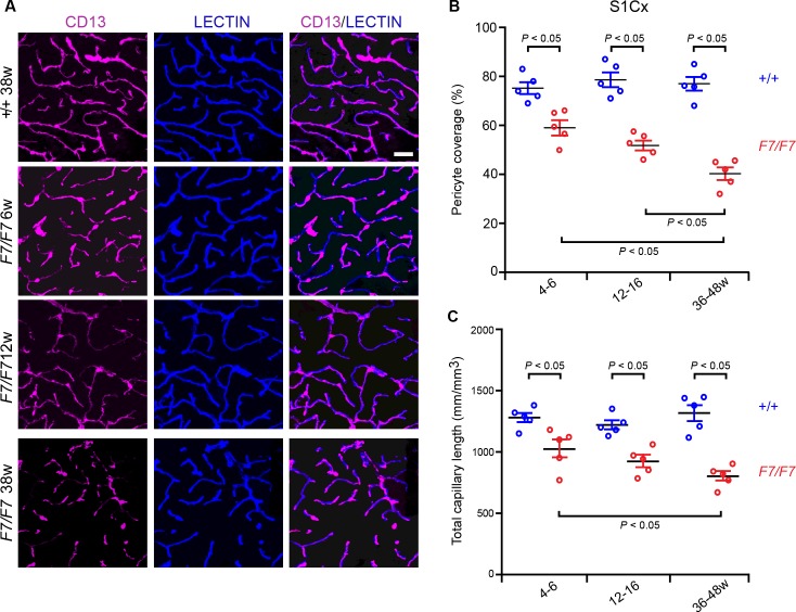
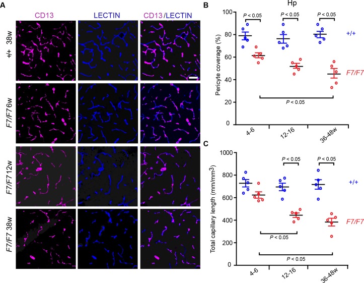
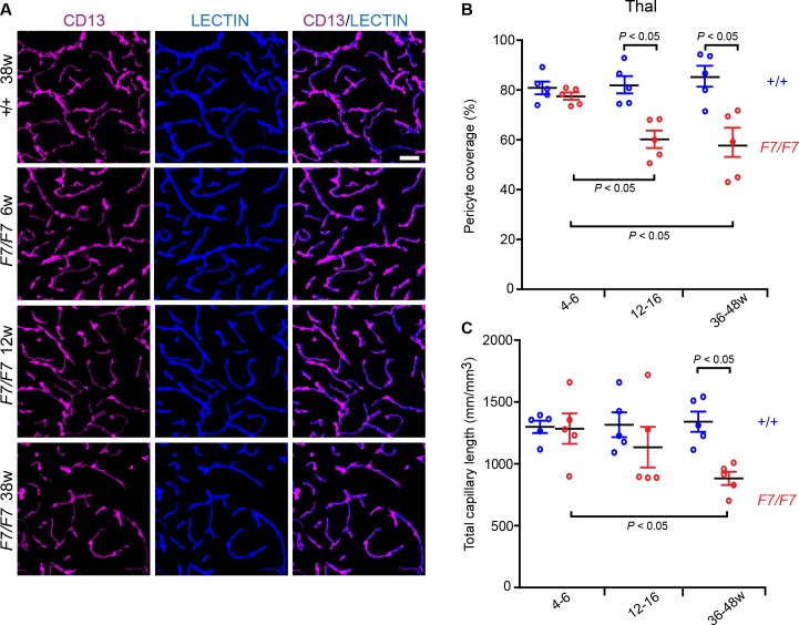
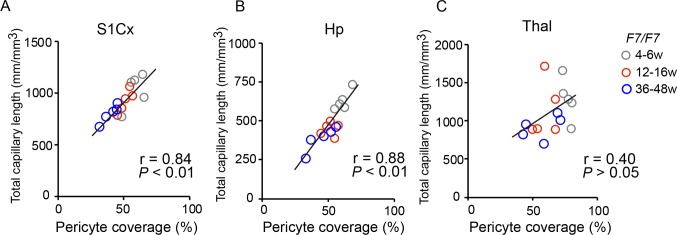
Verification
Claims the paper makes
The claims and statistics of this paper haven’t been checked yet.
Run the check — free, no account needed. It reads the open-access text and checks the sources it cites.
Claims other papers make about this one
Each is checked against this paper’s own text: does it say what the citing paper says it does?
The cited source says what the claim attributes to it.
In the citing paper
“Pericytedeficient mouse models caused by aberrant PDGF-BB/ PDGFR𝛃 signaling (365, 419, 436, 570, 575) exhibit impaired BBB formation (28, 59, 130) and maintenance (59, 436).”
From Blood-Brain Barrier: From Physiology to Disease and Back (2019) · cites this paper as [436]
Self-check
Percentages, sums and reported differences re-computed from the paper’s own numbers.
The self-check needs the paper’s full text.
Citations
Cites
1 retracted
RETRACTED ARTICLE: Pericyte loss influences Alzheimer-like neurodegeneration in mice
Sagare, A. P., Bell, R. D., Zhao, Z., Ma, Q., Winkler, E. A., Ramanathan, A., & Zlokovic, B. V. (2013) · Nature Communications · doi:10.1038/ncomms3932 · Cited by 589
Self-citationRetractedA Threshold Selection Method from Gray-Level Histograms
Otsu, N. (1979) · IEEE Transactions on Systems, Man and Cybernetics · doi:10.1109/tsmc.1979.4310076 · Cited by 44,708
Cahoy, J. D., Emery, B., Kaushal, A., Foo, L. C., Zamanian, J. L., Christopherson, K. S., Xing, Y., Lubischer, J. L., Krieg, P. A., Krupenko, S. A., et al. (2008) · The Journal of Neuroscience · doi:10.1523/jneurosci.4178-07.2008 · Cited by 3,129
Pericytes regulate the blood–brain barrier
Armulik, A., Genové, G., Mäe, M., Nisancioglu, M. H., Wallgard, E., Niaudet, C., He, L., Norlin, J., Lindblom, P., Strittmatter, K., et al. (2010) · Nature · doi:10.1038/nature09522 · Cited by 2,843
Pericytes: Developmental, Physiological, and Pathological Perspectives, Problems, and Promises
Armulik, A., Genové, G., & Betsholtz, C. (2011) · Developmental Cell · doi:10.1016/j.devcel.2011.07.001 · Cited by 2,653
Pericyte Loss and Microaneurysm Formation in PDGF-B-Deficient Mice
Lindahl, P., Johansson, B. R., Levéen, P., & Betsholtz, C. (1997) · Science · doi:10.1126/science.277.5323.242 · Cited by 2,226
Pericytes are required for blood–brain barrier integrity during embryogenesis
Daneman, R., Zhou, L., Kebede, A. A., & Barres, B. A. (2010) · Nature · doi:10.1038/nature09513 · Cited by 2,128
Blood-brain barrier breakdown in the aging human hippocampus
Montagne, A., Barnes, S. R., Sweeney, M. D., Halliday, M. R., Sagare, A. P., Zhao, Z., Toga, A. W., Jacobs, R. E., Liu, C. Y., Amezcua, L., et al. (2015) · Neuron · doi:10.1016/j.neuron.2014.12.032 · Cited by 2,102
Self-citationCapillary pericytes regulate cerebral blood flow in health and disease
Hall, C. N., Reynell, C., Gesslein, B., Hamilton, N. B., Mishra, A., Sutherland, B. A., O’Farrell, F. M., Buchan, A. M., Lauritzen, M., & Attwell, D. (2014) · Nature · doi:10.1038/nature13165 · Cited by 1,870
Hellström, M., Kalén, M., Lindahl, P., Abramsson, A., & Betsholtz, C. (1999) · Development · doi:10.1242/dev.126.14.3047 · Cited by 1,734
Cited by
Sorted by citations
Items 101–109 of 109
Wu, Y., Wu, H., Zeng, J., Pluimer, B. R., Dong, S., Xie, X., Guo, X., Ge, T., Liang, X., Feng, S., et al. (2021) · Figshare · doi:10.6084/m9.figshare.14478665
Self-citationLi, J., Li, M., Ge, Y., Chen, J., Ma, J., Wang, C., Sun, M., Wang, L., Yao, S., & Yao, C. (2022) · Figshare · doi:10.6084/m9.figshare.19906252
Li, L., Kong, J., Fan, R., Yuan, Y. Z., & Zhu, L. (2025) · Molecular Neurobiology · doi:10.1007/s12035-025-05603-z
“The resulting neurovascular uncoupling, decreased brain oxygenation and metabolic stress accelerate neurodegeneration [116], a process triggered by altered PDGFRβ signaling in the brain in humans with AD [117].”
Hu, S. X., Lopez-Robles, Y., Liu, E., Lin, Y., Chen, W., Manjunath, C., Zhao, Z., Zhang, L., & Ma, Q. (2026) · Fluids and Barriers of the CNS · doi:10.1186/s12987-025-00755-5
“Pericyte coverage was assessed by thresholding projected images with CD13 and CD31 signals separately using Otsu’s thresholding plugin of NIH Image J software 1.54e [41, 42].”
Self-citationCentral nervous system pericytes express soluble ST2 in inflammation and injury
Jansson, D., Highet, B., Li, S., Stevenson, T. J., Smyth, L. C. D., Rustenhoven, J., Feng, S. X., Daneman, R., Dorrier, C., Clarkson, A. N., et al. (2026) · Molecular Brain · doi:10.1186/s13041-026-01291-5
Rôle des péricytes et des cellules endothéliales dans l'insuffisance cardiaque
Cornuault, L. (2022) · theses.fr (ABES) · doi:10.70675/902151b1zd809z4b09z9562zd16a859cc3f2
Tsai, S., Liew, H., Wang, M.-J., Hung, W.-T., & Huang, H.-Y. (2026) · Tzu Chi Medical Journal · doi:10.4103/tcmj.tcmj-d-25-00173
“Adult mice show a regional and progressive loss of pericyte coverage (typically 40%–60% remains in the cortex and hippocampus), while VSMCs are relatively preserved [56].”
Mitochondrial control of blood-brain barrier homeostasis in neuroinflammatory psychiatric disorders
Pérez-López, L. M., Camacho-Morales, A., Álvarez, I. H., & Pérez‐Maya, A. A. (2026) · Frontiers in Molecular Neuroscience · doi:10.3389/fnmol.2026.1924581
“Importantly, oxidative and mitochondrial stress can activate matrix-remodeling pathways and alter pericyte– endothelial communication, suggesting that mitochondrial dysfunction may indirectly destabilize the ECM and amplify BBB disruption (Nikolakopoulou et al., 2017; Sun et al., 2021).”
Delgado-Minjares, K. M., Flores-Maldonado, C., Campos-Blázquez, J. P., Díaz-Cintra, S. Y., Andrade-Guerrero, J., Colín-García, W., León‐Arcia, K., Martínez‐Fong, D., Contreras, R. G., & Soto-Rojas, L. O. (2026) · International Journal of Molecular Sciences · doi:10.3390/ijms27198705
“We did not assess regional and zonation-related heterogeneity of pericytes and endothelial cells [44,45], and pericyte impairment and reduced PDGF-BB/PDGFR-β signaling have been identified in AD [46–48].”
Metadata
- Authors
- Nikolakopoulou, A. M., Zhao, Z., Montagne, A., & Zlokovic, B. V.
- Affiliations
- University of Southern California (US)
- Venue
- PLOS ONE
- Publisher
- Public Library of Science (PLoS)
- Published
- 2017-04-25
- Volume
- 12
- Issue
- 4
- Pages
- e0176225
- ISSN
- 1932-6203 (ISSN-L)
- PMC
- PMC5404855
- OpenAlex
- W2607556181
- Cited by
- 109
- References
- 44
- Citations by year
- 2026: 7 · 2025: 12 · 2024: 12 · 2023: 10 · 2022: 17 · 2021: 13 · 2020: 11 · 2019: 14 · 2018: 12 · 2017: 1
- FWCI
- 5.18
- Type
- journal article
- Language
- en
- Open access
- gold
- doi.org
- Licence
- Locations
- PLoS ONE (publishedVersion, cc-by)
- PubMed Central (publishedVersion, cc-by)
- PubMed (publishedVersion)
- DOAJ (DOAJ: Directory of Open Access Journals) (submittedVersion, cc-by-sa)
- Figshare (submittedVersion, cc-by)
- Funders
- National Institutes of Health (AG039452, AG023084, NS034467, NS100459)
- Topics
- Life Sciences › Neuroscience › Neurology › Barrier Structure and Function Studies
- Life Sciences › Biochemistry, Genetics and Molecular Biology › Molecular Biology › Angiogenesis and VEGF in Cancer
- Life Sciences › Neuroscience › Neurology › Neuroinflammation and Neurodegeneration Mechanisms
- Keywords
- brain pericytes, vascular smooth muscle cells, PDGFRβ signaling, PDGF-BB, pericyte loss, blood-brain barrier disruption, cerebral microvasculature, neurovascular unit
- MeSH terms
- Animals, Blood-Brain Barrier, Brain, Mice, Transgenic, Muscle, Smooth, Vascular, Signal Transduction, Pericytes, Receptor, Platelet-Derived Growth Factor beta, Myocytes, Smooth Muscle, Mice
About these checks
Registry, retraction, Cites and statistics results are computed, not judged, with one exception: a printed reference that no index lists is read once by a model before it counts as a citation error. Claims are extracted by a model and kept only when their quote appears verbatim in the text; each cited source is then checked, with a model’s help, for whether it says what the claim relies on. Those verdicts can be wrong — read the claim’s quote and the cited source before relying on one.
Statistics and arithmetic are recomputed, not judged.
Confidence bands are shown only for fields calibrated on our annotated test set. None is calibrated yet, so no band is shown.
What counts as a claim ›How we measure confidence ›How we check this ›
Are you an author, or do you see a problem with this page? Request a review
