

The DOI matches its Crossref record; 97 of the 97 references it cites were found in a registry or an index.
Differential Mechanisms of CRF1 and CRF2 Receptor Functions in the Amygdala in Pain-Related Synaptic Facilitation and Behavior
Open accessAbstract
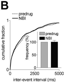
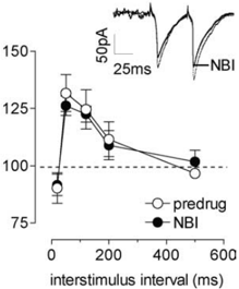
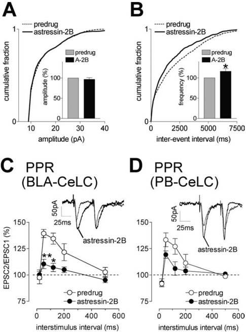
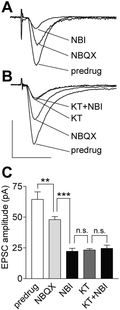
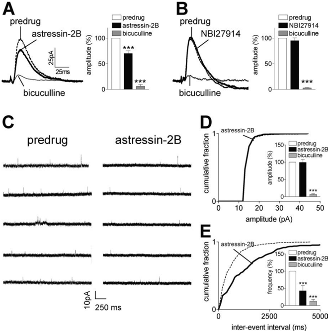
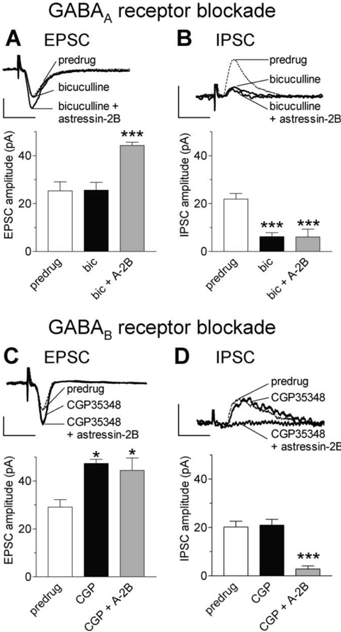
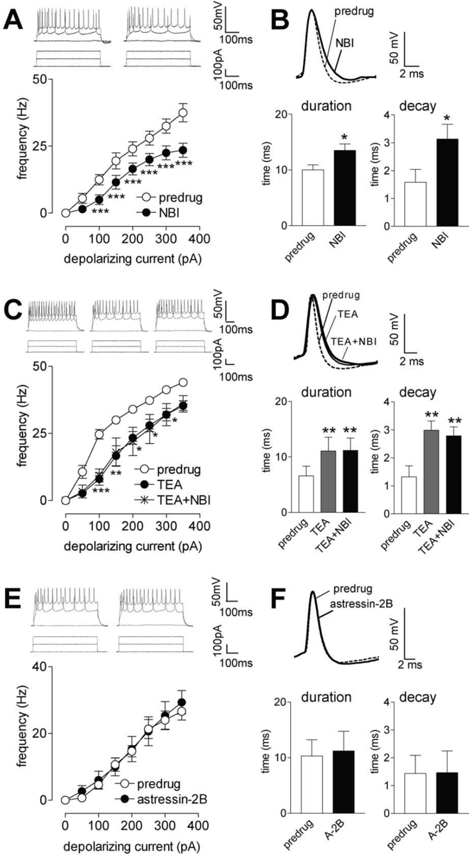
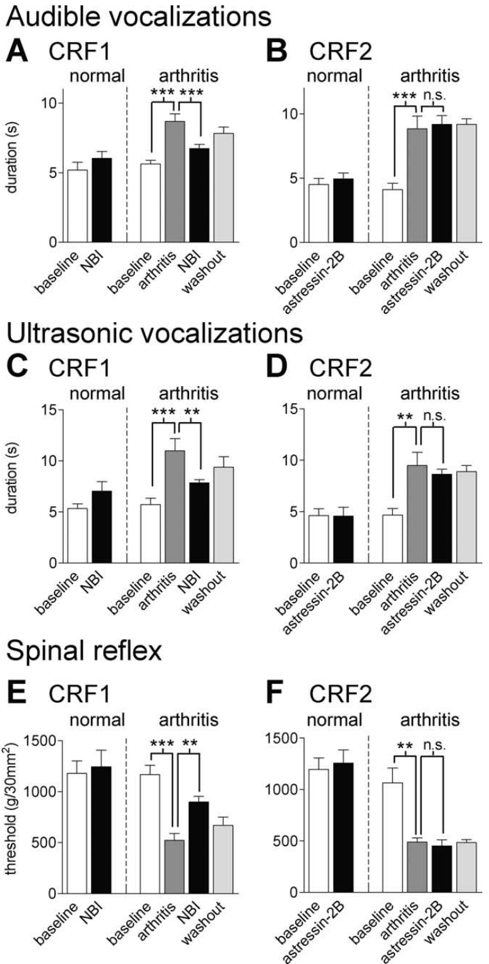
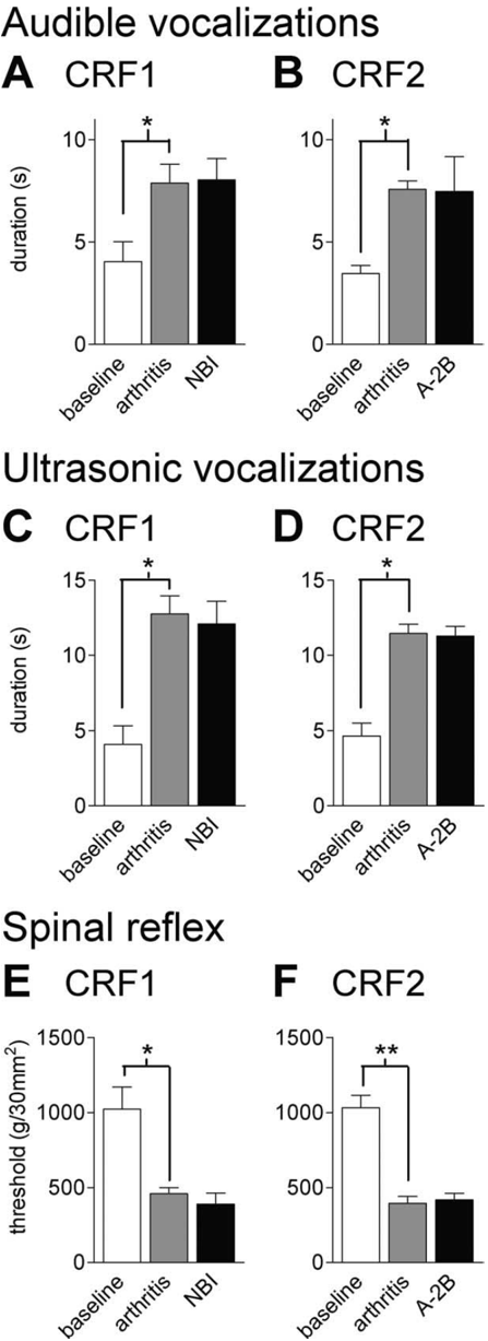
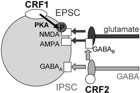
A major site of extrahypothalamic expression of corticotropin-releasing factor (CRF) and its G-protein-coupled CRF1 and CRF2 receptors is the amygdala, a key player in emotions and affective disorders. Pain-related plasticity in the laterocapsular division of the central nucleus of the amygdala (CeLC) generates emotional-affective responses and anxiety-like behavior. CRF1 receptor antagonists have anxiolytic effects. Although both CRF1 and CRF2 receptors couple positively to adenylyl cyclase, they can have opposite effects, but the underlying mechanism is unknown. This study addressed CRF1 and CRF2 receptor functions and mechanisms in the amygdala in a model of arthritic pain. Using whole-cell patch-clamp recordings of CeLC neurons, we found that a selective CRF1 receptor antagonist (NBI27914 [5-chloro-4-(N-(cyclopropyl)methyl-N-propylamino)-2-methyl-6-(2,4,6-trichlorophenyl)]) amino-pyridine inhibited synaptic facilitation in brain slices from arthritic rats through a postsynaptic mechanism. Inhibition of the NMDA receptor-mediated synaptic component was occluded by a protein kinase A (PKA) inhibitor, consistent with our previous demonstration of PKA-dependent increased NMDA receptor function in arthritis pain-related plasticity. NBI27914 also decreased neuronal excitability through inhibition of highly tetraethylammonium (TEA)-sensitive ion channels that contribute to action potential repolarization and firing rate. In contrast, a CRF2 receptor antagonist (astressin-2B [cyclo(31–34) [d-Phe11,His12,CαMeLeu13,39, Nle17, Glu31, Lys34] Ac-Sauvagine(8–40)]) facilitated synaptic transmission through presynaptic inhibition of GABAergic transmission (disinhibition). NBI27914 inhibited arthritis pain-related behaviors (audible and ultrasonic vocalizations and hindlimb withdrawal reflexes). Astressin-2B had no significant behavioral effect. The data suggest that endogenous CRF1 receptor activation in the amygdala contributes to pain-related synaptic facilitation, increased excitability, and pain behavior through a postsynaptic mechanism involving activation of PKA and highly TEA-sensitive K+-currents. Presynaptic CRF2 receptor-mediated inhibition does not reach behavioral significance.
Overview
The claims and statistics of this paper haven’t been checked yet.
Run the check — free, no account needed. It reads the open-access text and checks the sources it cites.
Integrity signals
Registry
Cites
Statistics
Figures
Verification
Claims the paper makes
The claims and statistics of this paper haven’t been checked yet.
Run the check — free, no account needed. It reads the open-access text and checks the sources it cites.
Claims other papers make about this one
Each is checked against this paper’s own text: does it say what the citing paper says it does?
In the citing paper
“In a model of arthritic pain, the blockade of CRF 1 receptors in the CeA inhibited both pain-and anxiety-like behaviors (Ji et al., 2007;Fu and Neugebauer, 2008).”
From Drug Addiction: Hyperkatifeia/Negative Reinforcement as a Framework for Medications Development (2021) · cites this paper as Fu and Neugebauer, 2008
Self-check
Percentages, sums and reported differences re-computed from the paper’s own numbers.
The self-check needs the paper’s full text.
Citations
Cites
The Rat Brain in Stereotaxic Coordinates
Paxinos, G., & Watson, C. (1983) · Elsevier eBooks · doi:10.1016/c2009-0-63235-9 · Cited by 28,585
AChE-stained horizontal sections of the rat brain in stereotaxic coordinates
Paxinos, G., WATSON, C. R. R., & Emson, P. C. (1980) · Journal of Neuroscience Methods · doi:10.1016/0165-0270(80)90021-7 · Cited by 4,514
CRF and CRF receptors: role in stress responsivity and other behaviors
Bale, T. L., & Vale, W. W. (2004) · Annual Review of Pharmacology and Toxicology · doi:10.1146/annurev.pharmtox.44.101802.121410 · Cited by 1,270
Protein kinase C reduces Mg2+ block of NMDA-receptor channels as a mechanism of modulation
Chen, L., & Huang, L.-Y. M. (1992) · Nature · doi:10.1038/356521a0 · Cited by 974
New Vistas on Amygdala Networks in Conditioned Fear
Paré, D., Quirk, G. J., & Ledoux, J. E. (2004) · Journal of Neurophysiology · doi:10.1152/jn.00153.2004 · Cited by 841 · cited 14×
“CRF mRNA expression in the CeA increased in models of colitis pain (Greenwood-Van Meerveld et al., 2006) and neuropathic pain (Ulrich-Lai et al., 2006).”
BackgroundInfluentialKv3 channels: voltage-gated K+ channels designed for high-frequency repetitive firing
Rudy, B., & McBain, C. J. (2001) · Trends in Neurosciences · doi:10.1016/s0166-2236(00)01892-0 · Cited by 781
Fear conditioning induces a lasting potentiation of synaptic currents in vitro
McKernan, M. G., & Shinnick-Gallagher, P. (1997) · Nature · doi:10.1038/37605 · Cited by 771
The amygdala and persistent pain
Neugebauer, V., Li, W., Bird, G. C., & Han, J. S. (2004) · The Neuroscientist · doi:10.1177/1073858403261077 · Cited by 719
Self-citationZobel, A. W., Nickel, T., Künzel, H. E., Ackl, N., Sonntag, A., Ising, M., & Holsboer, F. (2000) · Journal of Psychiatric Research · doi:10.1016/s0022-3956(00)00016-9 · Cited by 705
Presynaptic inhibition of elicited neurotransmitter release
Wu, L., & Saggau, P. (1997) · Trends in Neurosciences · doi:10.1016/s0166-2236(96)01015-6 · Cited by 627
Pain pathways and parabrachial circuits in the rat.
Gauriau, C., & Bernard, J. (2002) · Experimental Physiology · doi:10.1113/eph8702357 · Cited by 572
The CRF peptide family and their receptors: yet more partners discovered
Dautzenberg, F. M., & Hauger, R. L. (2002) · Trends in Pharmacological Sciences · doi:10.1016/s0165-6147(02)01946-6 · Cited by 534
Corticotropin-releasing factor receptors 1 and 2 in anxiety and depression
Reul, J. M. H. M. (2002) · Current Opinion in Pharmacology · doi:10.1016/s1471-4892(01)00117-5 · Cited by 515
Misgeld, U., Bijak, M., & Jarolimek, W. (1995) · Progress in Neurobiology · doi:10.1016/0301-0082(95)00012-k · Cited by 490
Erisir, A., Lau, D., Rudy, B., & Leonard, C. S. (1999) · Journal of Neurophysiology · doi:10.1152/jn.1999.82.5.2476 · Cited by 458
Corticotrophin-releasing factor receptors: from molecular biology to drug design
Chalmers, D. T., Lovenberg, T. W., Grigoriadis, D. E., Behan, D. P., & Souza, E. B. D. (1996) · Trends in Pharmacological Sciences · doi:10.1016/0165-6147(96)81594-x · Cited by 398
Martina, M., Schultz, J. H., Ehmke, H., Monyer, H., & Jonas, P. (1998) · The Journal of Neuroscience · doi:10.1523/jneurosci.18-20-08111.1998 · Cited by 396
Regulation of NMDA receptors by phosphorylation
Chen, B.-S., & Roche, K. W. (2007) · Neuropharmacology · doi:10.1016/j.neuropharm.2007.05.018 · Cited by 386
Prefrontal Control of the Amygdala
Likhtik, E., Pelletier, J. G., Paz, R., & Paré, D. (2005) · The Journal of Neuroscience · doi:10.1523/jneurosci.2314-05.2005 · Cited by 361
Inhibition of the Amygdala: Key to Pathological States?
Quirk, G. J., & Gehlert, D. R. (2003) · Annals of the New York Academy of Sciences · doi:10.1111/j.1749-6632.2003.tb07087.x · Cited by 314
CGP 35348: a centrally active blocker of GABAB receptors
Olpe, H.-R., Karlsson, G., Pozza, M. F., Brugger, F., Steinmann, M., van Riezen, H., Fagg, G. E., Hall, R. G., Froestl, W., & Bittiger, H. (1990) · European Journal of Pharmacology · doi:10.1016/0014-2999(90)90337-6 · Cited by 306
Contributions of Kv3 Channels to Neuronal Excitability
RUDY, B., CHOW, A., LAU, D., AMARILLO, Y., OZAITA, A., SAGANICH, M., MORENO, H., NADAL, M. S., HERNANDEZ‐PINEDA, R., HERNANDEZ‐CRUZ, A., et al. (1999) · Annals of the New York Academy of Sciences · doi:10.1111/j.1749-6632.1999.tb11295.x · Cited by 305
Neuroanatomical circuits modulating fear and anxiety behaviors
Charney, D. S. (2003) · Acta Psychiatrica Scandinavica · doi:10.1034/j.1600-0447.108.s417.3.x · Cited by 293
Rainnie, D. G., Bergeron, R., Sajdyk, T. J., Patil, M. M., Gehlert, D. R., & Shekhar, A. (2004) · Journal of Neuroscience · doi:10.1523/jneurosci.5740-03.2004 · Cited by 293
Amygdaloid CRF Pathways: Role in Autonomic, Neuroendocrine, and Behavioral Responses to Stress
Gray, T. S. (1993) · Annals of the New York Academy of Sciences · doi:10.1111/j.1749-6632.1993.tb49922.x · Cited by 288
Stress‐induced visceral hypersensitivity to rectal distension in rats: role of CRF and mast cells
Gué, M., Rio-Lacheze, C. D., Eutamène, H., Théodorou, V., Fioramonti, J., & Bueno, L. P. (1997) · Neurogastroenterology & Motility · doi:10.1046/j.1365-2982.1997.d01-63.x · Cited by 282
Microdialysis and SPECT measurements of amphetamine-induced dopamine release in nonhuman primates
Laruelle, M. A., Iyer, R. N., Al‐Tikriti, M. S., Zea‐Ponce, Y., Malison, R. T., Zoghbi, S. S., Baldwin, R. M., Kung, H. F., Charney, D. S., Hoffer, P. B., et al. (1997) · Synapse · doi:10.1002/(sici)1098-2396(199701)25:1 · Cited by 265
Neugebauer, V., Li, W., Bird, G. C., Bhave, G. B., & Gereau, R. W. (2003) · Journal of Neuroscience · doi:10.1523/jneurosci.23-01-00052.2003 · Cited by 249
Self-citationIkeda, R., Takahashi, Y., Inoue, K., & Kato, F. (2006) · Pain · doi:10.1016/j.pain.2006.09.003 · Cited by 243
The role of corticotropin-releasing factor in pain and analgesia
Lariviere, W. R., & Melzack, R. (2000) · Pain · doi:10.1016/s0304-3959(99)00193-1 · Cited by 230
Taché, Y., Martı́nez, V., Wang, L., & Million, M. (2004) · British Journal of Pharmacology · doi:10.1038/sj.bjp.0705760 · Cited by 228
Activation of the Extracellular Signal-Regulated Kinase in the Amygdala Modulates Pain Perception
Carrasquillo, Y., & Gereau, R. W. (2007) · Journal of Neuroscience · doi:10.1523/jneurosci.3536-06.2007 · Cited by 219
Networks of Parvalbumin-Positive Interneurons in the Basolateral Amygdala
Woodruff, A. R., & Sah, P. (2007) · Journal of Neuroscience · doi:10.1523/jneurosci.3686-06.2007 · Cited by 205
Blank, T., Nijholt, I. M., Grammatopoulos, D. K., Randeva, H. S., Hillhouse, E. W., & Spiess, J. (2003) · Journal of Neuroscience · doi:10.1523/jneurosci.23-02-00700.2003 · Cited by 204
Role of CRF 1 and CRF 2 receptors in fear and anxiety
Takahashi, L. K. (2001) · Neuroscience & Biobehavioral Reviews · doi:10.1016/s0149-7634(01)00046-x · Cited by 186
Techniques for Assessing Knee Joint Pain in Arthritis
Neugebauer, V., Han, J. S., Adwanikar, H., Fu, Y., & Ji, G. (2007) · Molecular Pain · doi:10.1186/1744-8069-3-8 · Cited by 180
Self-citationHan, J. S., Li, W., & Neugebauer, V. (2005) · Journal of Neuroscience · doi:10.1523/jneurosci.4112-05.2005 · Cited by 175
Self-citationDifferential Sensitization of Amygdala Neurons to Afferent Inputs in a Model of Arthritic Pain
Neugebauer, V., & Li, W. (2003) · Journal of Neurophysiology · doi:10.1152/jn.00799.2002 · Cited by 174
Self-citationThe role of emotion in pain modulation.
Rhudy, J. L., & Meagher, M. W. (2001) · Current Opinion in Psychiatry · doi:10.1097/00001504-200105000-00012 · Cited by 170
Rivier, J., Gulyás, J., Kirby, D., Low, W., Perrin, M. H., Kunitake, K. S., DiGruccio, M. R., Vaughan, J. M., Reubi, J. C., Waser, B., et al. (2002) · Journal of Medicinal Chemistry · doi:10.1021/jm0202122 · Cited by 160
Tanimoto, S., Nakagawa, T., Yamauchi, Y., Minami, M., & Satoh, M. (2003) · European Journal of Neuroscience · doi:10.1046/j.1460-9568.2003.02952.x · Cited by 159
Rome, H. P., & Rome, J. D. (2000) · Pain Medicine · doi:10.1046/j.1526-4637.2000.99105.x · Cited by 158
Cognitive-Behavioral Approach to the Treatment of Chronic Pain Patients
Turk, D. (2003) · Regional Anesthesia & Pain Medicine · doi:10.1016/s1098-7339(03)00392-4 · Cited by 155
Limbic and HPA axis function in an animal model of chronic neuropathic pain
Ulrich-Lai, Y. M., Xie, W., Meij, J. T. A., Dolgas, C. M., Yu, L., & Herman, J. P. (2006) · Physiology & Behavior · doi:10.1016/j.physbeh.2006.03.012 · Cited by 146
Schrader, L. A., & Tasker, J. G. (1997) · Journal of Neurophysiology · doi:10.1152/jn.1997.77.2.527 · Cited by 144
McNally, G. P., & Akil, H. (2002) · Neuroscience · doi:10.1016/s0306-4522(02)00105-7 · Cited by 141
Pain-Related Anxiety-Like Behavior Requires CRF1 Receptors in the Amygdala
Ji, G., Fu, Y., Ruppert, K. A., & Neugebauer, V. (2007) · Molecular Pain · doi:10.1186/1744-8069-3-13 · Cited by 139
Self-citationLiu, J., Yu, B., Neugebauer, V., Grigoriadis, D. E., Rivier, J., Vale, W., Shinnick‐Gallagher, P., & Gallagher, J. P. (2004) · Journal of Neuroscience · doi:10.1523/jneurosci.5531-03.2004 · Cited by 134
Self-citationBird, G. C., Lash, L. L., Han, J. S., Zou, X., Willis, W. D., & Neugebauer, V. (2005) · The Journal of Physiology · doi:10.1113/jphysiol.2005.084780 · Cited by 132
Self-citationSchwaber, J. S., Sternini, C., Brecha, N. C., Rogers, W. T., & Card, J. P. (1988) · The Journal of Comparative Neurology · doi:10.1002/cne.902700310 · Cited by 132
Han, J. S., Bird, G. C., Li, W., Jones, J., & Neugebauer, V. (2004) · Journal of Neuroscience Methods · doi:10.1016/j.jneumeth.2004.07.005 · Cited by 130
Self-citationNegative affect: effects on an evaluative measure of human pain
Rhudy, J. L., & Meagher, M. W. (2003) · Pain · doi:10.1016/s0304-3959(03)00119-2 · Cited by 129
Ji, G., & Neugebauer, V. (2007) · Journal of Neurophysiology · doi:10.1152/jn.00135.2007 · Cited by 125
Self-citationFaber, E. S. L., & Sah, P. (2003) · The Journal of Physiology · doi:10.1113/jphysiol.2003.050120 · Cited by 121
Pedersen, L. H., Scheel‐Krüger, J., & Blackburn-Munro, G. (2006) · Pain · doi:10.1016/j.pain.2006.06.036 · Cited by 120
Han, J. S., & Neugebauer, V. (2004) · Pain · doi:10.1016/j.pain.2004.10.022 · Cited by 117
Self-citationMillion, M., Wang, L., Wang, Y., Adelson, D. W., Yuan, P.-Q., Maillot, C., Coutinho, S. V., Mcroberts, J. A., Bayati, A., Mattsson, H., et al. (2005) · Gut · doi:10.1136/gut.2004.051391 · Cited by 115
Wyllie, D. J. A., Manabe, T., & Nicoll, R. A. (1994) · Neuron · doi:10.1016/0896-6273(94)90158-9 · Cited by 112
Meerveld, B. G., Johnson, A. C., Cochrane, S. W., Schulkin, J. A., & Myers, D. A. (2005) · Neurogastroenterology & Motility · doi:10.1111/j.1365-2982.2005.00648.x · Cited by 111
Li, W., & Neugebauer, V. (2004) · Journal of Neurophysiology · doi:10.1152/jn.00485.2003 · Cited by 109
Self-citationMillion, M., Grigoriadis, D. E., Sullivan, S., Crowe, P. D., McRoberts, J. A., Zhou, H., Saunders, P. R., Maillot, C., Mayer, E. A., & Taché, Y. (2003) · Brain Research · doi:10.1016/s0006-8993(03)03027-0 · Cited by 107
Urocortins and the regulation of gastrointestinal motor function and visceral pain
Martínez, V., Wang, L., Million, M., Rivier, J., & Taché, Y. (2004) · Peptides · doi:10.1016/j.peptides.2004.05.025 · Cited by 106
Porter, J. T., & Nieves, D. (2004) · Journal of Neurophysiology · doi:10.1152/jn.00196.2004 · Cited by 99
CRF signaling: molecular specificity for drug targeting in the CNS
ARZT, E. S., & Holsboer, F. (2006) · Trends in Pharmacological Sciences · doi:10.1016/j.tips.2006.08.007 · Cited by 96
Asan, E., Yilmazer‐Hanke, D., Eliava, M., Hantsch, M., Lesch, K., & Schmitt, A. G. (2005) · Neuroscience · doi:10.1016/j.neuroscience.2004.11.040 · Cited by 95
Firing Properties and Connectivity of Neurons in the Rat Lateral Central Nucleus of the Amygdala
de Armentia, M. L., & Sah, P. (2004) · Journal of Neurophysiology · doi:10.1152/jn.00211.2004 · Cited by 91
Kv3 K+ channels enable burst output in rat cerebellar Purkinje cells
McKay, B. E., & Turner, R. W. (2004) · European Journal of Neuroscience · doi:10.1111/j.1460-9568.2004.03539.x · Cited by 89
Li, W., & Neugebauer, V. (2004) · Pain · doi:10.1016/j.pain.2004.03.015 · Cited by 88
Self-citationDifferences between presynaptic and postsynaptic GABAB mechanisms in rat hippocampal pyramidal cells
Pitler, T. A., & Alger, B. E. (1994) · Journal of Neurophysiology · doi:10.1152/jn.1994.72.5.2317 · Cited by 86
Pollandt, S., Liu, J., Orozco-Cabal, L. F., Grigoriadis, D. E., Vale, W., Gallagher, J. P., & Shinnick‐Gallagher, P. (2006) · European Journal of Neuroscience · doi:10.1111/j.1460-9568.2006.05049.x · Cited by 82
Physiological properties of central amygdala neurons: species differences
Dumont, É. C., Martina, M., Samson, R. D., Drolet, G., & Paré, D. (2002) · European Journal of Neuroscience · doi:10.1046/j.0953-816x.2001.01879.x · Cited by 78
Cabell, L., & Audesirk, G. J. (1993) · International Journal of Developmental Neuroscience · doi:10.1016/0736-5748(93)90007-z · Cited by 73
Myers, D. A., Gibson, M. S., Schulkin, J. A., & van-Meerveld, B. G. (2005) · Behavioural Brain Research · doi:10.1016/j.bbr.2005.03.001 · Cited by 69
Enhanced group III mGluR-mediated inhibition of pain-related synaptic plasticity in the amygdala
Han, J. S., Bird, G. C., & Neugebauer, V. (2004) · Neuropharmacology · doi:10.1016/j.neuropharm.2004.01.006 · Cited by 65
Self-citationHarrigan, E. A., Magnuson, D. J., Thunstedt, G. M., & Gray, T. S. (1994) · Brain Research Bulletin · doi:10.1016/0361-9230(94)90079-5 · Cited by 64
Mechanism of Corticotropin-Releasing Factor Type I Receptor Regulation by Nonpeptide Antagonists
Hoare, S. R. J., Sullivan, S., LING, N. C., Crowe, P. D., & Grigoriadis, D. E. (2003) · Molecular Pharmacology · doi:10.1124/mol.63.3.751 · Cited by 63
Hoare, S. R. J., Sullivan, S. K., Fan, J. Y., Khongsaly, K., & Grigoriadis, D. E. (2004) · Peptides · doi:10.1016/j.peptides.2004.10.019 · Cited by 62
Kasten, M. R., Rudy, B., & Anderson, M. P. (2007) · The Journal of Physiology · doi:10.1113/jphysiol.2007.141135 · Cited by 59
Li, W., & Neugebauer, V. (2006) · Journal of Neurophysiology · doi:10.1152/jn.00495.2006 · Cited by 58
Self-citationDivergent role for CRF1 and CRF2 receptors in the modulation of visceral pain
Nijsen, M., Ongenae, N., Meulemans, A., & Coulie, B. J. (2005) · Neurogastroenterology & Motility · doi:10.1111/j.1365-2982.2005.00644.x · Cited by 52
Grachev, I. D., Fredickson, B. E., & Apkarian, A. V. (2001) · Molecular Psychiatry · doi:10.1038/sj.mp.4000834 · Cited by 52
Vit, J., Clauw, D. J., Moallem, T. M., Boudah, A., Ohara, P. T., & Jasmin, L. (2006) · Pain · doi:10.1016/j.pain.2005.12.024 · Cited by 49
The effect of amygdala lesions on conditional and unconditional vocalizations in rats
Borszcz, G. S., & Leaton, R. N. (2003) · Neurobiology of Learning and Memory · doi:10.1016/s1074-7427(03)00002-9 · Cited by 47
Meerveld, B. G., Johnson, A. C., Schulkin, J. A., & Myers, D. A. (2006) · Brain Research · doi:10.1016/j.brainres.2005.11.071 · Cited by 47
Properties and expression of Kv3 channels in cerebellar Purkinje cells
Sacco, T., Luca, A. D., & Tempia, F. (2006) · Molecular and Cellular Neuroscience · doi:10.1016/j.mcn.2006.07.006 · Cited by 45
Bidirectional synaptic plasticity at nociceptive afferents in the rat central amygdala
de Armentia, M. L., & Sah, P. (2007) · The Journal of Physiology · doi:10.1113/jphysiol.2006.121822 · Cited by 44
Chapter 11 Subcortical processing of nociceptive information: basal ganglia and amygdala
Neugebauer, V. (2006) · Handbook of clinical neurology · doi:10.1016/s0072-9752(06)80015-1 · Cited by 42
Self-citationStressin1-A, a Potent Corticotropin Releasing Factor Receptor 1 (CRF1)-Selective Peptide Agonist
Rivier, J., Gulyás, J., Kunitake, K. S., DiGruccio, M. R., Cantle, J. P., Perrin, M. H., Donaldson, C., Vaughan, J. M., Million, M., Gourcerol, G., et al. (2007) · Journal of Medicinal Chemistry · doi:10.1021/jm0613875 · Cited by 42
Joanny, P., Steinberg, J., Oliver, C., & Grino, M. (1997) · Journal of Neuroendocrinology · doi:10.1046/j.1365-2826.1997.00548.x · Cited by 41
Enhanced Group II mGluR-Mediated Inhibition of Pain-Related Synaptic Plasticity in the Amygdala
Han, J. S., Fu, Y., Bird, G. C., & Neugebauer, V. (2006) · Molecular Pain · doi:10.1186/1744-8069-2-18 · Cited by 40
Self-citationMcDonald, A. J., & Mascagni, F. (2006) · Neuroscience · doi:10.1016/j.neuroscience.2005.11.047 · Cited by 38
Potes, C. S., Neto, F. L. M., & Castro‐Lopes, J. M. (2006) · Brain Research · doi:10.1016/j.brainres.2006.07.089 · Cited by 34
Sinniger, V., Porcher, C., Mouchet, P., Juhem, A., & Bonaz, B. L. (2004) · Pain · doi:10.1016/j.pain.2004.05.014 · Cited by 33
Kunzel, H. E., Ising, M., Zobel, A. W., Nickel, T., Ackl, N., Sonntag, A., Holsboer, F., & Uhr, M. (2004) · Journal of Psychiatric Research · doi:10.1016/j.jpsychires.2004.06.006 · Cited by 31
Microdialysis in pain research
Stiller, C., Taylor, B. K., Linderoth, B., Gustafsson, H., Afrah, A. W., & Brodin, E. (2003) · Advanced Drug Delivery Reviews · doi:10.1016/s0169-409x(03)00104-2 · Cited by 31
Hirakura, Y., Lin, M.-C., & Kagan, B. (1999) · Journal of Neuroscience Research · doi:10.1002/(sici)1097-4547(19991201)58:5 · Cited by 25
Pain-Modulating Neurons and Behavioral State
Heinricher, M. M., & McGaraughty, S. (2019) · CRC Press eBooks · doi:10.1201/9780429114373-31 · Cited by 3
Research works printed but not linked: 1
- Gallagher, 2004, Mood and anxiety disorders in chronic pain, Prog Pain Res Management, 27, 139
Cited by
Sorted by citations
Central mechanisms of pathological pain
Kuner, R. (2010) · Nature Medicine · doi:10.1038/nm.2231 · Cited by 795
Strang, J., Volkow, N. D., Degenhardt, L., Hickman, M., Johnson, K., Koob, G. F., Marshall, B. D. L., Tyndall, M., & Walsh, S. L. (2020) · Nature Reviews Disease Primers · doi:10.1038/s41572-019-0137-5 · Cited by 639
Role of Corticotropin-Releasing Factor in Drug Addiction
Logrip, M. L., Koob, G. F., & Zorrilla, E. P. (2011) · CNS Drugs · doi:10.2165/11587790-000000000-00000 · Cited by 474
Neurobiology of opioid addiction: Opponent process, hyperkatifeia, and negative reinforcement
Koob, G. F. (2020) · Biological Psychiatry · doi:10.1016/j.biopsych.2019.05.023 · Cited by 477
Neugebauer, V. (2015) · Handbook of experimental pharmacology · doi:10.1007/978-3-662-46450-2_13 · Cited by 419
Self-citation
Metadata
- Authors
- Fu, Y., & Neugebauer, V.
- Affiliations
- The University of Texas Medical Branch at Galveston (US)
- Venue
- The Journal of Neuroscience
- Publisher
- Society for Neuroscience
- Published
- 2008-04-09
- Volume
- 28
- Issue
- 15
- Pages
- 3861-3876
- ISSN
- 0270-6474 (ISSN-L)
- 1529-2401
- PMC
- PMC2557030
- OpenAlex
- W2050750919
- Cited by
- 184
- References
- 97
- Citations by year
- 2026: 5 · 2025: 6 · 2024: 7 · 2023: 4 · 2022: 7 · 2021: 9 · 2020: 13 · 2019: 11 · 2018: 12 · 2017: 8 · 2016: 11 · 2015: 17 · 2014: 16 · 2013: 9 · 2012: 7
- FWCI
- 6.06
- Type
- journal article
- Language
- en
- Open access
- hybrid
- doi.org
- Licence
- Locations
- Journal of Neuroscience (publishedVersion, cc-by-nc-sa)
- PubMed Central (publishedVersion)
- PubMed (publishedVersion)
- Funders
- Northwestern University
- National Institutes of Health
- National Institute of Neurological Disorders and Stroke (NS-38261, NS11255, P01NS011255, R01NS038261)
- Topics
- Life Sciences › Neuroscience › Cellular and Molecular Neuroscience › Neuropeptides and Animal Physiology
- Life Sciences › Neuroscience › Cellular and Molecular Neuroscience › Neuroscience and Neuropharmacology Research
- Health Sciences › Medicine › Physiology (medical) › Pain Mechanisms and Treatments
- Keywords
- corticotropin-releasing factor, CRF1 receptor, CRF2 receptor, amygdala, central amygdala, arthritic pain, protein kinase A, NMDA receptor, neuronal excitability, presynaptic inhibition, GABAergic neurotransmission, anxiety-like behavior, ultrasonic vocalizations
- MeSH terms
- CRF Receptor, Type 1, Amygdala, Aniline Compounds, Animals, Arthritis, Behavior, Animal, Electrophysiology, Enzyme Activation, Hindlimb, Neurons, Pain, Peptide Fragments, Peptides, Cyclic, Pyrimidines, Reflex, Spinal Cord, Synapses, Vocalization, Animal, Potassium Channels, Rats, Sprague-Dawley, Cyclic AMP-Dependent Protein Kinases, Receptors, Corticotropin-Releasing Hormone, Patch-Clamp Techniques, Tetraethylammonium, Rats, In Vitro Techniques
About these checks
Registry, retraction, Cites and statistics results are computed, not judged, with one exception: a printed reference that no index lists is read once by a model before it counts as a citation error. Claims are extracted by a model and kept only when their quote appears verbatim in the text; each cited source is then checked, with a model’s help, for whether it says what the claim relies on. Those verdicts can be wrong — read the claim’s quote and the cited source before relying on one.
Statistics and arithmetic are recomputed, not judged.
Confidence bands are shown only for fields calibrated on our annotated test set. None is calibrated yet, so no band is shown.
What counts as a claim ›How we measure confidence ›How we check this ›
Are you an author, or do you see a problem with this page? Request a review
