

The DOI matches its Crossref record; 10 of the 10 references it cites were found in a registry or an index.
Inhibition of p25/Cdk5 Attenuates Tauopathy in Mouse and iPSC Models of Frontotemporal Dementia
Abstract
Increased p25, a proteolytic fragment of the regulatory subunit p35, is known to induce aberrant activity of cyclin-dependent kinase 5 (Cdk5), which is associated with neurodegenerative disorders, including Alzheimer's disease. Previously, we showed that replacing endogenous p35 with the noncleavable mutant p35 (Δp35) attenuated amyloidosis and improved cognitive function in a familial Alzheimer's disease mouse model. Here, to address the role of p25/Cdk5 in tauopathy, we generated double-transgenic mice by crossing mice overexpressing mutant human tau (P301S) with Δ p35KI mice. We observed significant reduction of phosphorylated tau and its seeding activity in the brain of double transgenic mice compared with the P301S mice. Furthermore, synaptic loss and impaired LTP at hippocampal CA3 region of P301S mice were attenuated by blocking p25 generation. To further validate the role of p25/Cdk5 in tauopathy, we used frontotemporal dementia patient-derived induced pluripotent stem cells (iPSCs) carrying the Tau P301L mutation and generated P301L:Δp35KI isogenic iPSC lines using CRISPR/Cas9 genome editing. We created cerebral organoids from the isogenic iPSCs and found that blockade of p25 generation reduced levels of phosphorylated tau and increased expression of synaptophysin. Together, these data demonstrate a crucial role for p25/Cdk5 in mediating tau-associated pathology and suggest that inhibition of this kinase can remedy neurodegenerative processes in the presence of pathogenic tau mutation. SIGNIFICANCE STATEMENT Accumulation of p25 results in aberrant Cdk5 activation and induction of numerous pathological phenotypes, such as neuroinflammation, synaptic loss, Aβ accumulation, and tau hyperphosphorylation. However, it was not clear whether p25/Cdk5 activity is necessary for the progression of these pathological changes. We recently developed the Δ p35KI transgenic mouse that is deficient in p25 generation and Cdk5 hyperactivation. In this study, we used this mouse model to elucidate the role of p25/Cdk5 in FTD mutant tau-mediated pathology. We also used a frontotemporal dementia patient-derived induced pluripotent stem cell carrying the Tau P301L mutation and generated isogenic lines in which p35 is replaced with noncleavable mutant Δp35. Our data suggest that p25/Cdk5 plays an important role in tauopathy in both mouse and human model systems.
Overview
The claims and statistics of this paper haven’t been checked yet.
Run the check — free, no account needed. It reads the open-access text and checks the sources it cites.
Integrity signals
Registry
Cites
Statistics
Figures
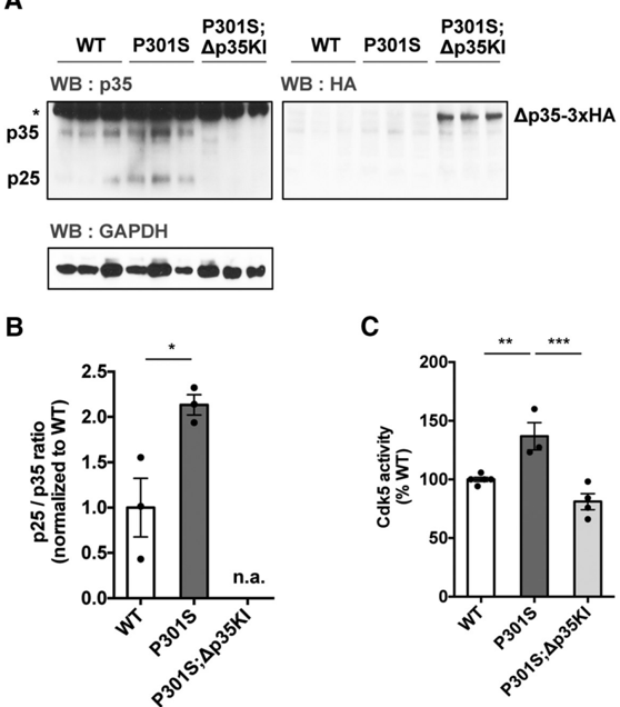
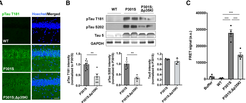
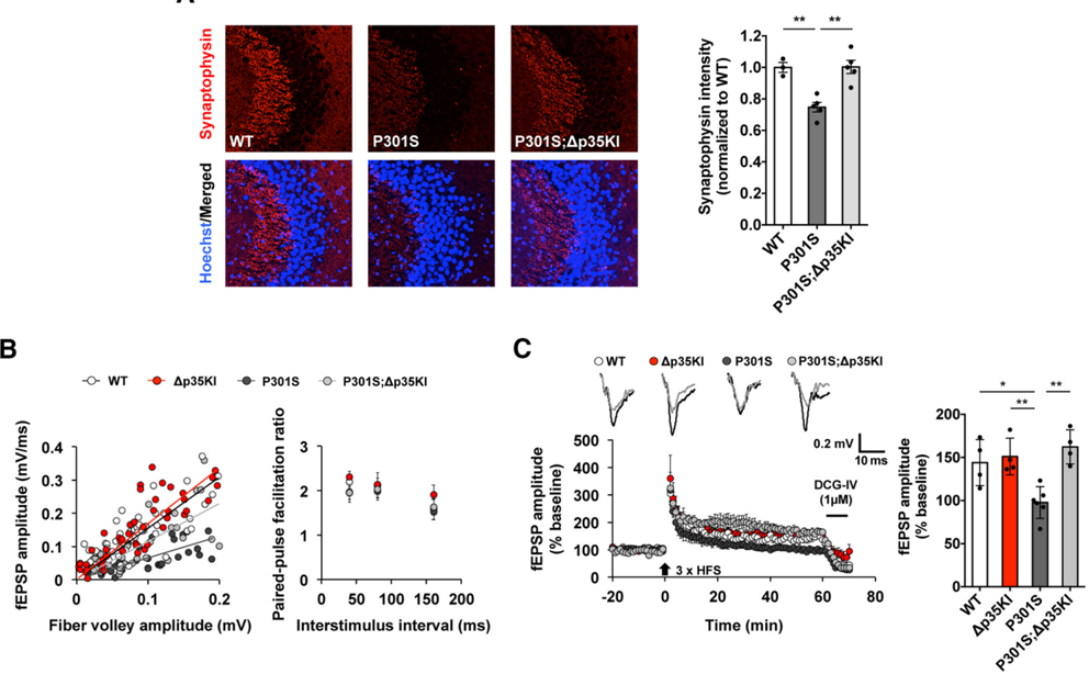
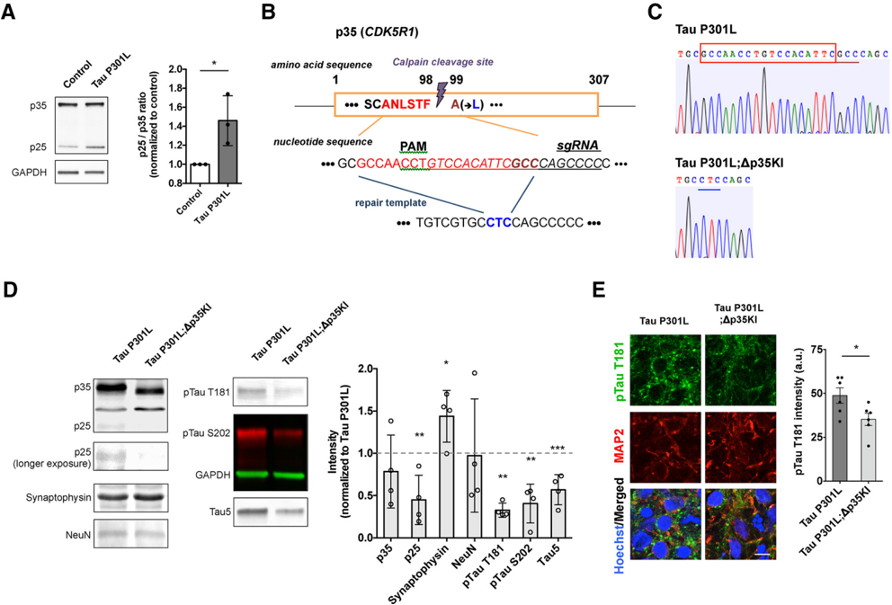
Verification
Claims the paper makes
The claims and statistics of this paper haven’t been checked yet.
Run the check — free, no account needed. It reads the open-access text and checks the sources it cites.
Claims other papers make about this one
Each is checked against this paper’s own text: does it say what the citing paper says it does?
In the citing paper
“The HNO model has been shown to recapitulate some of the key pathological features of AD and offers greater complexity and translational potential as compared to conventional cell and animal models92–95.”
From GRAMD1B is a regulator of lipid homeostasis, autophagic flux and phosphorylated tau (2025) · cites this paper as [92]
Self-check
Percentages, sums and reported differences re-computed from the paper’s own numbers.
The self-check needs the paper’s full text.
Citations
Cites
Genome engineering using the CRISPR-Cas9 system
Ran, F. A., Hsu, P. D., Wright, J., Agarwala, V., Scott, D. A., & Zhang, F. (2013) · Nature Protocols · doi:10.1038/nprot.2013.143 · Cited by 11,133
Hypothetical model of dynamic biomarkers of the Alzheimer's pathological cascade
Jack, C. R., Knopman, D. S., Jagust, W. J., Shaw, L. M., Aisen, P. S., Weiner, M. W., Petersen, R. C., & Trojanowski, J. Q. (2010) · The Lancet Neurology · doi:10.1016/s1474-4422(09)70299-6 · Cited by 4,806
Synapse Loss and Microglial Activation Precede Tangles in a P301S Tauopathy Mouse Model
Yoshiyama, Y., Higuchi, M., Zhang, B., Huang, S.-M., Iwata, N., Saido, T. C., Maeda, J., Suhara, T., Trojanowski, J. Q., & Lee, V. M.-Y. (2007) · Neuron · doi:10.1016/j.neuron.2007.01.010 · Cited by 2,208
Transmission and spreading of tauopathy in transgenic mouse brain
Clavaguera, F., Bolmont, T., Crowther, R. A., Abramowski, D., Frank, S., Probst, A., Fraser, G., Stalder, A. K., Beibel, M., Staufenbiel, M., et al. (2009) · Nature Cell Biology · doi:10.1038/ncb1901 · Cited by 1,803
Propagation of Tau Pathology in a Model of Early Alzheimer's Disease
de Calignon, A., Polydoro, M., Suárez-Calvet, M., William, C., Adamowicz, D. H., Kopeikina, K. J., Pitstick, R., Sahara, N., Ashe, K. H., Carlson, G. A., et al. (2012) · Neuron · doi:10.1016/j.neuron.2011.11.033 · Cited by 1,427
Probing sporadic and familial Alzheimer’s disease using induced pluripotent stem cells
Israel, M. A., Yuan, S. H., Bardy, C., Reyna, S. M., Mu, Y., Herrera, C., Hefferan, M. P., Van Gorp, S., Nazor, K. L., Boscolo, F. S., et al. (2012) · Nature · doi:10.1038/nature10821 · Cited by 1,253
Propagation of Tau Misfolding from the Outside to the Inside of a Cell
Frost, B., Jacks, R. L., & Diamond, M. I. (2009) · Journal of Biological Chemistry · doi:10.1074/jbc.m808759200 · Cited by 1,252
Raja, W. K., Mungenast, A. E., Lin, Y.-T., Ko, T., Abdurrob, F., Seo, J., & Tsai, L.-H. (2016) · PLOS ONE · doi:10.1371/journal.pone.0161969 · Cited by 569
Self-citationUntangling tau hyperphosphorylation in drug design for neurodegenerative diseases
Mazanetz, M. P., & Fischer, P. M. (2007) · Nature Reviews Drug Discovery · doi:10.1038/nrd2111 · Cited by 459
Downregulation of MicroRNA-9 in iPSC-Derived Neurons of FTD/ALS Patients with TDP-43 Mutations
Zhang, Z., Almeida, S., Lu, Y., Nishimura, A. L., Peng, L., Sun, D., Wu, B., Karydas, A. M., Tartaglia, M. C., Fong, J. C., et al. (2013) · PLoS ONE · doi:10.1371/journal.pone.0076055 · Cited by 142
Research works printed but not linked: 27
- 10.1371/journal.pone.0010810 · DOI
- 10.1073/pnas.040577797 · DOI
- 10.1016/S0896-6273(03)00627-5 · DOI · Paper page
- 10.1016/j.stemcr.2013.08.001 · DOI · Paper page
- 10.1074/jbc.M207426200 · DOI
- 10.1073/pnas.1411649111 · DOI · Paper page
- 10.1016/j.cell.2010.06.036 · DOI · Paper page
- 10.3389/fnmol.2014.00065 · DOI
- 10.1155/2012/731526 · DOI
- 10.1016/j.cell.2016.10.044 · DOI
- 10.1002/embj.201387009 · DOI
- 10.1074/jbc.M907757199 · DOI
- 10.1038/35012636 · DOI
- 10.1523/JNEUROSCI.3716-14.2015 · DOI
- 10.1186/scrt105 · DOI
- 10.1093/hmg/ddu064 · DOI · Paper page
- 10.1006/bbrc.2000.3070 · DOI
- 10.1016/S0896-6273(03)00259-9 · DOI
- 10.1038/45159 · DOI
- 10.1523/JNEUROSCI.3637-10.2010 · DOI
- 10.1523/JNEUROSCI.1132-14.2014 · DOI
- 10.1016/j.neuron.2016.04.042 · DOI · Paper page
- 10.1016/j.cell.2014.01.065 · DOI
- 10.1038/nn.4023 · DOI · Paper page
- 10.1016/j.stemcr.2016.08.001 · DOI · Paper page
- 10.1006/nbdi.2000.0333 · DOI
- 10.1074/jbc.M112.436402 · DOI
Cited by
Sorted by citations
Building Models of Brain Disorders with Three-Dimensional Organoids
Amin, N. D., & Paşca, S. P. (2018) · Neuron · doi:10.1016/j.neuron.2018.10.007 · Cited by 312
Targeted degradation of aberrant tau in frontotemporal dementia patient-derived neuronal cell models
Silva, M. C., Ferguson, F. M., Cai, Q., Donovan, K. A., Nandi, G., Patnaik, D., Zhang, T., Huang, H.-T., Lucente, D. E., Dickerson, B. C., et al. (2019) · eLife · doi:10.7554/elife.45457 · Cited by 283
Self-citationBioengineering Approaches for the Advanced Organoid Research
Yi, S. A., Zhang, Y., Rathnam, C., Pongkulapa, T., & Lee, K. (2021) · Advanced Materials · doi:10.1002/adma.202007949 · Cited by 218
Silva, M. C., Nandi, G. A., Tentarelli, S., Gurrell, I. K., Jamier, T., Lucente, D., Dickerson, B. C., Brown, D. G., Brandon, N. J., & Haggarty, S. J. (2020) · Nature Communications · doi:10.1038/s41467-020-16984-1 · Cited by 157
Self-citationAlzheimer’s disease: Ablating single master site abolishes tau hyperphosphorylation
Stefanoska, K., Gajwani, M., Tan, A. R. P., Ahel, H. I., Asih, P. R., Volkerling, A., Poljak, A., & Ittner, A. (2022) · Science Advances · doi:10.1126/sciadv.abl8809 · Cited by 128
A comprehensive resource for induced pluripotent stem cells from patients with primary tauopathies
Karch, C. M., Kao, A. W., Karydas, A., Onanuga, K., Martinez, R., Argouarch, A., Wang, C., Huang, C., Sohn, P. D., Bowles, K. R., et al. (2019) · Stem Cell Reports · doi:10.1016/j.stemcr.2019.09.006 · Cited by 119
Self-citationMahaman, Y. A. R., Huang, F., Wu, M., Wang, Y., Wei, Z., Bao, J., Salissou, M. T. M., Ke, D., Wang, Q., Liu, R., et al. (2018) · Journal of Alzheimer s Disease · doi:10.3233/jad-180091 · Cited by 93
Yang, Y., & Zhang, L. (2020) · Food & Function · doi:10.1039/c9fo02611h · Cited by 57
Quantitative phosphoproteomics uncovers dysregulated kinase networks in Alzheimer’s disease
Morshed, N., Lee, M. J., Rodriguez, F. H., Lauffenburger, D. A., Mastroeni, D., & White, F. M. (2021) · Nature Aging · doi:10.1038/s43587-021-00071-1 · Cited by 58
The Role of Amyloid-Beta and Tau in the Early Pathogenesis of Alzheimer’s Disease
Yin, X., Qiu, Y., Zhao, C., Zhou, Z., Bao, J., & Qian, W. (2021) · Medical Science Monitor · doi:10.12659/msm.933084 · Cited by 48
Tarawneh, R. (2023) · Biomolecules · doi:10.3390/biom13050830 · Cited by 29
Molecular mechanisms of Alzheimer’s disease: From therapeutic targets to promising drugs
Alan, E., Kerry, Z., & Sevin, G. (2023) · Fundamental & Clinical Pharmacology · doi:10.1111/fcp.12861 · Cited by 17
GRAMD1B is a regulator of lipid homeostasis, autophagic flux and phosphorylated tau
Acosta Ingram, D., Turkes, E., Kim, T. Y., Vo, S., Sweeney, N., Bonte, M.-A., Rutherford, R., Julian, D. L., Pan, M., Marsh, J., et al. (2025) · Nature Communications · doi:10.1038/s41467-025-58585-w · Cited by 12
Metadata
- Authors
- Seo, J., Kritskiy, O., Watson, L. A., Barker, S. J., Dey, D., Raja, W. K., Lin, Y.-T., Ko, T., Cho, S., Penney, J., Silva, M. C., Sheridan, S. D., Lucente, D., Gusella, J. F., Dickerson, B. C., Haggarty, S. J., & Tsai, L.-H.
- Venue
- The Journal of Neuroscience
- Publisher
- Society for Neuroscience
- Published
- 2017-09-14
- Volume
- 37
- Issue
- 41
- Pages
- 9917-9924
- ISSN
- 0270-6474
- 1529-2401
- Cited by
- 156
- References
- 10
- Language
- en
About these checks
Registry, retraction, Cites and statistics results are computed, not judged, with one exception: a printed reference that no index lists is read once by a model before it counts as a citation error. Claims are extracted by a model and kept only when their quote appears verbatim in the text; each cited source is then checked, with a model’s help, for whether it says what the claim relies on. Those verdicts can be wrong — read the claim’s quote and the cited source before relying on one.
Statistics and arithmetic are recomputed, not judged.
Confidence bands are shown only for fields calibrated on our annotated test set. None is calibrated yet, so no band is shown.
What counts as a claim ›How we measure confidence ›How we check this ›
Are you an author, or do you see a problem with this page? Request a review
