Cited by 80

The DOI matches its Crossref record; 5 of the 5 references it cites were found in a registry or an index.

Deletion of CPEB3 Enhances Hippocampus-Dependent Memory via Increasing Expressions of PSD95 and NMDA Receptors

Chao, Hsu-Wen, Tsai, Li-Yun, Lu, Yi-Ling, Lin, Pei-Yi, Huang, Wen-Hsuan, Chou, Hsin-Jung, Lu, Wen-Hsin, Lin, Hsiu-Chen, Lee, Ping-Tao, Huang, Yi-Shuian·2013·The Journal of Neuroscience

Abstract

Long-term memory requires activity-dependent synthesis of plasticity-related proteins (PRPs) to strengthen synaptic efficacy and consequently consolidate memory. Cytoplasmic polyadenylation element binding protein (CPEB)3 is a sequence-specific RNA-binding protein that regulates translation of several PRP RNAs in neurons. To understand whether CPEB3 plays a part in learning and memory, we generated CPEB3 knock-out (KO) mice and found that the null mice exhibited enhanced hippocampus-dependent, short-term fear memory in the contextual fear conditioning test and long-term spatial memory in the Morris water maze. The basal synaptic transmission of Schaffer collateral-CA1 neurons was normal but long-term depression evoked by paired-pulse low-frequency stimulation was modestly facilitated in the juvenile KO mice. Molecular and cellular characterizations revealed several molecules in regulating plasticity of glutamatergic synapses are translationally elevated in the CPEB3 KO neurons, including the scaffolding protein PSD95 and the NMDA receptors along with the known CPEB3 target, GluA1. Together, CPEB3 functions as a negative regulator to confine the strength of glutamatergic synapses by downregulating the expression of multiple PRPs and plays a role underlying certain forms of hippocampus-dependent memories.

Overview

The claims and statistics of this paper haven’t been checked yet.

Run the check — free, no account needed. It reads the open-access text and checks the sources it cites.

A few free checks a day · papers already verified open instantly · results are public

Integrity signals

Registry

Crossref · 10.1523/jneurosci.3043-13.2013

Cites

5 of 5 references verified0 citation errors · 0 not checkable

Statistics

Figures

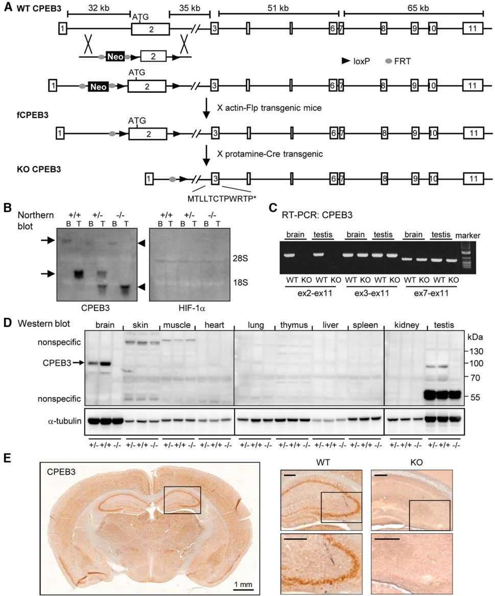
Figure 1 Generation and characterization of CPEB3-null mice. A , B , Schematic illustration of the targeting strategy. The cpeb3 gene consists of 11 exons (numbered boxes) and spans a region of 185kb.Thetargetingvectorcontainingthefliprecombinasetarget(FRT)-flankedphosphoglyceratekinase(PGK)promoter-drivenneomycin-resistancegene(Neo)andloxP-flankedexon2cassette wasinserted into the cpeb3 genebyhomologousrecombination.AftertheexcisionofthePGK-NeocassettebycrossingwithaC57BL/6mouselineexpressingFlprecombinasedrivenbyaubiquitous 𝛃 -actin promoter (actin-flp), the resulting female progenies carrying the floxed CPEB3 allele CPEB3 (fCPEB3) were mated with C57BL/6 males containing a Cre recombinase transgene under the control of protamine promoter (protamine-cre) to excise exon 2 in sperms to produce the KO allele. Male offspring carrying WT ( + / + ), heterozygous ( + / -) or KO ( -/ -) alleles from heterozygous matings were selected for ( B ) Northern blot analysis using a probe against exon 3- 6 of CPEB3 RNA or HIF-1 𝛂 RNA(loading control). The arrows denote two CPEB3 transcripts derived from alternative usage of the polyadenylation signals in the 3 ′ -UTR. The arrowheads denote the truncated CPEB3 transcripts without the exon 2 sequence. C , RT-PCR analysis using RNAs isolated from the brain and testes of WT and KO littermates confirmed the presence of exon 2-deleted CPEB3 transcript in the KO tissues. D , E , Western blotting ( D ) and immunohistochemistry ( E ) of coronal brain slices using the affinity-purified polyclonal CPEB3 antibody revealed no immunostained signal in the KO tissue. Scale bar, 0.25 mm unless specified.
Figure 1 Generation and characterization of CPEB3-null mice. A , B , Schematic illustration of the targeting strategy. The cpeb3 gene consists of 11 exons (numbered boxes) and spans a region of 185kb.Thetargetingvectorcontainingthefliprecombinasetarget(FRT)-flankedphosphoglyceratekinase(PGK)promoter-drivenneomycin-resistancegene(Neo)andloxP-flankedexon2cassette wasinserted into the cpeb3 genebyhomologousrecombination.AftertheexcisionofthePGK-NeocassettebycrossingwithaC57BL/6mouselineexpressingFlprecombinasedrivenbyaubiquitous 𝛃 -actin promoter (actin-flp), the resulting female progenies carrying the floxed CPEB3 allele CPEB3 (fCPEB3) were mated with C57BL/6 males containing a Cre recombinase transgene under the control of protamine promoter (protamine-cre) to excise exon 2 in sperms to produce the KO allele. Male offspring carrying WT ( + / + ), heterozygous ( + / -) or KO ( -/ -) alleles from heterozygous matings were selected for ( B ) Northern blot analysis using a probe against exon 3- 6 of CPEB3 RNA or HIF-1 𝛂 RNA(loading control). The arrows denote two CPEB3 transcripts derived from alternative usage of the polyadenylation signals in the 3 ′ -UTR. The arrowheads denote the truncated CPEB3 transcripts without the exon 2 sequence. C , RT-PCR analysis using RNAs isolated from the brain and testes of WT and KO littermates confirmed the presence of exon 2-deleted CPEB3 transcript in the KO tissues. D , E , Western blotting ( D ) and immunohistochemistry ( E ) of coronal brain slices using the affinity-purified polyclonal CPEB3 antibody revealed no immunostained signal in the KO tissue. Scale bar, 0.25 mm unless specified.CC-BY-NC-SA · From the PDF
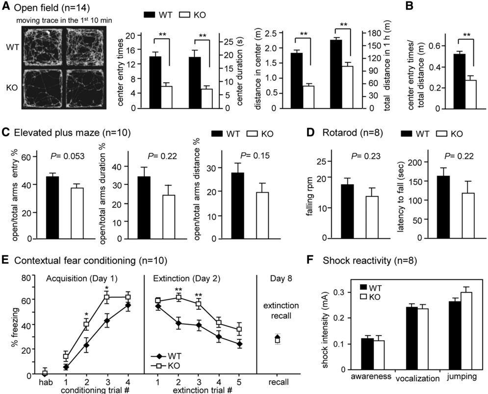
Figure2 Behavioral characterization. Anxiety level and exploratory behavior of WT and KO mice were assessed using open field and EPM tasks. A , Representative moving traces in the open arena are shown. The entry times, staying duration, and traveling distance in the center during the first 10 min and the total 1 h moving distance in the arena were presented as bar graphs. B , The center entry times were normalized against the moving distance in the first 10 min. C , The percentage of entry and staying duration into the open arms versus the total arms in the EPM was analyzed. D , Rotarod. The rotating speed that caused the mouse to fall and the latency in which the mouse could stay on the rotating rod were analyzed. E , Contextual fear conditioning. The freezing response during habituation (hab) and acquisition was analyzed for 2 min per trial. A foot shock was given at the end of habituation and the first three conditioning trials. During extinction, the mouse was placed in the chamber for 30 min without reinforcing the shock. The freezing level was analyzed every 6 min for five segments. Consolidated extinction memory was recalled a week later by monitoringthefreezingbehaviorfor6minintheoriginalchamber. F , Shock reactivity. The current intensities required to evoke various responses in the mice were recorded. The data were expressed as mean ± SEM; * p < 0.05 and ** p < 0.01, Student's t test.
Figure2 Behavioral characterization. Anxiety level and exploratory behavior of WT and KO mice were assessed using open field and EPM tasks. A , Representative moving traces in the open arena are shown. The entry times, staying duration, and traveling distance in the center during the first 10 min and the total 1 h moving distance in the arena were presented as bar graphs. B , The center entry times were normalized against the moving distance in the first 10 min. C , The percentage of entry and staying duration into the open arms versus the total arms in the EPM was analyzed. D , Rotarod. The rotating speed that caused the mouse to fall and the latency in which the mouse could stay on the rotating rod were analyzed. E , Contextual fear conditioning. The freezing response during habituation (hab) and acquisition was analyzed for 2 min per trial. A foot shock was given at the end of habituation and the first three conditioning trials. During extinction, the mouse was placed in the chamber for 30 min without reinforcing the shock. The freezing level was analyzed every 6 min for five segments. Consolidated extinction memory was recalled a week later by monitoringthefreezingbehaviorfor6minintheoriginalchamber. F , Shock reactivity. The current intensities required to evoke various responses in the mice were recorded. The data were expressed as mean ± SEM; * p < 0.05 and ** p < 0.01, Student's t test.CC-BY-NC-SA · From the PDF
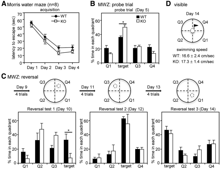
Figure 3 CPEB3 KO mice showed enhanced consolidated spatial memory in the MWZ test. A , The WT and KO mice normally learned in the hidden platform version of MWZ within four trials per day for 4 consecutive days. B , In the probe trial, the platform was removed, and the percentage of time that the mouse spent navigating in each quadrant was analyzed. Both of the groups of mice searched closer to the target quadrant where the platform had been previously placed. C , Reversal learning. On day 9, the mice were trained for four trials to locate the hidden platform in a new position. The reversal probe test was conducted 24 h later on day 10. The reversal learning and probe test were repeated two more times with an altered platform position. D , The motor ability and visual acuity of the mice were assessed by the swimming speed in the cued version of MWZ, in which the platform was indicated by a visible flag. All of the data were expressed as the mean ± SEM; * p < 0.05 (Student's t test).
Figure 3 CPEB3 KO mice showed enhanced consolidated spatial memory in the MWZ test. A , The WT and KO mice normally learned in the hidden platform version of MWZ within four trials per day for 4 consecutive days. B , In the probe trial, the platform was removed, and the percentage of time that the mouse spent navigating in each quadrant was analyzed. Both of the groups of mice searched closer to the target quadrant where the platform had been previously placed. C , Reversal learning. On day 9, the mice were trained for four trials to locate the hidden platform in a new position. The reversal probe test was conducted 24 h later on day 10. The reversal learning and probe test were repeated two more times with an altered platform position. D , The motor ability and visual acuity of the mice were assessed by the swimming speed in the cued version of MWZ, in which the platform was indicated by a visible flag. All of the data were expressed as the mean ± SEM; * p < 0.05 (Student's t test).CC-BY-NC-SA · From the PDF
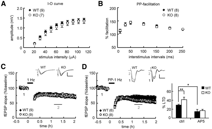
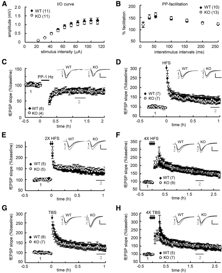
Figure 4 Genetic ablation of CPEB3-facilitated PP-LFS-induced LTD in the SC pathway of juvenile (3- to 4-week-old) hippocampal slices. Basal synaptic transmission. A , B , Input- output (I-O) curve and ( B ) paired-pulse (PP) facilitation did not show a significant difference between the WT and KO mice ( p = 0.66 and 0.97, respectively, two-way ANOVA). C , D , Normal LFS-induced LTD (WT: 76.82 ± 2.84%; KO: 75.37 ± 3.94%, p = 0.96, Student's t test, at 50 -70 min after stimulation) and ( D ) increased PP-LFS-elicited LTD in the KO hippocampal slices (WT: 70.34 ± 3.70%; KO: 59.81 ± 5.48%, p < 0.01, Student's t test, at 50 -70 min after stimulation). PP-LFS-evoked LTD was blocked by the NMDAR antagonist, 50 𝛍 M AP5. Numbers in parentheses represent the number of slices isolated from 4 to 5 male mice that were used for recordings. All of the data were expressed as the mean ± SEM. Statistics from C to D were analyzed using Student's t test. Traces represent baseline (black line, 1) and 50 -70 min after stimulation (gray line, 2). Calibration, 0.5 mV, 20 ms.
Figure 4 Genetic ablation of CPEB3-facilitated PP-LFS-induced LTD in the SC pathway of juvenile (3- to 4-week-old) hippocampal slices. Basal synaptic transmission. A , B , Input- output (I-O) curve and ( B ) paired-pulse (PP) facilitation did not show a significant difference between the WT and KO mice ( p = 0.66 and 0.97, respectively, two-way ANOVA). C , D , Normal LFS-induced LTD (WT: 76.82 ± 2.84%; KO: 75.37 ± 3.94%, p = 0.96, Student's t test, at 50 -70 min after stimulation) and ( D ) increased PP-LFS-elicited LTD in the KO hippocampal slices (WT: 70.34 ± 3.70%; KO: 59.81 ± 5.48%, p < 0.01, Student's t test, at 50 -70 min after stimulation). PP-LFS-evoked LTD was blocked by the NMDAR antagonist, 50 𝛍 M AP5. Numbers in parentheses represent the number of slices isolated from 4 to 5 male mice that were used for recordings. All of the data were expressed as the mean ± SEM. Statistics from C to D were analyzed using Student's t test. Traces represent baseline (black line, 1) and 50 -70 min after stimulation (gray line, 2). Calibration, 0.5 mV, 20 ms.CC-BY-NC-SA · From the PDF
Figure 5 Normal LTP and LTD in the SC pathway of adult (9- to 12-week-old) KO hippocampal slices. Basal synaptic transmission. A , B , Input- output (I-O) curve and ( B ) paired-pulse (PP) facilitation were normal in the KO slices ( p = 0.09 and 0.72, respectively, two-way ANOVA). C-H , LTD induced by PP-LFS showed no significant difference (WT: 83.61 ± 3.71%,KO:81.64 ± 4.74%, p = 0.12 at 50 -70 min after stimulation) between the WT and KO groups. No significant difference was observed between the WT and KO slices in LTP evoked by ( D ) one train (1X) of HFS (WT: 136.67 ± 12.78%, KO: 128.77 ± 10.8%, p = 0.10 at 50 - 60 min), ( E ) two trains (2X) of HFS (WT: 128.6 ± 5.05%, KO: 126.39 ± 4.26%, p = 0.43 at 50 - 60 min), ( F ) four trains (4X) of HFS (WT: 165.84 ± 9.01%, KO: 162.8 ± 16.08%, p = 0.43 at 100 -120 min after stimulation), ( G ) 1X TBS (WT: 126.07 ± 7.35%; KO, 126.32 ± 14.42%, p = 0.96 at 50 - 60 min), and ( H ) 4X TBS (WT: 145.42 ± 10.02%, KO: 151.42 ± 13.13%, p = 0.08 at 100 -120 min after stimulation). Numbers in parentheses represent the number of recorded slices isolated from 4 to 5 male mice. All of the data were expressed as the mean ± SEM. Statistics from C to H were analyzed using Student's t test. Traces represent baseline (black line, 1) and the indicated time after stimulation (gray line, 2). Calibration: 0.5 mV, 20 ms.
Figure 5 Normal LTP and LTD in the SC pathway of adult (9- to 12-week-old) KO hippocampal slices. Basal synaptic transmission. A , B , Input- output (I-O) curve and ( B ) paired-pulse (PP) facilitation were normal in the KO slices ( p = 0.09 and 0.72, respectively, two-way ANOVA). C-H , LTD induced by PP-LFS showed no significant difference (WT: 83.61 ± 3.71%,KO:81.64 ± 4.74%, p = 0.12 at 50 -70 min after stimulation) between the WT and KO groups. No significant difference was observed between the WT and KO slices in LTP evoked by ( D ) one train (1X) of HFS (WT: 136.67 ± 12.78%, KO: 128.77 ± 10.8%, p = 0.10 at 50 - 60 min), ( E ) two trains (2X) of HFS (WT: 128.6 ± 5.05%, KO: 126.39 ± 4.26%, p = 0.43 at 50 - 60 min), ( F ) four trains (4X) of HFS (WT: 165.84 ± 9.01%, KO: 162.8 ± 16.08%, p = 0.43 at 100 -120 min after stimulation), ( G ) 1X TBS (WT: 126.07 ± 7.35%; KO, 126.32 ± 14.42%, p = 0.96 at 50 - 60 min), and ( H ) 4X TBS (WT: 145.42 ± 10.02%, KO: 151.42 ± 13.13%, p = 0.08 at 100 -120 min after stimulation). Numbers in parentheses represent the number of recorded slices isolated from 4 to 5 male mice. All of the data were expressed as the mean ± SEM. Statistics from C to H were analyzed using Student's t test. Traces represent baseline (black line, 1) and the indicated time after stimulation (gray line, 2). Calibration: 0.5 mV, 20 ms.CC-BY-NC-SA · From the PDF
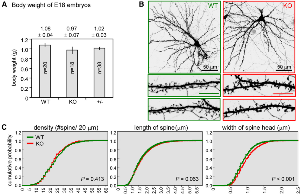
Figure 6 Enlarged dendritic spine heads in cultured CPEB3 KO neurons. A , The body weight of E18 embryos isolated from heterozygous CPEB3 matings. B , The WT and KO neurons were transfected with the GFP plasmid on 14 DIV and fixed on 18 DIV for GFP immunostaining. Representative images of the whole neurons and dendritic spine area are shown. Scale bars: 10 𝛍 munless denoted. C , Quantification analysis of dendritic spine morphology. Approximately 30 pyramidal neurons in each group (WT, green; KO, red) were collected from three independent cultures and analyzed using the MetaMorph software. The cumulative probability curves of density, length, and width of dendritic spines in each group ( ∼ 3000 spines) were plotted and analyzed for the statistical difference between groups using Student's t test.
Figure 6 Enlarged dendritic spine heads in cultured CPEB3 KO neurons. A , The body weight of E18 embryos isolated from heterozygous CPEB3 matings. B , The WT and KO neurons were transfected with the GFP plasmid on 14 DIV and fixed on 18 DIV for GFP immunostaining. Representative images of the whole neurons and dendritic spine area are shown. Scale bars: 10 𝛍 munless denoted. C , Quantification analysis of dendritic spine morphology. Approximately 30 pyramidal neurons in each group (WT, green; KO, red) were collected from three independent cultures and analyzed using the MetaMorph software. The cumulative probability curves of density, length, and width of dendritic spines in each group ( ∼ 3000 spines) were plotted and analyzed for the statistical difference between groups using Student's t test.CC-BY-NC-SA · From the PDF
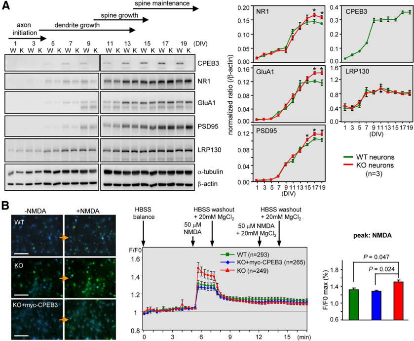
Figure 7 Elevated NMDAR-induced calcium influx in CPEB3 KO neurons. A , Developmental expression of various synaptic proteins in WT and KO neurons. CPEB3 WT and KO neurons obtained at different DIV were harvested for Western blotting. The protein levels of NR1, GluA1, PSD95, CPEB3, and leucine-rich protein (LRP)130 were normalized against 𝛃 -actin and plotted against DIV. The results from three independent cultures were quantified and displayed as the mean ± SEM. B , WT and KO neurons at 16 -17 DIV were subjected to calcium imaging. Several of the KO neurons were infected with the lentivirus expressing myc-CPEB3 on 12-13 DIV for the rescue experiment (KO + myc-CPEB3). The Fura2-AM-loaded neurons were treated as indicated. Representative calcium imagesbeforeorafterNMDAtreatmentareshownontheleft.Thechangein[Ca 2 + ] influx was monitored by the fluorescence ratio at 340/380 nm ( F / F 0 ) and plotted against time. The data obtained from three independent cultures with ∼ 250-300 neurons in each group were expressed as the mean ± SEM. The statistical difference in [Ca 2 + ] influx induced by NMDA between groups was analyzed using Student's t test. Scale bars: 100 mm.
Figure 7 Elevated NMDAR-induced calcium influx in CPEB3 KO neurons. A , Developmental expression of various synaptic proteins in WT and KO neurons. CPEB3 WT and KO neurons obtained at different DIV were harvested for Western blotting. The protein levels of NR1, GluA1, PSD95, CPEB3, and leucine-rich protein (LRP)130 were normalized against 𝛃 -actin and plotted against DIV. The results from three independent cultures were quantified and displayed as the mean ± SEM. B , WT and KO neurons at 16 -17 DIV were subjected to calcium imaging. Several of the KO neurons were infected with the lentivirus expressing myc-CPEB3 on 12-13 DIV for the rescue experiment (KO + myc-CPEB3). The Fura2-AM-loaded neurons were treated as indicated. Representative calcium imagesbeforeorafterNMDAtreatmentareshownontheleft.Thechangein[Ca 2 + ] influx was monitored by the fluorescence ratio at 340/380 nm ( F / F 0 ) and plotted against time. The data obtained from three independent cultures with ∼ 250-300 neurons in each group were expressed as the mean ± SEM. The statistical difference in [Ca 2 + ] influx induced by NMDA between groups was analyzed using Student's t test. Scale bars: 100 mm.CC-BY-NC-SA · From the PDF
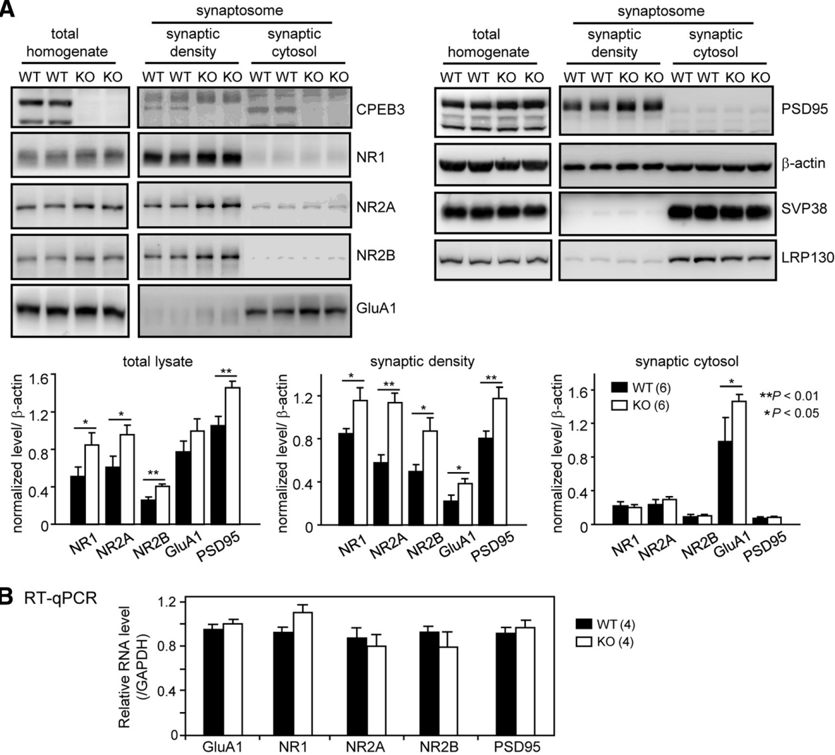
Figure 8 Deletion of CPEB3 resulted in increased total and synaptic protein levels of PSD95 and NMDAR. A , The total homogenates and synaptosomal fractions (synaptic density and synaptic cytosol) were isolated from 3-month-old WT and KO mouse brains ( n = 6 in each group) and used for Western blotting analysis. The protein levels of NR1, NR2A, NR2B, GluA1, and PSD95 were normalized against 𝛃 -actin. Synaptophysin (SVP38) and leucine-rich protein (LRP)130 served as nontarget controls. The quantified data were expressed as the mean ± SEM in the bar graphs. Significant differences between the WT and KO groups are denoted as * p < 0.05 or ** p < 0.01, Student's t test. B , The relative expression levels of various RNAs in WT and KO brains were similar. The cortices and hippocampi isolated from the 3-month-old WT and KO mice (4 animals in each genotype) were extracted for RNAs. The total RNAs were then reverse-transcribed and processed for RT-qPCR.
Figure 8 Deletion of CPEB3 resulted in increased total and synaptic protein levels of PSD95 and NMDAR. A , The total homogenates and synaptosomal fractions (synaptic density and synaptic cytosol) were isolated from 3-month-old WT and KO mouse brains ( n = 6 in each group) and used for Western blotting analysis. The protein levels of NR1, NR2A, NR2B, GluA1, and PSD95 were normalized against 𝛃 -actin. Synaptophysin (SVP38) and leucine-rich protein (LRP)130 served as nontarget controls. The quantified data were expressed as the mean ± SEM in the bar graphs. Significant differences between the WT and KO groups are denoted as * p < 0.05 or ** p < 0.01, Student's t test. B , The relative expression levels of various RNAs in WT and KO brains were similar. The cortices and hippocampi isolated from the 3-month-old WT and KO mice (4 animals in each genotype) were extracted for RNAs. The total RNAs were then reverse-transcribed and processed for RT-qPCR.CC-BY-NC-SA · From the PDF
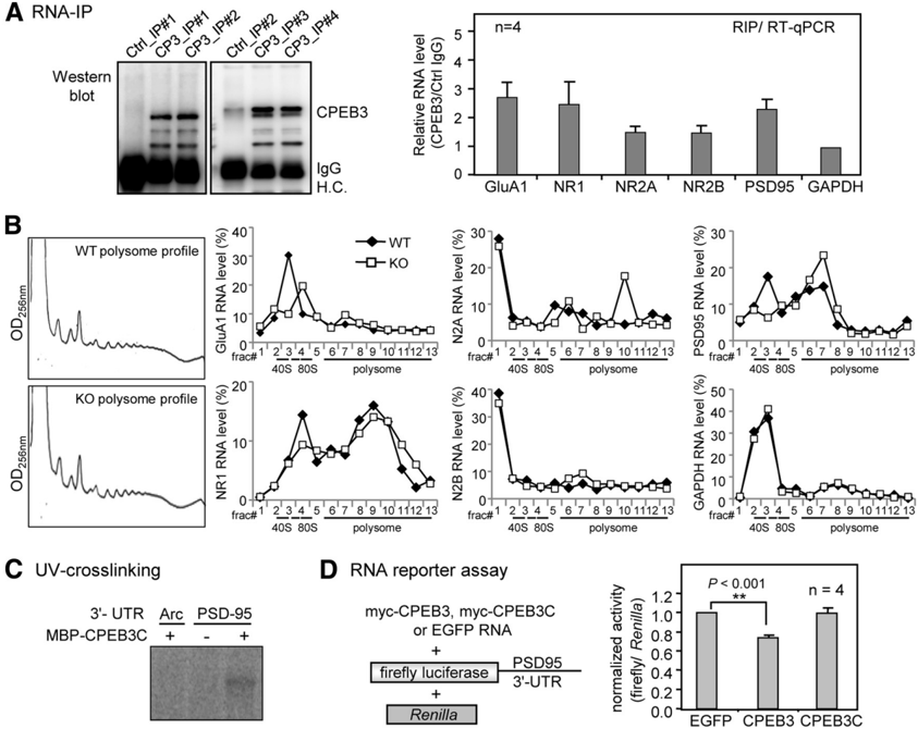
Figure 9 CPEB3 binds to the PSD95 RNA and represses its translation. A , RIP. The mouse brain lysate was immunoprecipitated with control or CPEB3 IgG. The precipitated RNAs were reverse transcribed followed by RT-qPCR to determine the relative RNA levels between control and CPEB3 immunoprecipitates. B , The polysomal profiles of WT and CPEB3 KO neurons are shownontheleft. The polysomal distribution of GluA1, NR1, NR2A, NR2B, PSD95, and the control GAPDH RNAs in WT and KO neurons was determined by RT-qPCR using RNAs isolated from each fraction and expressed as the percentage of total RNAs summed from all fractions. C , CPEB3 bound to PSD95 3 ′ -UTR. The recombinant maltose binding protein (MBP) fused to the C terminus of CPEB3 RBD (MBP-CPEB3C) was UV-cross-linked with 32 P-labeled 3 ′ -UTRs of Arc and PSD95 RNAs, RNase treated, and then analyzed by SDS-PAGE. D , RNA reporter assay. The cultured neurons were transfected with RNAs encoding myc-CPEB3, myc-CPEB3C, or EGFP, in combination with the firefly luciferase appended to the PSD95 3 ′ -UTR and Renilla luciferase. The normalized luciferase activity (firefly/ Renilla ) was calculated. Four independent results were analyzed and expressed as the mean ± SEM. Asterisks mark the significant difference (Student's t test).
Figure 9 CPEB3 binds to the PSD95 RNA and represses its translation. A , RIP. The mouse brain lysate was immunoprecipitated with control or CPEB3 IgG. The precipitated RNAs were reverse transcribed followed by RT-qPCR to determine the relative RNA levels between control and CPEB3 immunoprecipitates. B , The polysomal profiles of WT and CPEB3 KO neurons are shownontheleft. The polysomal distribution of GluA1, NR1, NR2A, NR2B, PSD95, and the control GAPDH RNAs in WT and KO neurons was determined by RT-qPCR using RNAs isolated from each fraction and expressed as the percentage of total RNAs summed from all fractions. C , CPEB3 bound to PSD95 3 ′ -UTR. The recombinant maltose binding protein (MBP) fused to the C terminus of CPEB3 RBD (MBP-CPEB3C) was UV-cross-linked with 32 P-labeled 3 ′ -UTRs of Arc and PSD95 RNAs, RNase treated, and then analyzed by SDS-PAGE. D , RNA reporter assay. The cultured neurons were transfected with RNAs encoding myc-CPEB3, myc-CPEB3C, or EGFP, in combination with the firefly luciferase appended to the PSD95 3 ′ -UTR and Renilla luciferase. The normalized luciferase activity (firefly/ Renilla ) was calculated. Four independent results were analyzed and expressed as the mean ± SEM. Asterisks mark the significant difference (Student's t test).CC-BY-NC-SA · From the PDF
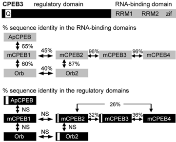
Figure 10 Sequence comparison of CPEB family proteins. All CPEB proteins have an N-terminal region (black box) and a C-terminal region (gray box) containing two RNA recognition motifs (RRM) and two zinc fingers (Zif). Among the RBDs, there is considerable identity ranging from 40 to 96%. Mouse CPEBs 2- 4 are nearly identical in the RBDs. Mouse CPEB1 (mCPEB1) is closer to Aplysia CPEB (ApCPEB) and Drosophila CPEB (Orb) than it is to mCPEB2; in addition, Drosophila Orb2 is more similar to mCPEBs 2- 4 than it is to Orb. Except for CPEBs 2-4, there is little identity (NS, not significant) among the N-terminal regions of the CPEB proteins. Moreover, the Q-rich sequence of variable length (white box) is present in some CPEB proteins.
Figure 10 Sequence comparison of CPEB family proteins. All CPEB proteins have an N-terminal region (black box) and a C-terminal region (gray box) containing two RNA recognition motifs (RRM) and two zinc fingers (Zif). Among the RBDs, there is considerable identity ranging from 40 to 96%. Mouse CPEBs 2- 4 are nearly identical in the RBDs. Mouse CPEB1 (mCPEB1) is closer to Aplysia CPEB (ApCPEB) and Drosophila CPEB (Orb) than it is to mCPEB2; in addition, Drosophila Orb2 is more similar to mCPEBs 2- 4 than it is to Orb. Except for CPEBs 2-4, there is little identity (NS, not significant) among the N-terminal regions of the CPEB proteins. Moreover, the Q-rich sequence of variable length (white box) is present in some CPEB proteins.CC-BY-NC-SA · From the PDF

Verification

Claims the paper makes

The claims and statistics of this paper haven’t been checked yet.

Run the check — free, no account needed. It reads the open-access text and checks the sources it cites.

A few free checks a day · papers already verified open instantly · results are public

Claims other papers make about this one

Each is checked against this paper’s own text: does it say what the citing paper says it does?

Self-check

Percentages, sums and reported differences re-computed from the paper’s own numbers.

The self-check needs the paper’s full text.

Citations

Cites

Research works printed but not linked: 48

  1. 10.1101/lm.72704 · DOI
  2. 10.1073/pnas.0608492103 · DOI · Paper page
  3. 10.1101/lm.73706 · DOI
  4. 10.1523/JNEUROSCI.0640-10.2010 · DOI · Paper page
  5. 10.1083/jcb.200712094 · DOI
  6. 10.1093/nar/gks598 · DOI
  7. 10.1038/emboj.2011.448 · DOI
  8. 10.1096/fj.08-106450 · DOI · Paper page
  9. 10.1016/j.neuron.2008.10.055 · DOI · Paper page
  10. Cui, 2013, Increased NR2A:NR2B ratio compresses long-term depression range and constrains long-term memory, Sci Rep, 3, 1036, 10.1038/srep01036 · DOI
  11. 10.1016/S0959-4388(00)00215-4 · DOI · Paper page
  12. 10.1101/cshperspect.a012344 · DOI
  13. 10.1523/JNEUROSCI.3722-04.2004 · DOI
  14. 10.1002/ana.23860 · DOI · Paper page
  15. 10.1073/pnas.0609307104 · DOI · Paper page
  16. 10.1101/lm.026849.112 · DOI
  17. 10.1073/pnas.1121288109 · DOI · Paper page
  18. 10.1038/sj.emboj.7601322 · DOI
  19. 10.1101/lm.2111311 · DOI
  20. 10.1016/S0896-6273(00)80225-1 · DOI
  21. 10.1038/nn1996 · DOI
  22. 10.1046/j.1460-9568.2000.00903.x · DOI
  23. 10.1016/j.neuron.2012.08.028 · DOI · Paper page
  24. 10.1016/j.cell.2012.01.004 · DOI
  25. 10.1073/pnas.1004433107 · DOI
  26. 10.1007/s00335-005-2430-8 · DOI
  27. 10.1038/24790 · DOI · Paper page
  28. 10.1093/nar/gkq635 · DOI · Paper page
  29. 10.1016/0165-0270(84)90007-4 · DOI · Paper page
  30. 10.1016/j.molcel.2011.05.006 · DOI · Paper page
  31. 10.1016/S0896-6273(00)80336-0 · DOI
  32. 10.1016/S0896-6273(01)00443-3 · DOI · Paper page
  33. 10.1016/j.cell.2011.09.056 · DOI
  34. 10.1093/nar/gkq634 · DOI · Paper page
  35. 10.1523/JNEUROSCI.3264-12.2013 · DOI
  36. 10.1101/gad.1735809 · DOI
  37. 10.1128/MCB.05896-11 · DOI · Paper page
  38. 10.1038/ni.2222 · DOI
  39. 10.1126/science.290.5494.1170 · DOI
  40. 10.1016/j.cell.2010.01.008 · DOI
  41. 10.1523/JNEUROSCI.0289-13.2013 · DOI
  42. 10.1038/43432 · DOI
  43. 10.1073/pnas.1133424100 · DOI
  44. 10.1016/j.molcel.2012.05.016 · DOI
  45. 10.1016/j.neuron.2008.09.039 · DOI · Paper page
  46. 10.1128/MCB.00296-12 · DOI
  47. 10.1038/sj.mp.4002056 · DOI
  48. 10.1038/nn1893 · DOI

Cited by

Sorted by citations

See all 80

Metadata

Authors
Chao, H.-W., Tsai, L.-Y., Lu, Y.-L., Lin, P.-Y., Huang, W.-H., Chou, H.-J., Lu, W.-H., Lin, H.-C., Lee, P.-T., & Huang, Y.-S.
Venue
The Journal of Neuroscience
Published
2013-10-23
Volume
33
Issue
43
Pages
17008-17022
ISSN
  • 0270-6474
  • 1529-2401
Cited by
80
References
5
Language
en

About these checks

Registry, retraction, Cites and statistics results are computed, not judged, with one exception: a printed reference that no index lists is read once by a model before it counts as a citation error. Claims are extracted by a model and kept only when their quote appears verbatim in the text; each cited source is then checked, with a model’s help, for whether it says what the claim relies on. Those verdicts can be wrong — read the claim’s quote and the cited source before relying on one.

Statistics and arithmetic are recomputed, not judged.

Confidence bands are shown only for fields calibrated on our annotated test set. None is calibrated yet, so no band is shown.

What counts as a claim ›How we measure confidence ›How we check this ›

Are you an author, or do you see a problem with this page? Request a review